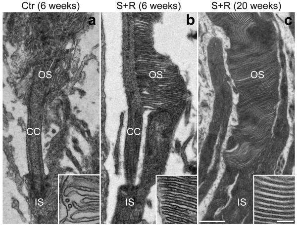
Figure 6.
Photoreceptor morphology rescue following combined suppression and replacement therapy.
The right eyes of P5 P347S mice were subretinally injected with a mixture of 6.0×10⁸ vp AAV-S and 1.8×10¹⁰ vp AAV-R (b and c; n=3 and n=1 respectively) while the left eyes were injected with 6.0×10⁸ vp AAV-C (a; n=4). Note that AAV-S and AAV-C co-express EGFP. Six (a and b) and twenty (c) weeks post-injection, eyes were fixed, whole mounted and transduced areas identified by EGFP fluorescence and excised. The excised retinal samples were processed for transmission electron microscopy (TEM). Combined suppression and replacement therapy resulted in the preservation of rod photoreceptor outer segments (OS) with correctly formed membrane disks (b and c). In contrast in control retinas the rod photoreceptor inner segments (IS) attached to truncated OS with disorganized disks. CC: connecting cilium. Scale bars: 500 nm and 100 nm (inserts).

