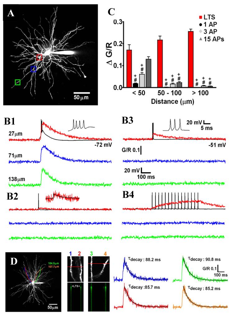
Figure 7. Intrinsic Ca2+ signalling in TC neuron dendrites.
(A) Maximum intensity projection of a dorsal lateral geniculate TC neuron illustrating the dendritic sites where linescans shown in B were performed. Proximal, intermediate and distal dendritic locations are colour coded red, blue and green respectively. (B) Dendritic Δ[Ca2+] evoked by (1) LTCPs, (2) single backpropagating action potentials, (3) Three backpropagating action potentials at 200 Hz (mimicking the high frequency burst elicited by an LTCP, and (4) 15 backpropagating action potentials at 30 Hz are shown overlaid onto somatically recorded voltage voltage traces (in black). A single action potential-elicited Ca2+ transient is shown enlarged for clarity in 2, with a mono-exponential fit shown in black. (C) Summary of Δ[Ca2+] amplitudes grouped by dendritic location. *: P < 0.05 (vs proximal Δ[Ca2+]LTCP), #: P < 0.001 (vs proximal Δ[Ca2+]LTCP), +: P <0.001 (vs Δ[Ca2+]LTCP for each dendritic location) (n = 6-11 cells). (D) Somatically evoked LTCPs produce synchronous Ca2+ transients throughout the entire dendritic tree. Two different pairs of distal dendrites (100-110 μm from the soma) lying in the same focal plane but originating from different stem dendrites (coloured tracings constructed from 3D Z series) were imaged separately during evoked LTCP activity. In both pairs of dendrites, the LTCP-eleicited Δ[Ca2+] occurred simultaneously and in all four dendrites (amplitude and τ of the decay of the evoked Ca2+ transients are identical). Reproduced with permission from Errington et al. (2010).

