Fig. 1.

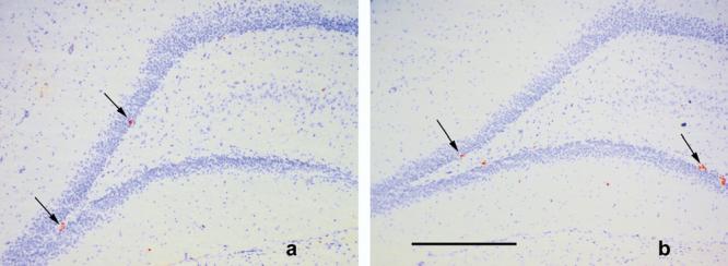
Photomicrographs of coronal sections (20 μm) through the dentate gyrus of (a) LH (b) SD strains of rats. Ki67-labeled cells are indicated by the arrows. Cresyl Violet background stain. Scale bar=500 μm.

Photomicrographs of coronal sections (20 μm) through the dentate gyrus of (a) LH (b) SD strains of rats. Ki67-labeled cells are indicated by the arrows. Cresyl Violet background stain. Scale bar=500 μm.