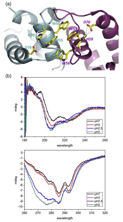
Fig. 4.
The internal salt bridges within HdeB. (a) In each monomer, a salt bridge between D76 and H59 is present. The residues are colored as in Fig. 2. (b) CD spectra of HdeB WT at different pH values: (top) far-UV CD and (bottom) near-UV CD. The CD spectrum of HdeB WT shows pH-dependent changes indicating changes in the secondary and tertiary structures, respectively.

