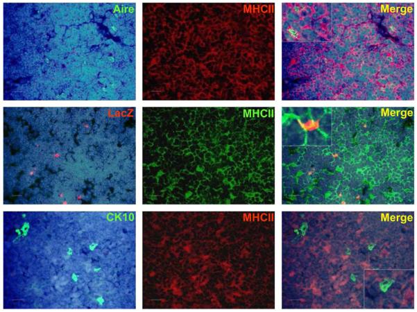
FIGURE A9. Co-staining of Aire, LacZ, and CK10 with MHCII in Aire+/− (LacZ) and WT (Aire, CK10) mouse thymus.
Aire+ cells co-stained strongly with MHCII, whereas there was a weak co-staining or no co-staining for LacZ+ cells and CK10+ HCs with MHCII. DAPI was used for nuclear staining. Shown are representatives of at least two experiments. Bars correspond to 40 μm.

