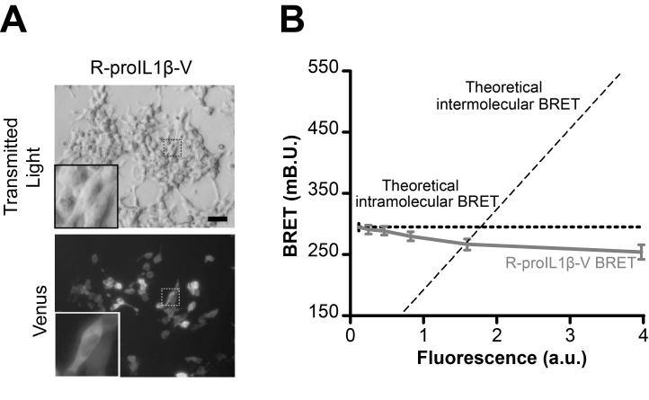
Fig. 3. In living HEK293 cells, BRET from R-proIL1β-V sensor is due to intramolecular energy transfer.
(A) Representative picture of HEK293 cells transfected with R-proIL1β-V. The sensor was detected after excitation of the Venus protein. Note the widespread localization of the protein in the higher magnification image as already described for native pro-IL-1β (8). Scale bar = 30 μM. (B) R-proIL1β-V BRET signal is due to intramolecular energy transfer. BRET signals were measured on HEK293 cells expressing increasing amounts of the R-proIL1β-V sensor and plotted as a function of the amount of protein. As expected for intramolecular energy transfer, BRET does not increase in cells expressing higher amounts of sensor. The curves shown represent the mean ± s.e.m. of n ≥ 6 wells.

