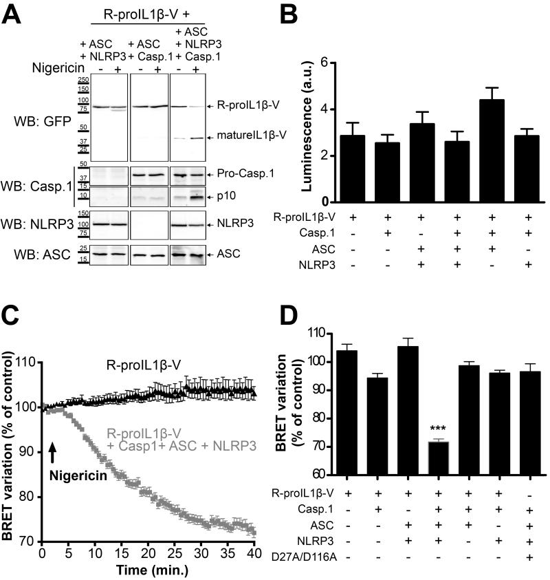
Fig. 4. R-proIL1β-V sensor allows real-time monitoring of IL-1β processing by BRET in living HEK293 cells.
(A) R-proIL1β-V is efficiently cleaved by active caspase-1 in a mature form. HEK293 cells overexpressing NLRP3 inflammasome components and R-proIL1β-V were stimulated with the K+ ionophore nigericin (5 μM for 40 min). Cleavage of the R-proIL1β-V sensor to a mature form (mIL1β-V) was detected by Western blot using a GFP antibody. Note that IL-1β processing is only observed in cells overexpressing all the NLRP3 inflammasome. The blot shown is representative of three independent experiments. (B) R-proIL1β-V displays similar expression level after coexpression with NRLP3 inflammasome components. HEK293 cells were transfected with the BRET sensor and different combination of ASC, NLRP3 and caspase-1. Luminescence signals were measured after addition of coelenterazine h. Results are mean ± s.e.m. of n ≥ 3 experiments and n ≥ 33 wells. (C) NLRP3 inflammasome activation results in a progressive decrease in BRET signal of the sensor. BRET signals were measured after stimulation with nigericin (5 μM, arrow) in HEK293 cells overexpressing R-proIL1β-V alone or in combination with NLRP3, ASC and caspase-1. Results are mean ± s.e.m. of n ≥ 3 experiments and n = 3 wells per experiment. (D) Histogram showing the BRET variation measured 30 min after nigericin stimulation in cells expressing R-proIL1β-V or R-proIL1β-D27A/D116A-V (D27A/D116A). Experiments were performed as described above with combination of transfection vectors for different inflammasome components. Note that decrease of the BRET signal is only observed in experimental conditions allowing activation of caspase-1 and IL-1β cleavage. Results are mean ± s.e.m. of n ≥ 3 experiments and n = 3 wells per experiment. *** P < 0.001 compared to the others conditions.

