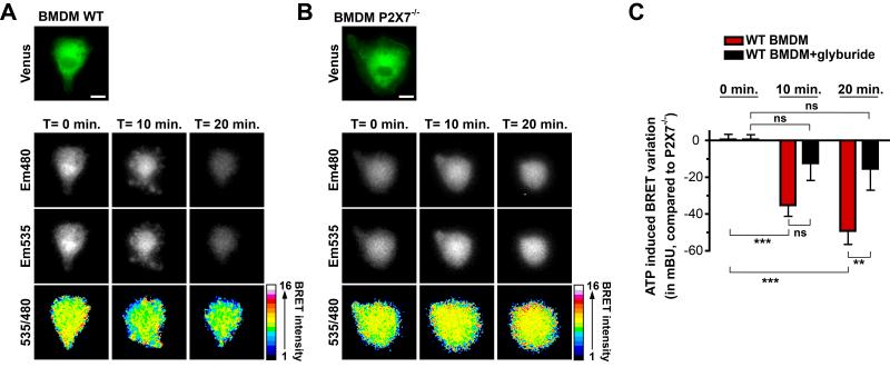
Fig. 7. Real-time monitoring of IL-1β processing in single primary macrophages.
(A and B) BRET imaging of R-proIL1β-V transfected in primary BMDM derived from wild type (WT; A) or P2X7 deficient mice (P2X7−/−; B). 24 hours after transfection with R-IL1β-V, cells were primed for 4 h with LPS (1 μg/ml). Transfected cells were identified after Venus excitation and subsequently coelenterazine h (20 μM) was added. Images were acquired at 480 and 535 nm under basal condition or after ATP stimulation (2 mM) every 1 min for 20 min. A 480 / 535 ratio picture was obtained and presented in pseudo-colors. Scale bar = 10 μM. Note that for the WT BMDM, membrane blebbing observed after ATP stimulation as a hallmark of P2X7 activation. (C) BRET variation measured on single WT or P2X7−/−-deficient macrophages as described above after incubated or not with glyburide (100 μM) and further stimulation with ATP. Results represent the BRET variation in WT macrophages compared to P2X7−/−-deficient macrophages. Results are mean ± s.e.m. of n ≥ 3 experiments and n ≥ 7 cells for each experimental condition. ** P < 0.01, *** P < 0.001. ns = non-significant.

