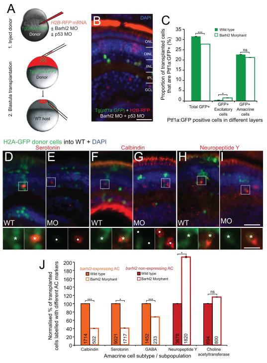
Figure 6.
Barhl2 knockdown causes a subtype fate switch towards alternate inhibitory subtypes. (A) The cell-autonomous effect of Barhl2 loss was assessed with transplantations: Donor Tg(ptf1a:GFP) embryos were injected with H2B-RFP mRNA and p53 MO (to aide cell survival (Robu et al., 2007)), with (knockdown) or without (control) Barhl2 MO. Donor cells were subsequently transplanted into unlabelled WT. (B) Some transplanted H2B-RFP cells express Ptf1a:GFP in both conditions. (C) Quantification reveals a small reduction in the proportion of Ptf1a:GFP labelled donor cells (32% WT to 27.8% morphant, p – value < 0.001) and a few mislocalised Ptf1a:GFP cells in excitatory layers (0.74% WT to 1.49% morphants, p – value = 0.012). Overall, the vast majority of Ptf1a:GFP remain as inhibitory ACs (22.96% WT to 21.2% morphants, p – value = 0.13). (D – I) Immunohistochemically labelled amacrine subtypes (red) in chimeric retinas arise from transplanted H2A-GFP labelled donor (e.g. asterisks for serotonin - D, calbindin - F, neuropeptide Y-H, I) and from unlabelled host cells (circles). (J) Quantification of the proportion of labelled transplanted cells shows varying degrees of loss in subtypes that usually express barhl2 (orange) and increases in AC subtypes that usually do not (red). ONL: outer nuclear layer; OINL: outer half of the inner nuclear layer; IINL: inner half of the inner nuclear layer; IPL: inner plexiform layer; GCL: ganglion cell layer; MO: morphant / morpholino; ns: not significant; *: p – value < 0.05; ***: p – value < 0.001; error bars indicate standard error of the mean. Scale bar B = 20 μm, scale bar I (for D – I) = 25 μm, scale bar I inset (for D – I insets) = 10 μm.

