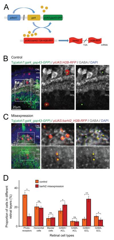
Figure 8.
Misexpression of barhl2 in atoh7-expressing progenitors drives the fate of amacrine subtypes that usually express barhl2. (A) Schematic showing misexpression design: The atoh7 promoter drives expression of Gal4 transcription factor. Gal4 activates the upstream activation sequence (pUAS) promoter to drive expression of gap43-GFP reporter by itself (control) or with Barhl2 and H2B-RFP reporter generated through in frame fusion with T2A peptide. After translation of the barhl2-T2A-H2B-RFP mRNA the T2A sequence will be cleaved and generate separate Barhl2 and H2B-RFP proteins. (B, C) Micrographs of 120 hpf Tg(atoh7:gal4/pUAS:gap43-GFP) retinas with pUAS driving H2B-RFP (control) or barhl2 and H2B-RFP (misexpression), and subsequently labelled for GABA (white). Boxes indicate higher power insets. Cell type and GABA colabeling of H2B-RFP-expressing cells was analysed. Examples of GABA-positive cells (yellow asterisks) and GABA-negative cells (green dots). (D) Quantification of cell fates in control (orange) or barhl2 misexpression (red). There is an increase in GABAergic cells in the INL (15.93% ± 4.86 SEM control to 22.2% ± 4.64 SEM misexpression, p – value = 0.0044) and particularly GCL (7.5% ± 4 SEM control to 28.32% ± 6.05 SEM misexpression, p – value < 0.0001 in GCL) with a concurrent loss in mainly photoreceptors (32.84% ± 5.76 SEM control to 10.12% ± 5.4 SEM misexpression, p – value = 0.01058) and presumed ganglion cells. ONL: outer nuclear layer; INL: inner nuclear layer; IPL: inner plexiform layer; GCL: ganglion cell layer; ns: not significant. Error bars indicate standard error of the mean. * p – value < 0.05, ** p – value < 0.001. Scale bar in B (for B, C) = 20 μm.

