

Abstract
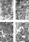
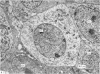
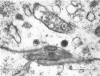
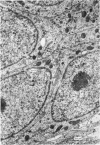
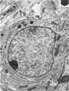
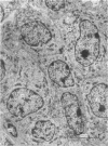
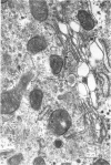
Definitive chief cells, characterized by the presence of specific secretory granules and typical membrane configurations, were present in the developing parathyroid glands of sheep embryos from 26 days of gestation (term = 150 days). During earlier stages of gestation developing chief cells contained lysosomal systems which appeared to be concerned with the autophagy of retained secretion product. The EM evidence suggests that physiologic release of the hormone does not occur until at least 30 days of gestation, which correlates well with the experimentally established time of onset of functional capacity in the parathyroid gland in the sheep. 'Bark cells' were found in the parathyroid primordia of embryos between 20 and 27 days of gestation. The significance of these "dark cells" is discussed with respect to glutaraldehyde fixation and to their possible relationship to chief cells.
Full text
PDF


Images in this article


Selected References
These references are in PubMed. This may not be the complete list of references from this article.
- Benedeczky I., Smith A. D. Ultrastructural studies on the adrenal medulla of golden hamster: origin and fate of secretory granules. Z Zellforsch Mikrosk Anat. 1972;124(3):367–386. doi: 10.1007/BF00355037. [DOI] [PubMed] [Google Scholar]
- Bloodworth J. M., Jr, Wers K. L. The ultrastructure of the normal dog adrenal. J Anat. 1968 Mar;102(Pt 3):457–476. [PMC free article] [PubMed] [Google Scholar]
- Carmichael S. W., Blair B. C. Normal ultrastructure of the day old dog adrenal medulla. J Anat. 1973 May;115(Pt 1):113–118. [PMC free article] [PubMed] [Google Scholar]
- Coupland R. E., Weakley B. S. Developing chromaffin tissue in the rabbit: an electron microscopic study. J Anat. 1968 Mar;102(Pt 3):425–455. [PMC free article] [PubMed] [Google Scholar]
- Coupland R. E., Weakley B. S. Electron microscopic observation on the adrenal medulla and extra-adrenal chromaffin tissue of the postnatal rabbit. J Anat. 1970 Mar;106(Pt 2):213–231. [PMC free article] [PubMed] [Google Scholar]
- Jordan R. K., McFarlane B., Scothorne R. J. An electron microscopic study of the histogenesis of the ultimobranchial body and of the C-cell system in the sheep. J Anat. 1973 Jan;114(Pt 1):115–136. [PMC free article] [PubMed] [Google Scholar]
- Romert P., Gauguin J. The early development of the median thyroid gland of the mouse. A light-, electron-microscopic and histochemical study. Z Anat Entwicklungsgesch. 1973 Apr 16;139(3):319–336. doi: 10.1007/BF00519971. [DOI] [PubMed] [Google Scholar]
- Roth S. I., Raisz L. G. The course and reversibility of the calcium effect on the ultrastructure of the rat parathyroid gland in organ culture. Lab Invest. 1966 Jul;15(7):1187–1211. [PubMed] [Google Scholar]
- SCOTHORNE R. J. FUNCTIONAL CAPACITY OF FETAL PARATHYROID GLANDS WITH REFERENCE TO THEIR CLINICAL USE AS HOMOGRAFTS. Ann N Y Acad Sci. 1964 Nov 30;120:669–676. doi: 10.1111/j.1749-6632.1964.tb34761.x. [DOI] [PubMed] [Google Scholar]
- Wessells N. K., Evans J. Ultrastructural studies of early morphogenesis and cytodifferentiation in the embryonic mammalian pancreas. Dev Biol. 1968 Apr;17(4):413–446. doi: 10.1016/0012-1606(68)90073-0. [DOI] [PubMed] [Google Scholar]