

Abstract
Aim:
The author discusses an alternative technique of segmental cervical spinal fixation.
Material and Methods:
The subtleties of the technique are discussed on the basis of experience with 3 cases with a follow-up of between 30 and 36 months.
Technique:
The technique involves debridement of facetal articular cartilage, distraction of facets, jamming of ‘Goel spacer’ into the articular cavity and fortification of the fixation by lateral mass plate and screw fixation. The ‘double-insurance’ method of fixation is safe for vertebral artery, nerve roots and spinal neural structures and the fixation is strong.
Conclusions:
The discussed technique is safe and provides a strong fixation and a ground for ultimate arthrodesis.
Keywords: Goel spacer, intraarticular, plate, screw, transarticular
INTRODUCTION
A number of methods of spinal stabilization using wires, rods, screws and spacers have been recently described. The technical revolution in instrumentation is continuing to evolve. These techniques make spinal stabilization safe and effective. We describe an alternative technique of spinal stabilization that involves a combination of previously described techniques and employs plates/rods, screws and spacers. The technique provides reassuring stability at the point of fulcrum of spinal movements and a ground for ultimate segmental arthrodesis.
MATERIALS AND METHODS
Three patients were treated with the described technique. The indication of treatment in two patients was the instability related to degenerative spondylosis and in one patient the instability was related to ossified posterior longitudinal ligament. In all the 3 cases, the fixation with the described technique was done only at one spinal level and at other levels the fixation was done by transarticular screw fixation or by intra-articular spacer impaction. The described technique was essentially employed where transarticular/interarticular method of fixation was either not possible or was unsuccessful, generally related to facetal shearing during the process of implant insertion.
Surgical technique
The patient was placed prone with the head end of the operating table elevated by 30°. Gardner-Wells traction was applied to stabilize the head during surgery and the direction of the traction resulted in a near-floating head position and avoided pressure on the face. A midline skin incision was made. The spinous process of the axis was exposed to identify the exact level of surgery. The facets on both sides were exposed after a subperiosteal dissection. The technique of impaction of intraarticular spacers has been detailed elsewhere and is summarized here.[1,2] The facets were distracted using varying-sized osteotomes ranging from 1.5 to 4 mm in thickness. The flat end of the osteotome was introduced into the facet joint and then turned 90° to make it vertical to effect distraction. The articular cartilage was widely removed using a screwing motion of the osteotome and when necessary a power-driven microdrill. The Goel cervical facet spacer was impacted into the joint by using gentle hammering over the base of the spacer impactor [Figure 1a–d]. The spacers varied in height from 2 to 4 mm, and the diameter was 8 mm. More often spacers with a height of 2.5-3 mm were used. The interspinous ligaments were widely removed in the treated spinal segments. Plate and screw fixation of the facets was then done. A 2-holed titanium plate (approximately 14 mm in length) was placed over the surface of the facets after appropriately flattening the region with microdrill. Facetal screws are then inserted through the holes in the plate by the technique described earlier by Roy-Camille and Saillant.[3] The screws used in the present series were of 2.6 mm thickness and 16 mm length. Bone graft was harvested from the iliac crest and was placed over the adequately prepared host bone area of laminae, facets and spinous processes. Post-operatively, the traction was discontinued and the patient was placed in a four-poster hard cervical collar for a period of 3 months, and all physical activities involving the neck were restricted during that period. After this period and after confirmation of spinal fusion, routine activities were permitted.
Figure 1a.

Images of a 43-year-old male patient. T2-weighted magnetic resonance imaging shows feature of multi-level ossified posterior longitudinal ligament, with evidence of compression related altered cord signals at C2-3 and C5-6 levels
Figure 1d.

Coronal computed tomography scan image showing the intraarticular spacers and end-on view of the screws traversing in the adjoining facets
Figure 1b.
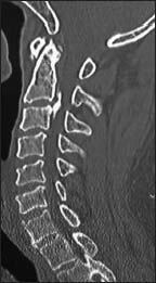
Sagittal image of computed tomography scan showing large C2-3 region ossified posterior longitudinal ligament
Figure 1c.

Computed tomography image through the facets showing plate and screw fixation and intraarticular spacer as described
RESULTS
The follow-up was 30, 33 and 36 months. In all the three patients, the fixation was successful and without any incidence. Successful segmental arthrodesis was achieved in all cases. There was no mechanical instrumental failure or infective complication.
DISCUSSION
The techniques in spinal instrumented fixation have remarkably evolved in the recent years. A number of techniques and instrumentation have been described that make the process of fixation not only safe but also easier to perform. Varieties of methods of facetal fixation have been described and are frequently employed. The firm cortical nature of bone material of the facets provides a firm ground for screw insertion. Transarticular screw fixation was described in 1972 by Roy-Camille and Saillant.[3] Facetal screw method and transarticular method of screw implantation have been frequently employed in the process of spinal stabilization. Appropriate angulation of the screws makes them safe as regards vertebral artery, nerve roots and spinal cord. In the year 2011, we described facetal distraction and intraarticular spacer insertion as a technique for spinal stabilization.[1] The technique provided distraction of the spinal segments and opportunity to stretch and restore the tautness of buckled posterior longitudinal ligament and the ligamentum flavum. The surgical technique was based on the concept that vertical instability of the spinal segments related to muscle weakness had a paramount role in the pathogenesis of spondylotic radiculopathy and myelopathy.[4,5,6] The technique provided spinal stability and restoration of the spinal canal and root canal dimensions and avoided the need for removal of any part of bone, ligaments, osteophytes or disc material. Earlier, we had described implantation or jamming of spacers within the atlantoaxial joint cavity for stabilization and for craniovertebral realignment by distraction.[7] The deployment of spacers in the atlantoaxial region was used as an additional measure of stabilization whilst employing plate and screw insertion technique into the facets of atlas and axis.[8,9,10]
The technique of fixation described in the present report is an extension of the technique used and described by us for craniovertebral atlantoaxial stabilization. Removal of articular cartilage, distraction of facets and insertion of spacers within the articular cavity provide a firm stabilization of the region. Plate and screw fixation provides reassuring stability. The entire construct provides a ground for segmental arthrodesis. Although our experience is rather limited, but our results and experience suggests that the technique has a great potential. The ease of conduct of the procedure and its safety are the highlights that can be exploited to provide segmental spinal stabilization.
Footnotes
Source of Support: Nil
Conflict of Interest: None declared.
REFERENCES
- 1.Goel A, Shah A. Facetal distraction as treatment for single- and multilevel cervical spondylotic radiculopathy and myelopathy: A preliminary report. J Neurosurg Spine. 2011;14:689–96. doi: 10.3171/2011.2.SPINE10601. [DOI] [PubMed] [Google Scholar]
- 2.Goel A, Shah A, Jadhav M, Nama S. Distraction of facets with intraarticular spacers as treatment for lumbar canal stenosis: Report on a preliminary experience with 21 cases. J Neurosurg Spine. 2013;19:672–7. doi: 10.3171/2011.8.SPINE11249. [DOI] [PubMed] [Google Scholar]
- 3.Roy-Camille R, Saillant G. Surgery of the cervical spine. 2. Dislocation. Fracture of the articular processes. Nouv Presse Med. 1972;1:2484–5. [PubMed] [Google Scholar]
- 4.Goel A. Facet distraction spacers for treatment of degenerative disease of the spine: Rationale and an alternative hypothesis of spinal degeneration. J Craniovertebr Junction Spine. 2010;1:65–6. doi: 10.4103/0974-8237.77669. [DOI] [PMC free article] [PubMed] [Google Scholar]
- 5.Goel A. Facet distraction-arthrodesis technique: Can it revolutionize spinal stabilization methods? J Craniovertebr Junction Spine. 2011;2:1–2. doi: 10.4103/0974-8237.85306. [DOI] [PMC free article] [PubMed] [Google Scholar]
- 6.Goel A. ‘Only fixation’ as rationale treatment for spinal canal stenosis. J Craniovertebr Junction Spine. 2011;2:55–6. doi: 10.4103/0974-8237.100049. [DOI] [PMC free article] [PubMed] [Google Scholar]
- 7.Goel A. Atlantoaxial joint jamming as a treatment for atlantoaxial dislocation: A preliminary report. Technical note. J Neurosurg Spine. 2007;7:90–4. doi: 10.3171/SPI-07/07/090. [DOI] [PubMed] [Google Scholar]
- 8.Goel A, Desai KI, Muzumdar DP. Atlantoaxial fixation using plate and screw method: A report of 160 treated patients. Neurosurgery. 2002;51:1351–6. [PubMed] [Google Scholar]
- 9.Goel A, Laheri V. Plate and screw fixation for atlanto-axial subluxation. Acta Neurochir (Wien) 1994;129:47–53. doi: 10.1007/BF01400872. [DOI] [PubMed] [Google Scholar]
- 10.Goel A. Treatment of basilar invagination by atlantoaxial joint distraction and direct lateral mass fixation. J Neurosurg Spine. 2004;1:281–6. doi: 10.3171/spi.2004.1.3.0281. [DOI] [PubMed] [Google Scholar]