PLOS One logoLink to PLOS One
. 2024 Jul 29;19(7):e0304135. doi: 10.1371/journal.pone.0304135

Timing matters in the use of renin-angiotensin system modulators and COVID-related cognitive and cerebrovascular dysfunction

Mackenzi Meier 1, Sara Becker 1, Erica Levine 1, Oriana DuFresne 1, Kaleigh Foster 2, Joshua Moore 2, Faith N Burnett 2, Veronica C Hermanns 2, Stan P Heath 2, Mohammed Abdelsaid 2, Maha Coucha 3,*
Editor: Michael Bader4
PMCID: PMC11285960  PMID: 39074114

Abstract

Renin-angiotensin system (RAS) modulators, including Angiotensin receptor blockers (ARB) and angiotensin-converting enzyme inhibitors (ACEI), are effective medications for controlling blood pressure. Cognitive deficits, including lack of concentration, memory loss, and confusion, were reported after COVID-19 infection. ARBs or ACEI increase the expression of angiotensin-converting enzyme-2 (ACE-2), a functional receptor that allows binding of SARS-CoV-2 spike protein for cellular invasion. To date, the association between the use of RAS modulators and the severity of COVID-19 cognitive dysfunction is still controversial. Purpose: This study addressed the following questions: 1) Does prior treatment with RAS modulator worsen COVID-19-induced cerebrovascular and cognitive dysfunction? 2) Can post-treatment with RAS modulator improve cognitive performance and cerebrovascular function following COVID-19? We hypothesize that pre-treatment exacerbates COVID-19-induced detrimental effects while post-treatment displays protective effects. Methods: Clinical study: Patients diagnosed with COVID-19 between May 2020 and December 2022 were identified through the electronic medical record system. Inclusion criteria comprised a documented medical history of hypertension treated with at least one antihypertensive medication. Subsequently, patients were categorized into two groups: those who had been prescribed ACEIs or ARBs before admission and those who had not received such treatment before admission. Each patient was evaluated on admission for signs of neurologic dysfunction. Pre-clinical study: Humanized ACE-2 transgenic knock-in mice received the SARS-CoV-2 spike protein via jugular vein injection for 2 weeks. One group had received Losartan (10 mg/kg), an ARB, in their drinking water for two weeks before the injection, while the other group began Losartan treatment after the spike protein injection. Cognitive functions, cerebral blood flow, and cerebrovascular density were determined in all experimental groups. Moreover, vascular inflammation and cell death were assessed. Results: Signs of neurological dysfunction were observed in 97 out of 177 patients (51%) taking ACEIs/ARBs prior to admission, compared to 32 out of 118 patients (27%) not receiving ACEI or ARBs. In animal studies, spike protein injection increased vascular inflammation, increased endothelial cell apoptosis, and reduced cerebrovascular density. In parallel, spike protein decreased cerebral blood flow and cognitive function. Our results showed that pretreatment with Losartan exacerbated these effects. However, post-treatment with Losartan prevented spike protein-induced vascular and neurological dysfunctions. Conclusion: Our clinical data showed that the use of RAS modulators before encountering COVID-19 can initially exacerbate vascular and neurological dysfunctions. Similar findings were demonstrated in the in-vivo experiments; however, the protective effects of targeting the RAS become apparent in the animal model when the treatment is initiated after spike protein injection.

Introduction

As of November 2023, there have been over 772 million confirmed cases of COVID-19 globally [World Health Organization, 2023]. Initially, COVID-19 was considered a severe acute respiratory syndrome with the potential for fatal pulmonary complications. However, studies have revealed that COVID-19 has a systemic effect impacting various organs and systems in the body, including the central nervous system. During the course of the disease, patients experienced several neurological complications such as headaches, disorientation, brain fog, and attention deficit [1,2]. Cognitive impairment was observed not only in severely affected COVID-19 patients but also in young individuals with mild to moderate disease cases [3]. As the disease is still virulent and threatening the quality of lives of people, greater attention should be focused on the short and long-term impact of COVID-19 on cognitive function and identify possible therapeutic interventions.

Patients with cardiovascular diseases such as diabetes, heart failure, obesity, or hypertension are at a higher risk for COVID-19 due to their advanced age and comorbid conditions [4,5]. Moreover, a significant association exists between hypertension and an increased likelihood of experiencing severe COVID-19 symptoms [6]. RAS modulators, including angiotensin-converting enzyme inhibitors (ACEI) and angiotensin receptor blockers (ARB), are first-line agents for the treatment of heart failure and hypertension. Multiple molecular mechanisms were investigated to explore the cardiovascular protective effects of the RAS modulators. One accepted mechanism is the RAS-induced upregulation of the angiotensin-converting enzyme-2 (ACE-2). ACE-2 degrades Ang II, the bioactive form of RAS, into Ang 1–7, which results in vasodilatory, antioxidant, and anti-inflammatory effects [7]. However, because ACE-2 acts as a binding receptor for SARS-CoV-2, facilitating its cell entry, a general concern was raised about whether RAS modulators will increase the risk of infection and disease progression [810].

Moreover, the contribution of RAS modulators to COVID-19-induced cognitive dysfunction is still a gap in knowledge. Therefore, this study aims to investigate the following: 1) Does prior treatment with RAS modulator worsen COVID-19-induced vascular and cognitive dysfunction? 2) Can post-treatment with RAS modulator improve cognitive performance and vascular function following COVID-19? To address these inquiries, a combination of clinical and preclinical research was conducted to shed light on the utility and potential risks associated with the use of ACEI or ARB in the context of COVID-19.

Methods

Clinical

The study population for this research was sourced from the St Joseph’s/Candler Meditech Electronic Medical Record system. Inclusion criteria were clearly defined as individuals who had been hospitalized for COVID-19 between 2020 and 2022, and who also had a comorbid condition of hypertension. Data was accessed from May 1, 2023 through June 30, 2023. Authors had access to patient information during the data collection period. These patients were categorized into two distinct cohorts: those who had been receiving ACEI/ARB medications before hospital admission and those who were not on ACEI/ARB therapy prior to their hospitalization. For each patient, we collected baseline demographic information, including age, gender, body mass index, comorbid conditions, concurrent antihypertensive therapy, and average blood pressure on admission. A summary of these baseline characteristics is presented in Table 1.

Table 1. Baseline characteristics of patients admitted to the hospital with a diagnosis of COVID19 and comorbid hypertension.

Characteristic ACEI/ARB prior to admission (n = 177) No ACEI/ARB prior to admission (n = 118)
Age 69 70
Sex (male) 85 (48%) 43 (43)
BMI kg/m2 32.8 30.9
Race    
Caucasian 97 (55%) 68 (58%)
African American 78 (44% 44 (37%)
Other 2 (1%) 6 (5%)
Concurrent anti-hypertensives    
CCB 73 (41%) 52 (44%)
Thiazides 52 (29%) 26 (22%)
Beta blockers 80 (45%) 56 (47%)
Major Comorbidities
Diabetes
Chronic kidney disease
Dementia
Hx of Stroke
Hyperlipidemia
Chronic heart failure

83 (43%)
22 (12%)
9 (5%)
17 (10%)
84 (47%)
19 (10%)

35 (30%)
12 (8%)
11 (9%)
17 (14%)
49 (41%)
10 (8%)
Average blood pressure on admission 137/76 138/79

Abbreviations: Body mass index (BMI, calculated as weight in kilogram divided by the square of height in meters), Angiotensin converting enzyme inhibitor (ACEI), angiotensin receptor blocker (ARB), calcium channel blocker (CCB).

Our assessment was primarily aimed at identifying indications of neurological dysfunction within the patient cohort. This evaluation encompassed a comprehensive examination of several key parameters, which included assessments for altered mental status, dizziness/vertigo, headache/migraine, loss of coordination, loss of consciousness, muscle weakness, confusion, slurred speech, and visual changes. Additionally, we evaluated patients who had undergone neurologic imaging to ascertain whether there were discernible distinctions between the two cohorts in terms of ischemic changes or atrophy.

Animals

Humanized ACE-2 Knock-in (ACE-2 KI) mice were purchased from Jackson Laboratory (Jax lab: Stock No: 035800, Ellsworth, Maine, USA) and underwent inbreeding within the animal facility at Mercer University. All animal protocols received approval from the Mercer University Institutional Animal Care and Use Committee (IACUC, accredited by The American Association for Accreditation of Laboratory Animal Care) and adhered to the current ARRIVE guidelines 2.0. Animals were provided with a standard mouse chow diet and unrestricted tap water access. Mice were kept on a 12-hour light-dark cycle. Animals were sacrificed using carbon dioxide and cervical dislocation. Buprenorphine 0.1mg/kg body weight was injected subcutaneously upon detecting any signs of animal distress.

Losartan treatment and SARS-CoV-2 spike protein injection

The study involved the administration of the SARS-CoV-2 spike protein through intravenous injection of SARS-CoV-2 nucleoprotein/spike protein recombinant (4ug/animal, Invitrogen, USA, Cat. No. RP-87706) into the jugular vein. Losartan (10 mg/kg body weight, Tokyo Chemical Industry, Tokyo, Japan, Cat. No. L0232) was introduced into the animals’ water supply. Losartan treatment commenced either two weeks before the spike protein injection or immediately after the injection. The mice were sacrificed two weeks after receiving the SARS-CoV-2 recombinant spike protein injection. Male and female humanized ACE-2 KI (hACE2 KI) mice were randomly divided blindly into four groups: 1) Control group, 2) SARS-CoV-2 spike protein injection, 3) Pre-Losartan treatment group with SARS-CoV-2 spike protein injection, and 4) SARS-CoV-2 spike protein injection group with post-injection Losartan treatment.

RT-qPCR

Triazole (Thermo-Fisher, USA, Cat. No. AC345480250) was used to isolate RNA from brain homogenate. The Thermo Scientific NanoDrop 2000C Spectrophotometer (Thermo Scientific, USA) was used to quantify RNA concentrations. The QuantStudio™ 3 Real-Time PCR System (Applied Biosystems, Thermo Scientific, USA) was utilized to run qRT-PCR. Table 2 shows forward and reverse primers used in the study. GAPDH was used for normalization in all experiments.

Table 2. Primers.

Gene Forward Reverse
ACE2 5’-TCC ATT GGT CTT
CTG CCA TCC G-3’
5’-AGA CCA TCC ACC
TCC ACT TCT C-3’
TNF-α 5’-GGT GCC TAT GTC
TCA GCC TCT T-3’
5’-GCC ATA GAA CTG
ATG AGA GGG AG-3’
Il-6 5’-TAC CAC TTC ACA
AGT CGG AGG C-3’
5’-CTG CAA GTG CAT
CAT CGT TGT TC-3’
GAPDH 5’-CCA AGA AGT GCT
CAG AGA GGT G-3’
5’-GTC CTT GAA CTT
CTT TTT GGT CTC-3’

Western blot analysis

The expression of ACE-2 and the apoptotic marker, cleaved caspase-3, was detected by western blot. Brain tissues were homogenized in RIPA buffer (Millipore, Billerica, MA, USA, Cat# 3P 20188) and then separated by a 10% SDS-polyacrylamide gel using the Mini PROTEAN Tetra Cell SDS-PAGE Gel electrophoresis kit (Biorad Laboratories Inc, Hercules, CA). Subsequently, the separated proteins were transferred onto nitrocellulose membranes. These membranes were blocked and incubated with primary antibodies overnight (Table 3). The primary antibodies were detected using horseradish peroxidase-conjugated secondary antibody (1:5000. The Western Blots were imaged, and band intensity was quantified using the Azure Biosystems c600 (Azure Biosystems Inc., Dublin, CA) and Image-J software, respectively.

Table 3. Antibodies.

Antibody Vendor Catalog number
ACE-2 Proteintech (Rosemont, IL, USA) 21115-1-AP
Β-Actin R&D (Minneapolis, MN, USA) MAB8929
Cl. Caspase-3 R&D (Minneapolis, MN, USA) MAB835

Vascular density assessment

Brain tissues isolated from the hACE2 KI were fixed and sectioned as described previously by our group [11]. Brain sections were stained with Lycopersicon Esculentum Lectin, DyLight™ 488 (Vector Laboratories, Burlingame, CA, USA, Cat. No. DL-1174-1). Imaging of the sections was performed using a Nikon Eclipse Ti-E Inverted Microscope (Nikon Instruments Inc., Melville, NY). FIJI software was employed to analyze the three-dimensional structures of the Z-stacked images. Vascular density was calculated by dividing the mean density of stained vasculature, as determined by FIJI, by the total number of planes in the Z-stack.

Cerebral blood flow assessment

Cerebral blood flow was assessed using the RFLSI III Laser Speckle Imaging System (RWD, San Diego, CA, USA). Cerebral blood flow was initially measured at baseline before administering the SARS-CoV-2 spike protein injection, and before sacrifice. Mice were anesthetized using isoflurane to facilitate the procedure, and a vertical incision was made to expose the skull. The surgical site was cleaned and sealed with a clip following the procedure. The percent change in the cerebral blood flow was compared among the groups.

Cognitive function assessment

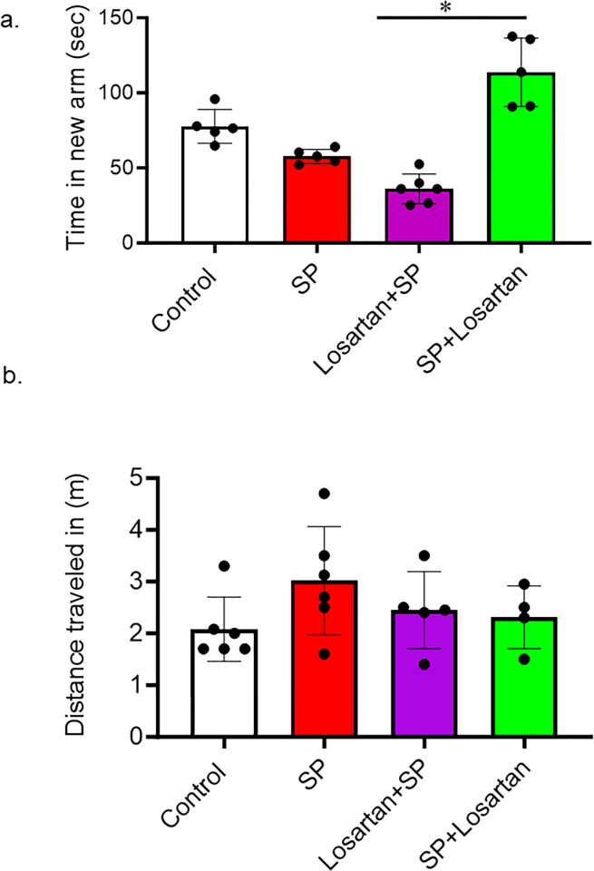
Using a Y-shape maze, memory and learning functions were evaluated at baseline and ten days after SARS-CoV-2 recombinant spike protein injection. Briefly, hACE2 KI mice were allowed to explore a Y-maze with only two open arms. On the test, the third arm was open. Using ANY-maze 6.1 tracking software, the time spent in the goal zone (new arm) and the number of entries were calculated. Total distance traveled by each animal is measured to exclude any motor dysfunction that could affect the cognitive functions assessment.

Statistical analysis

The clinical data analysis was performed employing Excel Data Analysis. Chi-square tests served as the primary statistical method for our analysis. Significance was determined at the conventional threshold of P <0.05, utilizing two-sided testing. Pre-clinical data analysis was conducted using GraphPad Prism version 10.1. For animal studies, the sample size was determined from our previous work. One-way ANOVA was used to assess the differences in the means between control, S-protein Pre-Losartan, and Post-Losartan treatment. Significance was determined at P<0.05. Data is presented as mean ± standard deviation. A Tukey’s post-hoc test was used to adjust for the multiple comparisons to assess significant interaction.

Results

The impact of receipt of an ACEI/ARB on neurologic dysfunction in patients admitted to the hospital with COVID-19

There were 295 patients included in our study who were admitted to the hospital with COVID-19 and concurrent hypertension. Of those, 177 patients were receiving ACEI/ARBs prior to their COVID-19 hospitalization, and 118 patients were not on ACEI/ARB therapy before admission. Our findings revealed a striking difference in the occurrence of neurologic dysfunction between the two groups. Among the patients who were taking ACEI/ARB medications prior to hospitalization, 91 individuals (51.4%) exhibited signs of neurologic dysfunction in contrast to 32 patients (27.1%) who were not on ACEI/ARB therapy before admission (P < 0.001) as reported in Table 4.

Table 4. The primary type of neurologic dysfunction in patients who were on ACEI/ARB prior to admission in contrast to the non-ACEI/ARB treated group.

Type of Neurological Dysfunction ACEI/ARB prior to admission
(n = 91)
No ACEI/ARB prior to admission
(n = 32)
Altered mental status 23 8
Dizziness/vertigo 7 3
Headache/migraine 9 2
Loss of coordination 4 0
Loss of consciousness 3 0
Muscle weakness 37 15
Confusion 4 4
Slurred speech 3 0
Visual changes 1 0

Abbreviations: Angiotensin-converting enzyme inhibitor (ACEI), angiotensin receptor blocker (ARB).

Among the patients experiencing neurological dysfunction, 42 individuals underwent neurologic imaging through either a magnetic resonance imaging (MRI) or computerized tomography (CT) scan, with 27 patients belonging to the ACEI/ARB group and 15 patients in the no ACEI/ARB group. Notably, 66.7% (18 out of 27) of the patients on ACEI/ARB therapy prior to admission displayed ischemic changes or atrophy on their neurologic imaging. In comparison, 53.3% (8 out of 15) of patients who were not on ACEI/ARB therapy before admission exhibited similar neuroimaging findings.

The impact of losartan treatment on ACE-2 expression

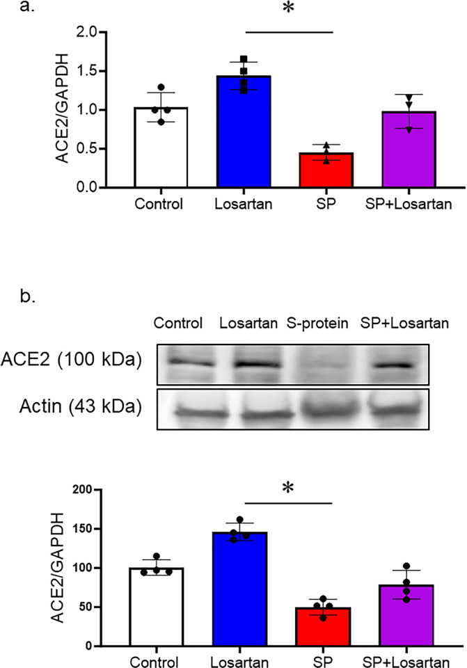
hACE2 KI mice received Losartan treatment for 2 weeks, other groups were intravenously injected with recombinant SARS-CoV-2 spike protein with or without Losartan treatment. To evaluate the impact of Losartan and spike protein on ACE-2 expression, we assessed ACE-2 gene and protein expression in brain homogenates using real-time PCR and Western blot analysis. The group that received Losartan only displayed an increase in ACE-2 expression. However, spike protein injection significantly reduced the ACE-2 gene and protein expressions, which were prevented by the post-Losartan treatment. (Fig 1A and 1B).

Fig 1. Losartan increased ACE-2 expression, while SARS-CoV-2 spike protein decreased ACE-2 expression in hACE2 brains.

Fig 1

hACE2 KI mice were treated only with Losartan or intravenously injected with SARS-CoV-2 spike protein (SP, 4 μg/ animal) with or without Losartan (10 mg/kg body weight) for 2 weeks. Brain homogenate was assessed for ACE-2 expression. a) RT-PCR analysis showing the effect of Losartan and spike protein on ACE-2 gene expression. Losartan treatment increased ACE-2 gene expression, while spike protein significantly reduced ACE-2 gene expression. b) Western blot analysis for ACE-2 expression in brain homogenate. Our results showed that Losartan treatment increased ACE-2 expression while spike protein decreased ACE2 expression. Treatment of Losartan after spike protein restored ACE-2 expression in hACE2 mice treated with spike protein. (One-way ANOVA, *P<0.05, n = 3–4).

The impact of pre and post-losartan treatment on inflammation and cell death

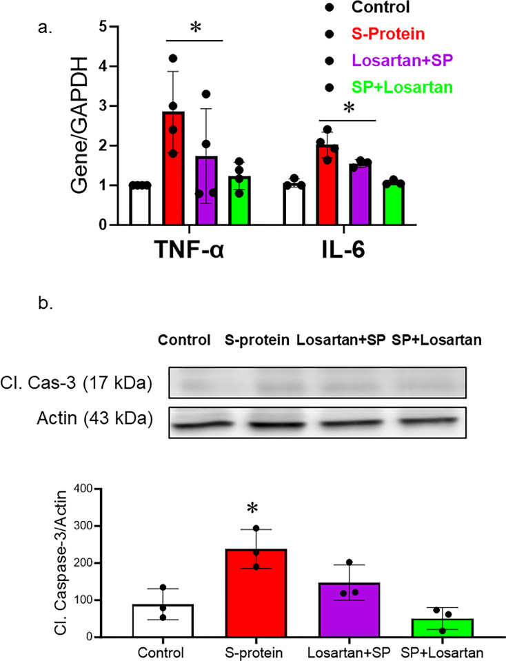
hACE2 KI mice were intravenously injected with recombinant SARS-CoV-2 spike protein. Losartan treatment began either two weeks before the spike protein injection or immediately after the injection. The mice were sacrificed two weeks after receiving the SARS-CoV-2 recombinant spike protein injection. We first evaluated the effect of SARS-CoV-2 spike protein on brain inflammation. RNA was isolated from brain homogenate and examined for inflammatory markers. Our results show that SARS-CoV-2 spike protein increased gene expression of inflammatory markers such as TNF-alpha and IL-6. (Fig 2A). Post-losartan treatment was more effective in reducing inflammatory markers compared to pre-losartan treatment. To evaluate the impact of pre and post-Losartan treatment on cell death, cleaved caspase-3 expression in brain homogenates was assessed using western blot. The expression of cleaved caspase-3, an apoptotic marker, was significantly enhanced following the spike protein injection. However, only post-Losartan treatment prevented SARS-CoV-2 spike protein-induced cell death (Fig 2B).

Fig 2. Only post-losartan treatment prevented SARS-CoV-2 spike protein-induced inflammation/apoptosis but not pre-losartan treatment.

Fig 2

hACE2 KI mice were injected with recombinant SARS-CoV-2 spike protein (4 mg/animal). Losartan treatment (10 mg/kg) began either two weeks before the spike protein injection (Losartan + SP) or immediately after the injection (SP + Losartan). Whole brain homogenate was assessed for inflammatory markers and apoptotic markers. a) RT-PCR analysis of inflammatory markers (TNF-α and Il-6) in hACE-2 brains. Our results show that spike protein injection caused a significant increase in TNF-α and Il-6 gene expression. Pre-Losartan treatment showed a similar increase in TNF-α and Il-6 gene expression. Post-Losartan treatment inhibited spike protein-induced increased inflammation. b) Western Blot analysis for apoptotic marker, cleaved caspase-3. Our results showed that spike protein and pre-losartan treatment increased caspase activation, while post-losartan treatment showed a protective effect against spike protein-induced cell death. (One-way ANOVA, *P<0.05, n = 4).

The impact of pre and post-losartan treatment on vascular density

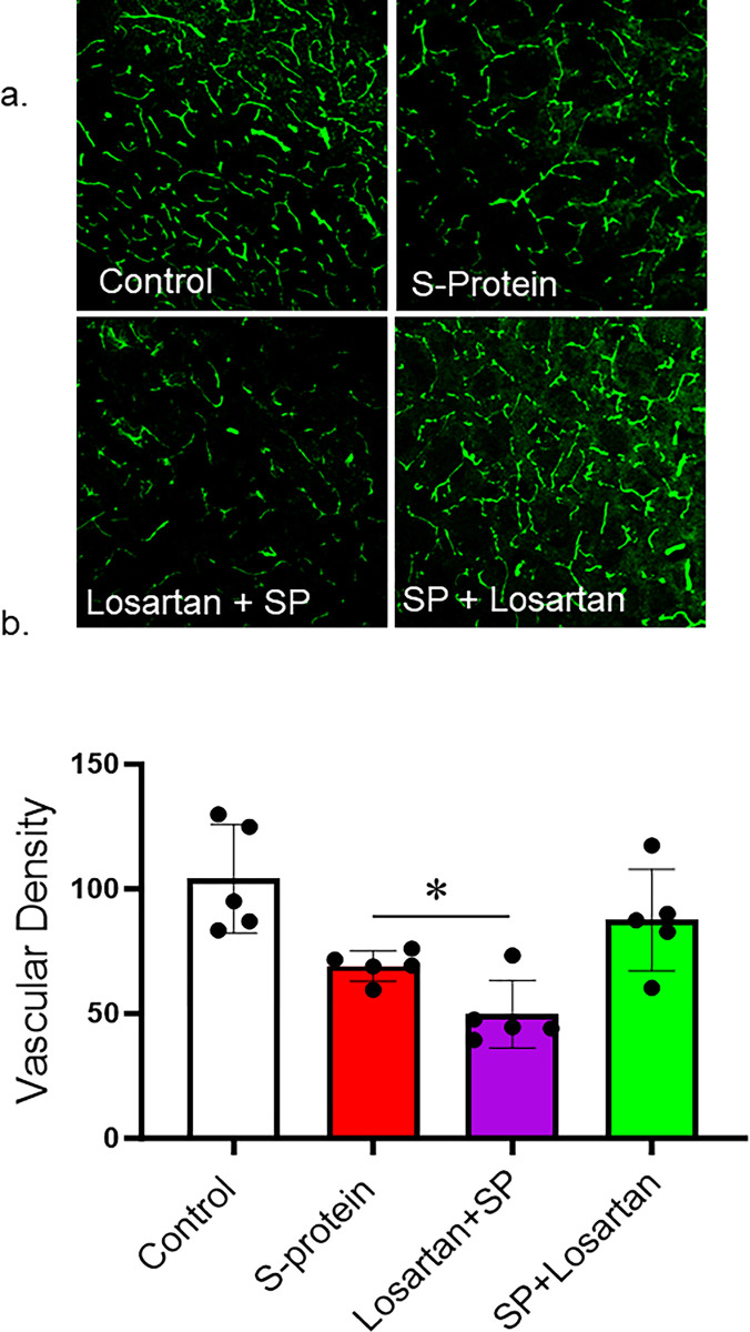
Our results showed that SARS-CoV-2 spike protein injection caused a vascular rarefaction as seen by decreased vascular density compared to control. In agreement with the immunoblotting results, only post-Losartan treatment prevented the reduction in vascular density. Interestingly, the decrease in vascular density was further enhanced in the mice who received Losartan two weeks before SARS-CoV-2 spike protein injection (Fig 3A and 3B).

Fig 3. Post-losartan treatment prevented SARS-CoV-2 Spike protein-induced vascular rarefaction but not pre-losartan treatment.

Fig 3

hACE2 KI mice were injected with recombinant SARS-CoV-2 spike protein (4 mg/animal). Losartan treatment (10 mg/kg) began either two weeks before the spike protein injection (Losartan + SP) or immediately after the injection (SP + Losartan). Brains were isolated and sectioned 30–40 um. The brain section was stained for vasculature. 3D confocal images were reconstructed to assess vascular density using FIJI software. a) Representative images of the brain cortex. b) Image analysis showing that SARS-CoV-2 caused vascular rarefaction and significantly reduced vascular density. Pre-Losartan treatment showed decreased vascular density. Only post-losartan treatment showed a protective effect and restored vascular density compared to control. (One-way ANOVA, *P<0.05, n = 5).

The impact of pre and post-losartan treatment on cerebral blood flow

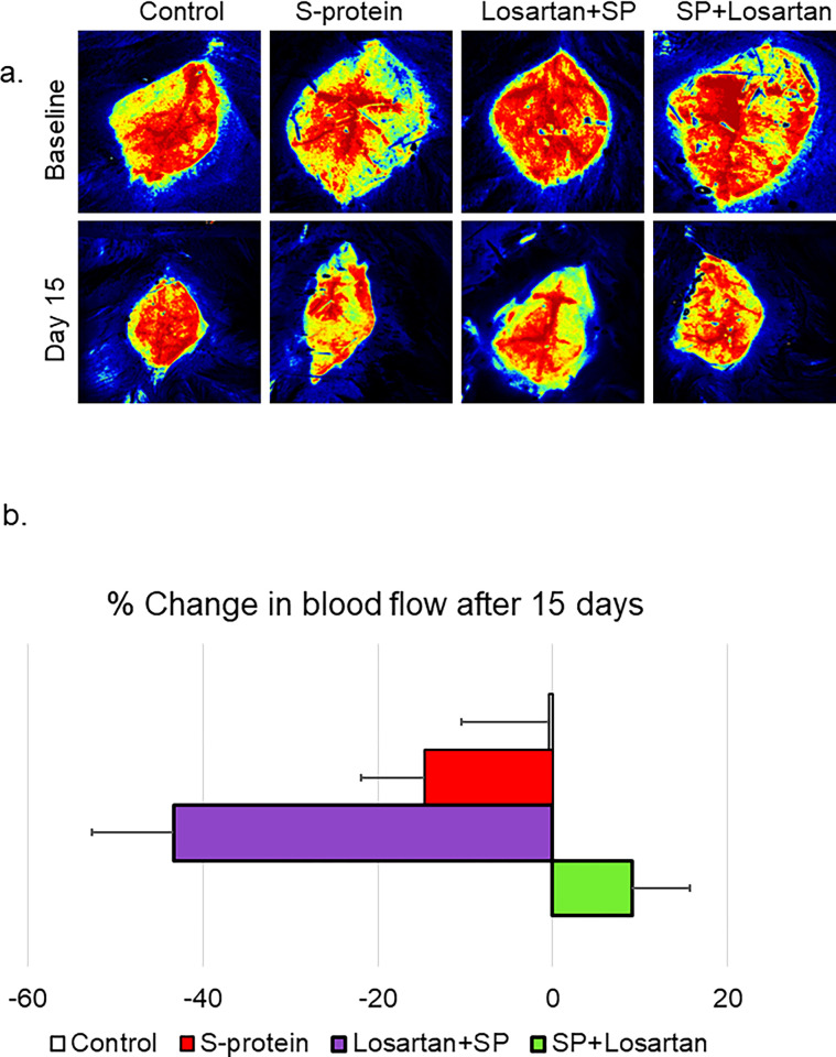
hACE2 KI started the Losartan treatment either two weeks before receiving the intravenous injection of the recombinant SARS-CoV-2 spike protein or immediately after the injection. The impact of the pre/post Losartan treatment on cerebral blood flow was assessed using laser speckle imaging at baseline and before sacrifice. Our results showed that pre-Losartan treatment caused a reduction in the cerebral blood flow, which was significant compared to the control (*P<0.05), and marginally non-significant compared to the spike protein group (P = 0.052). On the other hand, the percent change in the cerebral blood was similar in both the control and post-Losartan treatment groups (Fig 4A and 4B).

Fig 4. Pre-losartan treatment reduced cerebral blood flow after SARS-CoV-2 spike injection but not post-losartan treatment.

Fig 4

hACE2 KI mice were injected with recombinant SARS-CoV-2 spike protein (4 mg/animal). Losartan treatment (10 mg/kg) began either two weeks before the spike protein injection (Losartan + SP) or immediately after the injection (SP + Losartan). a) Representative images of cerebral blood flow were measured at baseline and 2 weeks after spike protein injection. b) Percent change in cerebral blood flow after 15 days compared to baseline. Our results showed cerebral blood flow was reduced in pre-Losartan treatment group compared to the control (*P<0.05). Post-Losartan treatment group was similar to the control. (One-way ANOVA, P<0.05, n = 3–7).

The impact of pre and post-losartan treatment on cognitive function

The Y-maze was used to assess short-term memory in hACE2 mice who received intravenous injections of SARS-CoV-2 spike protein. We also evaluated the different start times of Losartan treatment. Our results showed that SARS-CoV-2 spike protein injection caused a reduction in the time spent in the novel arm compared to the control. This reduction in time was further pronounced in the group of mice that had initiated Losartan treatment two weeks prior to SARS-CoV-2 spike protein injection. However, post-Losartan treatment prevented spike protein-induced cognitive dysfunction, as evidenced by the increased amount of time the mice spent exploring the arm they hadn’t visited before. Furthermore, we determined the total distance traveled by each animal to exclude any motor disability that could affect Y-maze results. We did not detect any motor disability due to losartan or spike protein injection (Fig 5A and 5B).

Fig 5. Post-losartan treatment prevented SARS-CoV-2 spike protein-induced decreased cognitive impairment but not pre-losartan treatment.

Fig 5

a) Learning and memory functions were assessed using Y-maze. Spike protein and pre-losartan treatment showed significant impairment in cognitive function, as seen by reduced time spent in the new arm. Only post-losartan treatment showed improvement in cognitive functions after spike protein injection. b) The total distance traveled was measured to detect any motor disability with spike protein injection. No significant results were observed in the total distance traveled between groups. (One-way ANOVA, *P<0.05, n = 5).

Discussion

The current study showed that individuals receiving ACEI/ARB therapy prior to their COVID-19 hospitalization experienced a significantly higher risk of experiencing neurologic dysfunction compared to those not on such therapy. In agreement, our animal studies showed that SARS-CoV-2 spike protein with prior treatment with Losartan, an ARB, had increased cerebrovascular inflammation and cell death, which were coupled with cognitive dysfunction. Remarkably, post-treatment with Losartan displayed a protective effect, preventing COVID-19-induced cerebrovascular and cognitive dysfunction. Our study underscores the importance of the de novo initiation of RAS modulators following COVID-19 infection.

The renin-angiotensin system plays a dual role in our body, exhibiting both protective and harmful effects depending on the abundance of angiotensin II and the activation of Angiotensin receptors [12,13]. Ang II binds with two main receptors, AT1R and AT2R, and induce different physiological responses. The augmented activation of AT1R by Ang II constitutes the detrimental arm of RAS that causes inflammation, apoptosis, and oxidative stress [14,15]. However, AT2R activation contributes to more vascular protective effects such as vasodilation and anti-inflammatory effects [16]. Moreover, ACE-2 is an enzyme that catalyzes the degradation of Ang II to Ang-(1–7), and the resultant molecules activate the Mas receptor, which results in protective actions [1719]. Therefore, Ang-(1–7)/Mas axis and the AT2R constitute the protective arm of the RAS. In COVID patients, studies have shown a shift in balance towards the RAS harmful arm due to the downregulation of ACE2 and the overactivation of AT1R by angiotensin II [20,21]. Therefore, using a RAS modulator as ACEI or ARB is a logical approach to restore the balance and enhance the protective arm of RAS. Our group has recently shown that using Losartan restored the RAS balance and reduced SARS-CoV-2-induced cerebrovascular dysfunction [22]. On the other side, ACE-2 also serves as a receptor that facilitates the entrance of SARS-COV-2 spike protein into the host cells [23]. Notably, experimental studies have shown that RAS modulators could upregulate ACE-2 expression [2426]. This has raised several concerns regarding whether pretreatment with RAS modulators could worsen COVID-19 outcomes.

Since the emergence of COVID-19, various studies have investigated factors linked to heightened disease severity, including the effects of ACEI/ARB medications. In 2020, Flacco et al. conducted a meta-analysis aiming to determine any potential connection between ACEIs or ARBs and severe or fatal COVID-19 outcomes. Their findings revealed no substantial correlation between the usage of these medications and an increased risk of severe or lethal outcomes in COVID-19 patients [27]. In 2021, Grover and Oberoi conducted a systematic review and meta-analysis with the objective of evaluating the clinical consequences for COVID-19 patients using ACEIs or ARBs. The study involved a comprehensive analysis of various clinical indicators among patients on these medications. The authors’ conclusion was that the utilization of ACEIs or ARBs did not significantly impact the clinical outcomes including severity of disease and mortality in patients with COVID-19 [28]. Also, in 2021, Singh et al. conducted a systematic review, meta-analysis, and meta-regression analysis to explore the connection between ACEIs, ARBs, and the severity and mortality of COVID-19 patients. Their study meticulously examined various datasets and research. The authors reached the conclusion that the utilization of ACEIs or ARBs did not lead to a significant increase in the severity or mortality of COVID-19 among patients [29]. In 2022, Gnanenthiran et al. published a meta-analysis to assess the safety and efficacy of RAS inhibition in adults with COVID‐19. The authors found no significant difference in all-cause mortality between patients receiving RAS inhibition therapy and those without it. However, there was a borderline decrease in acute myocardial infarction among patients on RAS inhibitors. On the other hand, there was an increased risk of acute kidney injury (AKI) associated with RAS inhibitors use, particularly in hospitalized patients. Despite this increased risk of AKI, there was no increase in the need for dialysis or other adverse outcomes such as congestive cardiac failure, stroke, or venous thromboembolism [30]. Most of the studies have focused on clinical outcomes, such as mortality and hospitalization duration. Given the intricate neurological implications of COVID-19, assessing the impact of ACEI/ARBs in these patients is of great importance, especially considering the well-established benefits of these medications in managing numerous chronic health conditions. In the clinical arm of our study, we assessed the correlation between the administration of a RAS modulators and the signs of neurological dysfunction, including but not limited to headache, altered mental status, dizziness, and loss of consciousness. A significant disparity was noted in the occurrence of neurologic dysfunction between the groups, with patients receiving ACEI/ARBs medication prior to admission showing more signs of neurologic dysfunction compared to those not on an ACEI/ARBs prior to admission. While these findings suggest a potential adverse effect of ACEI/ARBs, given their beneficial effects on various medical conditions, we aimed to delve deeper into the outcomes for patients who maintained ACEI/ARBs usage during and after COVID-19, as well as those who started ACEI/ARBs therapy around the same time as the diagnosis of COVID-19. Unfortunately, the data was not readily accessible in the electronic medical records.

To address the lack of a post-treatment arm in the clinical study, our research group used one of the well-established COVID-19 animal models [22,31] to test the impact of timing when administering RAS modulators. The in-vivo data showed that prior treatment with Losartan, an ARB, was associated with an upregulation in inflammation and cell death, in addition to a reduction in vascular density and cerebral blood flow. Those vascular and molecular changes were coupled with an increase in cognitive dysfunction. However, the de novo initiation of a Losartan subsequent to spike protein injection effectively prevented COVID-19-induced inflammation, apoptosis, and cognitive dysfunction. A plausible explanation lies in the ability of Losartan to block the AT1 receptor, thereby inhibiting the harmful arm and eventually restoring a balanced RAS equilibrium. Additionally, unopposed AT2R stimulation could be a mechanism underlying the protective effect of Losartan demonstrated in the study. In this study, we are proposing that timing matters when we administer a RAS modulator.

In the clinical arm of our study, we investigated the correlation between the administration of a RAS modulator and the presence of neurological dysfunction, encompassing symptoms such as headache, altered mental status, dizziness, and loss of consciousness. Our analysis revealed a notable discrepancy in the occurrence of neurological dysfunction between patient groups, with those receiving ACE/ARB medication prior to admission exhibiting a higher prevalence of neurological symptoms compared to individuals not on ACE/ARB therapy before hospitalization.

However, it is crucial to acknowledge the inherent limitations of the clinical aspect of our investigation. Firstly, the retrospective nature of our study introduces potential selection bias and may restrict access to comprehensive clinical data for all patients. Additionally, the lack of uniform neurological testing, such as CT scans or MRIs, for all individuals could impact the comprehensiveness of our findings. While we attempted to address this limitation by analyzing available clinical data and documenting subjective complaints, the variability in diagnostic evaluations remains a notable constraint. Moreover, the modest sample size and the disproportionate representation of African American patients in our study population may limit the generalizability of our results. Despite our efforts to contextualize our findings within the demographic framework of our study location, it is important to acknowledge the potential influence of demographic factors on our conclusions.

Regardless of these limitations, our study underscores the significance of recognizing neurological complications in COVID-19 patients undergoing RAAS inhibition, particularly considering the established therapeutic benefits of these medications in managing chronic health conditions. While our findings suggest a potential association between ACE/ARB use and neurological symptoms, further investigation into the complex relationship between ACE/ARB medications and neurological manifestations in COVID-19 patients is warranted.

In conclusion, this study contributes significant insights into the complex relationship between RAS modulators and COVID-19-induced cognitive and vascular dysfunction. The findings suggest that the timing of RAS modulator treatment plays a critical role in its effectiveness in mitigating cognitive and vascular damage in COVID-19. Future studies should address the limitation of our preclinical model, which exclusively utilized ARBs therapy, by incorporating ACEI to mimic the clinical arm where patients received either ARBs or ACEI. Additionally, further studies are needed to assess the reproducibility of the experimental data in a clinical setting. It is also essential to test whether the continuation of RAS modulators after COVID-19 could reverse the observed vascular and cognitive dysfunction. The current study highlights the importance of further investigation into the relationship between ACEI/ARBs medications and neurologic symptoms in COVID-19 patients, especially due to the profound benefits of these medications in multiple disease states.

Supporting information

S1 Raw images. Original blots.

(PDF)

pone.0304135.s001.pdf (2.6MB, pdf)

Data Availability

All relevant data are within the paper and its Supporting Information files.

Funding Statement

This study was supported by American Heart Association 23AIREA1045073 to MA WWW.heart.org The funder did not play any role in the study design, data collection and analysis.

References

  • 1.Hensley MK, Markantone D, Prescott HC. Neurologic Manifestations and Complications of COVID-19. Annu Rev Med. 2022;73:113–27. doi: 10.1146/annurev-med-042320-010427 [DOI] [PubMed] [Google Scholar]
  • 2.Zawilska JB, Lagodzinski A, Berezinska M. COVID-19: from the structure and replication cycle of SARS-CoV-2 to its disease symptoms and treatment. J Physiol Pharmacol. 2021;72(4). doi: 10.26402/jpp.2021.4.01 [DOI] [PubMed] [Google Scholar]
  • 3.Woo MS, Malsy J, Pottgen J, Seddiq Zai S, Ufer F, Hadjilaou A, et al. Frequent neurocognitive deficits after recovery from mild COVID-19. Brain Commun. 2020;2(2):fcaa205. doi: 10.1093/braincomms/fcaa205 [DOI] [PMC free article] [PubMed] [Google Scholar]
  • 4.John KJ, Mishra AK, Ramasamy C, George AA, Selvaraj V, Lal A. Heart failure in COVID-19 patients: Critical care experience. World J Virol. 2022;11(1):1–19. doi: 10.5501/wjv.v11.i1.1 [DOI] [PMC free article] [PubMed] [Google Scholar]
  • 5.Zhou Y, Yang Q, Chi J, Dong B, Lv W, Shen L, et al. Comorbidities and the risk of severe or fatal outcomes associated with coronavirus disease 2019: A systematic review and meta-analysis. Int J Infect Dis. 2020;99:47–56. doi: 10.1016/j.ijid.2020.07.029 [DOI] [PMC free article] [PubMed] [Google Scholar]
  • 6.Ebinger JE, Driver M, Joung S, Tran T, Barajas D, Wu M, et al. Hypertension and Excess Risk for Severe COVID-19 Illness Despite Booster Vaccination. Hypertension. 2022;79(10):e132–e4. doi: 10.1161/HYPERTENSIONAHA.122.19694 [DOI] [PMC free article] [PubMed] [Google Scholar]
  • 7.Carpenter RM, Young MK, Petri WAO, Lyons GR, Gilchrist C, Carey RM, et al. Repressed Ang 1–7 in COVID-19 Is Inversely Associated with Inflammation and Coagulation. mSphere. 2022;7(4):e0022022. doi: 10.1128/msphere.00220-22 [DOI] [PMC free article] [PubMed] [Google Scholar]
  • 8.Gallo G, Calvez V, Savoia C. Hypertension and COVID-19: Current Evidence and Perspectives. High Blood Press Cardiovasc Prev. 2022;29(2):115–23. doi: 10.1007/s40292-022-00506-9 [DOI] [PMC free article] [PubMed] [Google Scholar]
  • 9.Talebi-Taher M, Najafi MH, Behzad S. COVID-19 and RAAS inhibitors: is there a final conclusion? Iran J Microbiol. 2021;13(6):728–36. doi: 10.18502/ijm.v13i6.8071 [DOI] [PMC free article] [PubMed] [Google Scholar]
  • 10.El-Arif G, Farhat A, Khazaal S, Annweiler C, Kovacic H, Wu Y, et al. The Renin-Angiotensin System: A Key Role in SARS-CoV-2-Induced COVID-19. Molecules. 2021;26(22). doi: 10.3390/molecules26226945 [DOI] [PMC free article] [PubMed] [Google Scholar]
  • 11.Bailey J, Coucha M, Bolduc DR, Burnett FN, Barrett AC, Ghaly M, et al. GLP-1 receptor nitration contributes to loss of brain pericyte function in a mouse model of diabetes. Diabetologia. 2022. [DOI] [PMC free article] [PubMed] [Google Scholar]
  • 12.Ciulla MM. SARS-CoV-2 downregulation of ACE2 and pleiotropic effects of ACEIs/ARBs. Hypertens Res. 2020;43(9):985–6. doi: 10.1038/s41440-020-0488-z [DOI] [PMC free article] [PubMed] [Google Scholar]
  • 13.Angeli F, Zappa M, Reboldi G, Trapasso M, Cavallini C, Spanevello A, et al. The pivotal link between ACE2 deficiency and SARS-CoV-2 infection: One year later. Eur J Intern Med. 2021;93:28–34. doi: 10.1016/j.ejim.2021.09.007 [DOI] [PMC free article] [PubMed] [Google Scholar]
  • 14.Marshall RP, Gohlke P, Chambers RC, Howell DC, Bottoms SE, Unger T, et al. Angiotensin II and the fibroproliferative response to acute lung injury. Am J Physiol Lung Cell Mol Physiol. 2004;286(1):L156–64. doi: 10.1152/ajplung.00313.2002 [DOI] [PubMed] [Google Scholar]
  • 15.Zafari AM, Ushio-Fukai M, Akers M, Yin Q, Shah A, Harrison DG, et al. Role of NADH/NADPH oxidase-derived H2O2 in angiotensin II-induced vascular hypertrophy. Hypertension. 1998;32(3):488–95. doi: 10.1161/01.hyp.32.3.488 [DOI] [PubMed] [Google Scholar]
  • 16.Steckelings UM, Widdop RE, Sturrock ED, Lubbe L, Hussain T, Kaschina E, et al. The Angiotensin AT(2) Receptor: From a Binding Site to a Novel Therapeutic Target. Pharmacol Rev. 2022;74(4):1051–135. doi: 10.1124/pharmrev.120.000281 [DOI] [PMC free article] [PubMed] [Google Scholar]
  • 17.Raffai G, Durand MJ, Lombard JH. Acute and chronic angiotensin-(1–7) restores vasodilation and reduces oxidative stress in mesenteric arteries of salt-fed rats. Am J Physiol Heart Circ Physiol. 2011;301(4):H1341–52. doi: 10.1152/ajpheart.00202.2011 [DOI] [PMC free article] [PubMed] [Google Scholar]
  • 18.Santos RA, Campagnole-Santos MJ, Andrade SP. Angiotensin-(1–7): an update. Regul Pept. 2000;91(1–3):45–62. [DOI] [PubMed] [Google Scholar]
  • 19.Khajehpour S, Aghazadeh-Habashi A. Targeting the Protective Arm of the Renin-Angiotensin System: Focused on Angiotensin-(1–7). J Pharmacol Exp Ther. 2021;377(1):64–74. doi: 10.1124/jpet.120.000397 [DOI] [PubMed] [Google Scholar]
  • 20.Banu N, Panikar SS, Leal LR, Leal AR. Protective role of ACE2 and its downregulation in SARS-CoV-2 infection leading to Macrophage Activation Syndrome: Therapeutic implications. Life Sci. 2020;256:117905. doi: 10.1016/j.lfs.2020.117905 [DOI] [PMC free article] [PubMed] [Google Scholar]
  • 21.Rahmawati PL, Tini K, Susilawathi NM, Wijayanti IAS, Samatra DP. Pathomechanism and Management of Stroke in COVID-19: Review of Immunopathogenesis, Coagulopathy, Endothelial Dysfunction, and Downregulation of ACE2. J Clin Neurol. 2021;17(2):155–63. doi: 10.3988/jcn.2021.17.2.155 [DOI] [PMC free article] [PubMed] [Google Scholar]
  • 22.Burnett FN, Coucha M, Bolduc DR, Hermanns VC, Heath SP, Abdelghani M, et al. SARS-CoV-2 Spike Protein Intensifies Cerebrovascular Complications in Diabetic hACE2 Mice through RAAS and TLR Signaling Activation. Int J Mol Sci. 2023;24(22). doi: 10.3390/ijms242216394 [DOI] [PMC free article] [PubMed] [Google Scholar]
  • 23.St John MH, Barry AR. COVID-19 and the role of angiotensin-converting enzyme inhibitors and angiotensin receptor blockers. Can Pharm J (Ott). 2020;153(4):193–7. doi: 10.1177/1715163520929373 [DOI] [PMC free article] [PubMed] [Google Scholar]
  • 24.Abuohashish HM, Ahmed MM, Sabry D, Khattab MM, Al-Rejaie SS. ACE-2/Ang1-7/Mas cascade mediates ACE inhibitor, captopril, protective effects in estrogen-deficient osteoporotic rats. Biomed Pharmacother. 2017;92:58–68. doi: 10.1016/j.biopha.2017.05.062 [DOI] [PubMed] [Google Scholar]
  • 25.Liu CX, Hu Q, Wang Y, Zhang W, Ma ZY, Feng JB, et al. Angiotensin-converting enzyme (ACE) 2 overexpression ameliorates glomerular injury in a rat model of diabetic nephropathy: a comparison with ACE inhibition. Mol Med. 2011;17(1–2):59–69. doi: 10.2119/molmed.2010.00111 [DOI] [PMC free article] [PubMed] [Google Scholar]
  • 26.Vuille-dit-Bille RN, Camargo SM, Emmenegger L, Sasse T, Kummer E, Jando J, et al. Human intestine luminal ACE2 and amino acid transporter expression increased by ACE-inhibitors. Amino Acids. 2015;47(4):693–705. doi: 10.1007/s00726-014-1889-6 [DOI] [PubMed] [Google Scholar]
  • 27.Flacco ME, Acuti Martellucci C, Bravi F, Parruti G, Cappadona R, Mascitelli A, et al. Treatment with ACE inhibitors or ARBs and risk of severe/lethal COVID-19: a meta-analysis. Heart. 2020;106(19):1519–24. doi: 10.1136/heartjnl-2020-317336 [DOI] [PMC free article] [PubMed] [Google Scholar]
  • 28.Grover A, Oberoi M. A systematic review and meta-analysis to evaluate the clinical outcomes in COVID-19 patients on angiotensin-converting enzyme inhibitors or angiotensin receptor blockers. Eur Heart J Cardiovasc Pharmacother. 2021;7(2):148–57. doi: 10.1093/ehjcvp/pvaa064 [DOI] [PMC free article] [PubMed] [Google Scholar]
  • 29.Singh R, Rathore SS, Khan H, Bhurwal A, Sheraton M, Ghosh P, et al. Mortality and Severity in COVID-19 Patients on ACEIs and ARBs-A Systematic Review, Meta-Analysis, and Meta-Regression Analysis. Front Med (Lausanne). 2021;8:703661. doi: 10.3389/fmed.2021.703661 [DOI] [PMC free article] [PubMed] [Google Scholar]
  • 30.Gnanenthiran SR, Borghi C, Burger D, Caramelli B, Charchar F, Chirinos JA, et al. Renin-Angiotensin System Inhibitors in Patients With COVID-19: A Meta-Analysis of Randomized Controlled Trials Led by the International Society of Hypertension. J Am Heart Assoc. 2022;11(17):e026143. doi: 10.1161/JAHA.122.026143 [DOI] [PMC free article] [PubMed] [Google Scholar]
  • 31.Munoz-Fontela C, Dowling WE, Funnell SGP, Gsell PS, Riveros-Balta AX, Albrecht RA, et al. Animal models for COVID-19. Nature. 2020;586(7830):509–15. doi: 10.1038/s41586-020-2787-6 [DOI] [PMC free article] [PubMed] [Google Scholar]

Decision Letter 0

Michael Bader

29 Feb 2024

PONE-D-24-03599Timing Matters in the Use of Renin-Angiotensin System Modulators and COVID-Related Cognitive and Cerebrovascular DysfunctionPLOS ONE

Dear Dr. Coucha,

Thank you for submitting your manuscript to PLOS ONE. After careful consideration, we feel that it has merit but does not fully meet PLOS ONE’s publication criteria as it currently stands. Therefore, we invite you to submit a revised version of the manuscript that addresses the points raised during the review process.

Please submit your revised manuscript by Apr 14 2024 11:59PM. If you will need more time than this to complete your revisions, please reply to this message or contact the journal office at plosone@plos.org. When you're ready to submit your revision, log on to https://www.editorialmanager.com/pone/ and select the 'Submissions Needing Revision' folder to locate your manuscript file.

Please include the following items when submitting your revised manuscript:

  • A rebuttal letter that responds to each point raised by the academic editor and reviewer(s). You should upload this letter as a separate file labeled 'Response to Reviewers'.

  • A marked-up copy of your manuscript that highlights changes made to the original version. You should upload this as a separate file labeled 'Revised Manuscript with Track Changes'.

  • An unmarked version of your revised paper without tracked changes. You should upload this as a separate file labeled 'Manuscript'.

If you would like to make changes to your financial disclosure, please include your updated statement in your cover letter. Guidelines for resubmitting your figure files are available below the reviewer comments at the end of this letter.

If applicable, we recommend that you deposit your laboratory protocols in protocols.io to enhance the reproducibility of your results. Protocols.io assigns your protocol its own identifier (DOI) so that it can be cited independently in the future. For instructions see: https://journals.plos.org/plosone/s/submission-guidelines#loc-laboratory-protocols. Additionally, PLOS ONE offers an option for publishing peer-reviewed Lab Protocol articles, which describe protocols hosted on protocols.io. Read more information on sharing protocols at https://plos.org/protocols?utm_medium=editorial-email&utm_source=authorletters&utm_campaign=protocols.

We look forward to receiving your revised manuscript.

Kind regards,

Michael Bader

Academic Editor

PLOS ONE

Journal Requirements:

When submitting your revision, we need you to address these additional requirements.

1. Please ensure that your manuscript meets PLOS ONE's style requirements, including those for file naming. The PLOS ONE style templates can be found at 

https://journals.plos.org/plosone/s/file?id=wjVg/PLOSOne_formatting_sample_main_body.pdf and 

https://journals.plos.org/plosone/s/file?id=ba62/PLOSOne_formatting_sample_title_authors_affiliations.pdf

2. To comply with PLOS ONE submissions requirements, in your Methods section, please provide additional information regarding the experiments involving animals and ensure you have included details on (1) methods of sacrifice, and (2) efforts to alleviate suffering.

3. We note that the grant information you provided in the ‘Funding Information’ and ‘Financial Disclosure’ sections do not match. 

When you resubmit, please ensure that you provide the correct grant numbers for the awards you received for your study in the ‘Funding Information’ section.

4. Thank you for stating the following in the Competing Interests section: 

"Some data were presented as an abstract at the International Stroke Conference 2023 and the MIDYEAR 2023 Clinical Meeting & Exhibition.

The authors have declared that no competing interests exist.

Funding:This study was supported by American Heart Association 23AIREA1045073 to MA."

Please confirm that this does not alter your adherence to all PLOS ONE policies on sharing data and materials, by including the following statement: ""This does not alter our adherence to  PLOS ONE policies on sharing data and materials.” (as detailed online in our guide for authors http://journals.plos.org/plosone/s/competing-interests).  

If there are restrictions on sharing of data and/or materials, please state these. Please note that we cannot proceed with consideration of your article until this information has been declared. 

Please include your updated Competing Interests statement in your cover letter; we will change the online submission form on your behalf.

5. In the online submission form you indicate that your data is not available for proprietary reasons and have provided a contact point for accessing this data. Please note that your current contact point is a co-author on this manuscript. According to our Data Policy, the contact point must not be an author on the manuscript and must be an institutional contact, ideally not an individual. Please revise your data statement to a non-author institutional point of contact, such as a data access or ethics committee, and send this to us via return email. Please also include contact information for the third party organization, and please include the full citation of where the data can be found.

6. PLOS ONE now requires that authors provide the original uncropped and unadjusted images underlying all blot or gel results reported in a submission’s figures or Supporting Information files. This policy and the journal’s other requirements for blot/gel reporting and figure preparation are described in detail at https://journals.plos.org/plosone/s/figures#loc-blot-and-gel-reporting-requirements and https://journals.plos.org/plosone/s/figures#loc-preparing-figures-from-image-files. When you submit your revised manuscript, please ensure that your figures adhere fully to these guidelines and provide the original underlying images for all blot or gel data reported in your submission. See the following link for instructions on providing the original image data: https://journals.plos.org/plosone/s/figures#loc-original-images-for-blots-and-gels. 


In your cover letter, please note whether your blot/gel image data are in Supporting Information or posted at a public data repository, provide the repository URL if relevant, and provide specific details as to which raw blot/gel images, if any, are not available. Email us at plosone@plos.org if you have any questions.

7. We note that you have included the phrase “data not shown” in your manuscript. Unfortunately, this does not meet our data sharing requirements. PLOS does not permit references to inaccessible data. We require that authors provide all relevant data within the paper, Supporting Information files, or in an acceptable, public repository. Please add a citation to support this phrase or upload the data that corresponds with these findings to a stable repository (such as Figshare or Dryad) and provide and URLs, DOIs, or accession numbers that may be used to access these data. Or, if the data are not a core part of the research being presented in your study, we ask that you remove the phrase that refers to these data.

[Note: HTML markup is below. Please do not edit.]

Reviewers' comments:

Reviewer's Responses to Questions

Comments to the Author

1. Is the manuscript technically sound, and do the data support the conclusions?

The manuscript must describe a technically sound piece of scientific research with data that supports the conclusions. Experiments must have been conducted rigorously, with appropriate controls, replication, and sample sizes. The conclusions must be drawn appropriately based on the data presented.

Reviewer #1: No

Reviewer #2: Yes

Reviewer #3: No

**********

2. Has the statistical analysis been performed appropriately and rigorously?

Reviewer #1: No

Reviewer #2: Yes

Reviewer #3: I Don't Know

**********

3. Have the authors made all data underlying the findings in their manuscript fully available?

The PLOS Data policy requires authors to make all data underlying the findings described in their manuscript fully available without restriction, with rare exception (please refer to the Data Availability Statement in the manuscript PDF file). The data should be provided as part of the manuscript or its supporting information, or deposited to a public repository. For example, in addition to summary statistics, the data points behind means, medians and variance measures should be available. If there are restrictions on publicly sharing data—e.g. participant privacy or use of data from a third party—those must be specified.

Reviewer #1: No

Reviewer #2: No

Reviewer #3: No

**********

4. Is the manuscript presented in an intelligible fashion and written in standard English?

PLOS ONE does not copyedit accepted manuscripts, so the language in submitted articles must be clear, correct, and unambiguous. Any typographical or grammatical errors should be corrected at revision, so please note any specific errors here.

Reviewer #1: Yes

Reviewer #2: Yes

Reviewer #3: Yes

**********

5. Review Comments to the Author

Please use the space provided to explain your answers to the questions above. You may also include additional comments for the author, including concerns about dual publication, research ethics, or publication ethics. (Please upload your review as an attachment if it exceeds 20,000 characters)

Reviewer #1: 1. The authors must describe a technically sound piece of scientific research with data that supports the conclusions. Experiments must have been conducted rigorously, with appropriate controls, replication, and sample sizes

2. The figures lack legends that describe the statistical analyzes and sample sizes of each of the experimental groups.

3. The authors must validate that the preclinical model used emulates the recruited patients. In the preclinical model it is not specified whether they used only males or both males and females. The preclinical model used by the authors. Validation of the preclinical model is missing.

4. The patients received ACEI/ARB, however the mice received only ARB, losartan. The treatment with ACEi remains to be incorporated in the preclinical model.

Reviewer #2: The paper is well written and addresses an important question of the timing of RAS modulators in COVID-19 related cognitive dysfunction. They show statistically better outcomes in patients receiving the medication post infection. I think the paper is acceptable in its current form.

Reviewer #3: In this clinical/preclinical paper, the authors make an attempt to correlate RAS-inhibition before or post onset of Covid-19 with neurological dysfunction. While the preclinical part provides interesting results, appears to be well done and methodologically sound, I have major concerns with respect to the clinical part:

In general, the observed increase in neurological dysfunction in patients on RAS inhibition my be simply due to the fact that these patients were more severely ill, had higher BP levels to start with or cardio-renal problems etc. which afforded additional treatment with RAS inhibitors.

- The number of patients is relatively small. Meaningful statistics are difficult with such small numbers.

- The proportion of black patients is too high to be representative for the US population, let alone the rest of the world.

- Data on comorbidities ar lacking.

- No information on BP control

- No information on the methods to evaluate mental status

- Coincidence of neurological problems: The number (91) with neurological dysfunction in RAS inhibitor- treated patients does not distinguish between i) one dysfunction in one patient or ii) several neurological problems in one patient.

Minor problems:

- line 318: Ang II has the same affinity to AT1R and AT2R. The difference comes rather from the number of receptors in a given tissue under given circumstances.

- line 320: The AT2R-axis of the "protective RAS" needs to be referenced, e.g. Steckelings et al. Pharm. Reviews 2022.

- line 323: The protective RAS is not only constituted by the Ang 1-7/Mas axis but also via the AT2R. Stimulation of the unopposed AT2R could be a mechanism of the protective effects of Losartan post SP in the animals.

- line 348: Meta-analysis on Covid-19/ RAS inhibition by Gnanenthiran SR et al., J Am Heart Assoc. 2022

should be included.

**********

6. PLOS authors have the option to publish the peer review history of their article (what does this mean?). If published, this will include your full peer review and any attached files.

If you choose “no”, your identity will remain anonymous but your review may still be made public.

Do you want your identity to be public for this peer review? For information about this choice, including consent withdrawal, please see our Privacy Policy.

Reviewer #1: No

Reviewer #2: Yes: Abhinav Grover

Reviewer #3: No

**********

[NOTE: If reviewer comments were submitted as an attachment file, they will be attached to this email and accessible via the submission site. Please log into your account, locate the manuscript record, and check for the action link "View Attachments". If this link does not appear, there are no attachment files.]

While revising your submission, please upload your figure files to the Preflight Analysis and Conversion Engine (PACE) digital diagnostic tool, https://pacev2.apexcovantage.com/. PACE helps ensure that figures meet PLOS requirements. To use PACE, you must first register as a user. Registration is free. Then, login and navigate to the UPLOAD tab, where you will find detailed instructions on how to use the tool. If you encounter any issues or have any questions when using PACE, please email PLOS at figures@plos.org. Please note that Supporting Information files do not need this step.

PLoS One. 2024 Jul 29;19(7):e0304135. doi: 10.1371/journal.pone.0304135.r002

Author response to Decision Letter 0


2 Apr 2024

Response to Journal Requirements:

1. Please ensure that your manuscript meets PLOS ONE's style requirements, including those for file naming. The PLOS ONE style templates can be found at

https://journals.plos.org/plosone/s/file?id=wjVg/PLOSOne_formatting_sample_main_body.pdf and

https://journals.plos.org/plosone/s/file?id=ba62/PLOSOne_formatting_sample_title_authors_affiliations.pdf

The file naming was adjusted to follow PLOS ONE’s style requirements

2. To comply with PLOS ONE submissions requirements, in your Methods section, please provide additional information regarding the experiments involving animals and ensure you have included details on (1) methods of sacrifice, and (2) efforts to alleviate suffering.

We have updated the method section as directed with the following: “Animals were sacrificed using carbon dioxide and cervical dislocation. Buprenorphine 0.1 mg/kg body weight was injected subcutaneously upon detecting any signs of animal distress.”

3. We note that the grant information you provided in the ‘Funding Information’ and ‘Financial Disclosure’ sections do not match.

When you resubmit, please ensure that you provide the correct grant numbers for the awards you received for your study in the ‘Funding Information’ section.

We have updated the funding information.

4. Thank you for stating the following in the Competing Interests section:

"Some data were presented as an abstract at the International Stroke Conference 2023 and the MIDYEAR 2023 Clinical Meeting & Exhibition.

The authors have declared that no competing interests exist.

Funding:This study was supported by American Heart Association 23AIREA1045073 to MA."

Please confirm that this does not alter your adherence to all PLOS ONE policies on sharing data and materials, by including the following statement: "This does not alter our adherence to PLOS ONE policies on sharing data and materials.” (as detailed online in our guide for authors http://journals.plos.org/plosone/s/competing-interests).

If there are restrictions on sharing of data and/or materials, please state these. Please note that we cannot proceed with consideration of your article until this information has been declared.

Please include your updated Competing Interests statement in your cover letter; we will change the online submission form on your behalf.

“This does not alter our adherence to PLOS ONE policies on sharing data and materials.” was added to the manuscript and the cover letter as directed

5. In the online submission form you indicate that your data is not available for proprietary reasons and have provided a contact point for accessing this data. Please note that your current contact point is a co-author on this manuscript. According to our Data Policy, the contact point must not be an author on the manuscript and must be an institutional contact, ideally not an individual. Please revise your data statement to a non-author institutional point of contact, such as a data access or ethics committee, and send this to us via return email. Please also include contact information for the third party organization, and please include the full citation of where the data can be found.

All relevant data are within the manuscript and its Supporting Information files. The online submission was updated accordingly

6. PLOS ONE now requires that authors provide the original uncropped and unadjusted images underlying all blot or gel results reported in a submission’s figures or Supporting Information files. This policy and the journal’s other requirements for blot/gel reporting and figure preparation are described in detail at https://journals.plos.org/plosone/s/figures#loc-blot-and-gel-reporting-requirements and https://journals.plos.org/plosone/s/figures#loc-preparing-figures-from-image-files. When you submit your revised manuscript, please ensure that your figures adhere fully to these guidelines and provide the original underlying images for all blot or gel data reported in your submission. See the following link for instructions on providing the original image data: https://journals.plos.org/plosone/s/figures#loc-original-images-for-blots-and-gels.

In your cover letter, please note whether your blot/gel image data are in Supporting Information or posted at a public data repository, provide the repository URL if relevant, and provide specific details as to which raw blot/gel images, if any, are not available. Email us at plosone@plos.org if you have any questions.

The original uncropped images are submitted.

7. We note that you have included the phrase “data not shown” in your manuscript. Unfortunately, this does not meet our data sharing requirements. PLOS does not permit references to inaccessible data. We require that authors provide all relevant data within the paper, Supporting Information files, or in an acceptable, public repository. Please add a citation to support this phrase or upload the data that corresponds with these findings to a stable repository (such as Figshare or Dryad) and provide and URLs, DOIs, or accession numbers that may be used to access these data. Or, if the data are not a core part of the research being presented in your study, we ask that you remove the phrase that refers to these data.

The phrase was removed.

Response to Reviewers

Reviewer #1:

1. The authors must describe a technically sound piece of scientific research with data that supports the conclusions. Experiments must have been conducted rigorously, with appropriate controls, replication, and sample sizes

We thank the reviewer for their valuable comment. We assure the reviewer that we have adopted the NIH rigor policy and that we comply with transparent standards in our research. All our experiments have been rigorously designed. Our method section describes all experimental procedures in detail for transparency and reproducibility. Proper controls and animal randomization have been used to reduce bias. Our sample size was selected to ensure a power analysis at a significance level of P<0.05. Our animal studies used male and female h ACE-2 mice to avoid biological variables. Finally, all our antibodies and chemical reagent sources have been stated in our manuscript. These reagents have been authenticated by their vendor.

2. The figures lack legends that describe the statistical analyzes and sample sizes of each of the experimental groups.

We apologize for this misunderstanding. According to the PLOS One manuscript author guide, all the figure legends are included in the manuscript body and they include sample size and statistical method, as directed.

3. The authors must validate that the preclinical model used emulates the recruited patients. In the preclinical model, it is not specified whether they used only males or both males and females. The preclinical model used by the authors. Validation of the preclinical model is missing.

We apologize for the mistake and totally agree with the reviewer that the pre-clinical model must be validated. We and others have used hACE-2 mice in COVID-19 research. We have included the reference for studies using the animal model in our discussion. Both male and female hACE-2 mice have been used in our experimental design to avoid biological variables. We have updated our method section accordingly.

(1) Munoz-Fontela C, Dowling WE, Funnell SGP, Gsell PS, Riveros-Balta AX, Albrecht RA, et al. Animal models for COVID-19. Nature. 2020;586(7830):509-15.

(2) Burnett FN, Coucha M, Bolduc DR, Hermanns VC, Heath SP, Abdelghani M, et al. SARS-CoV-2 Spike Protein Intensifies Cerebrovascular Complications in Diabetic hACE2 Mice through RAAS and TLR Signaling Activation. Int J Mol Sci. 2023;24(22).

4. The patients received ACEI/ARB, however the mice received only ARB, losartan. The treatment with ACEi remains to be incorporated in the preclinical model.

We thank the reviewer for their constructive feedback. While patients received ACEI or ARB therapy, we exclusively used ARB, Losartan, in the preclinical study. This choice was based on our recent research demonstrating Losartan's efficacy in restoring RAS balance and mitigating spike protein-induced cerebrovascular dysfunction (2). We acknowledge that including ACE inhibitor therapy in our preclinical model would offer a more comprehensive reflection of the clinical arm. However, incorporating ACE inhibitors would require a complete repetition of the whole preclinical study, which is unfeasible. The following statement was included in the manuscript to reflect this limitation.

“Future studies should address the limitation of our preclinical model, which exclusively utilized ARB therapy, by incorporating ACEI to mimic the clinical arm where patients received either ARB or ACEI”

Reviewer #2: The paper is well written and addresses an important question of the timing of RAS modulators in COVID-19 related cognitive dysfunction. They show statistically better outcomes in patients receiving the medication post infection. I think the paper is acceptable in its current form.

Your feedback is greatly appreciated, and we're grateful for your time.

Reviewer #3: In this clinical/preclinical paper, the authors make an attempt to correlate RAS-inhibition before or post onset of Covid-19 with neurological dysfunction. While the preclinical part provides interesting results, appears to be well done and methodologically sound, I have major concerns with respect to the clinical part:

In general, the observed increase in neurological dysfunction in patients on RAS inhibition may be simply due to the fact that these patients were more severely ill, had higher BP levels to start with or cardio-renal problems etc. which afforded additional treatment with RAS inhibitors.

- The number of patients is relatively small. Meaningful statistics are difficult with such small numbers. - The proportion of black patients is too high to be representative for the US population, let alone the rest of the world.

- Data on comorbidities are lacking.

– No information on BP control

- No information on the methods to evaluate mental status - Coincidence of neurological problems: The number (91) with neurological dysfunction in RAS inhibitor- treated patients does not distinguish between i) one dysfunction in one patient or ii) several neurological problems in one patient.

We would like to extend our sincere gratitude for the time and effort the reviewer dedicated to reviewing our manuscript. The reviewer’s feedback has been instrumental in strengthening our manuscript. Below is our response to the reviewer’s comments

Firstly, we acknowledge that the clinical part of the study is a retrospective chart review, which inherently relies on electronic medical record documentation of subjective complaints. While we tried to identify objective measures of neurological dysfunction, such as CT scans or MRIs, not all patients underwent such evaluations due to various clinical reasons.

In response to your concern regarding the potential influence of patients' overall clinical condition, including severity of illness, baseline blood pressure levels, or cardiorenal problems, we have implemented several measures to address this issue. Specifically, we have incorporated additional data on comorbidities and blood pressure control into our analysis. These findings indicate that the two groups exhibit similarities in these parameters, thereby indicating that the observed disparities in neurological dysfunction are less likely to be solely attributable to baseline clinical characteristics.

We also acknowledge the high proportion of African American patients included in our study. As residents of the Southeast, where approximately half of the population is African American, it is not surprising that a significant number of African American patients were evaluated in our retrospective chart review. We appreciate your understanding of this demographic representation and assure you that it does not impact the validity of our findings.

Minor problems:

- line 318: Ang II has the same affinity to AT1R and AT2R. The difference comes rather from the number of receptors in a given tissue under given circumstances.

We apologize for this mistake, and it was corrected in the manuscript to the following “Ang II binds with two main receptors, AT¬1R and AT2R, and triggers different biological responses.”

- line 320: The AT2R-axis of the "protective RAS" needs to be referenced, e.g. Steckelings et al. Pharm. Reviews 2022.

The review is being referenced in the manuscript.

- line 323: The protective RAS is not only constituted by the Ang 1-7/Mas axis but also via the AT2R. Stimulation of the unopposed AT2R could be a mechanism of the protective effects of Losartan post SP in the animals.

The following challenges were made to enhance the clarity of the manuscript:

However, AT2R activation contributes to more vascular protective effects such as vasodilation and anti-inflammatory effects (3). Moreover, ACE-2 is an enzyme that catalyzes the degradation of Ang II to Ang-(1-7), and the resultant molecules activate the Mas receptor which results in protective actions (4-6). Therefore, Ang-(1-7)/Mas axis and the AT2R constitute the protective arm of the RAS.

Additionally, unopposed AT2R stimulation could be a mechanism underlying the protective effect of Losartan demonstrated in the study.

- line 348: Meta-analysis on Covid-19/ RAS inhibition by Gnanenthiran SR et al., J Am Heart Assoc. 2022 should be included.

The Meta-analysis was included in the manuscript.

In 2022, Gnanenthiran et al. published a meta-analysis to assess the safety and efficacy of RAS inhibiton in adults with COVID‐19. The authors found no significant difference in all-cause mortality between patients receiving RAS inhibition = therapy and those without it. However, there was a borderline decrease in acute myocardial infarction among patients on RASi. On the other hand, there was an increased risk of acute kidney injury (AKI) associated with RASi use, particularly in hospitalized patients. Despite this increased risk of AKI, there was no increase in the need for dialysis or other adverse outcomes such as congestive cardiac failure, stroke, or venous thromboembolism (7).

1. Munoz-Fontela C, Dowling WE, Funnell SGP, Gsell PS, Riveros-Balta AX, Albrecht RA, et al. Animal models for COVID-19. Nature. 2020;586(7830):509-15.

2. Burnett FN, Coucha M, Bolduc DR, Hermanns VC, Heath SP, Abdelghani M, et al. SARS-CoV-2 Spike Protein Intensifies Cerebrovascular Complications in Diabetic hACE2 Mice through RAAS and TLR Signaling Activation. Int J Mol Sci. 2023;24(22).

3. Steckelings UM, Widdop RE, Sturrock ED, Lubbe L, Hussain T, Kaschina E, et al. The Angiotensin AT(2) Receptor: From a Binding Site to a Novel Therapeutic Target. Pharmacol Rev. 2022;74(4):1051-135.

4. Raffai G, Durand MJ, Lombard JH. Acute and chronic angiotensin-(1-7) restores vasodilation and reduces oxidative stress in mesenteric arteries of salt-fed rats. Am J Physiol Heart Circ Physiol. 2011;301(4):H1341-52.

5. Santos RA, Campagnole-Santos MJ, Andrade SP. Angiotensin-(1-7): an update. Regul Pept. 2000;91(1-3):45-62.

6. Khajehpour S, Aghazadeh-Habashi A. Targeting the Protective Arm of the Renin-Angiotensin System: Focused on Angiotensin-(1-7). J Pharmacol Exp Ther. 2021;377(1):64-74.

7. Gnanenthiran SR, Borghi C, Burger D, Caramelli B, Charchar F, Chirinos JA, et al. Renin-Angiotensin System Inhibitors in Patients With COVID-19: A Meta-Analysis of Randomized Controlled Trials Led by the International Society of Hypertension. J Am Heart Assoc. 2022;11(17):e026143.

Attachment

Submitted filename: Response to Reviewers.docx

pone.0304135.s002.docx (43.8KB, docx)

Decision Letter 1

Michael Bader

26 Apr 2024

PONE-D-24-03599R1Timing Matters in the Use of Renin-Angiotensin System Modulators and COVID-Related Cognitive and Cerebrovascular DysfunctionPLOS ONE

Dear Dr. Coucha,

Thank you for submitting your manuscript to PLOS ONE. After careful consideration, we feel that it has merit but does not fully meet PLOS ONE’s publication criteria as it currently stands. Therefore, we invite you to submit a revised version of the manuscript in which the clinical part is deleted.

Please submit your revised manuscript by Jun 10 2024 11:59PM. If you will need more time than this to complete your revisions, please reply to this message or contact the journal office at plosone@plos.org. When you're ready to submit your revision, log on to https://www.editorialmanager.com/pone/ and select the 'Submissions Needing Revision' folder to locate your manuscript file.

Please include the following items when submitting your revised manuscript:

  • A rebuttal letter that responds to each point raised by the academic editor and reviewer(s). You should upload this letter as a separate file labeled 'Response to Reviewers'.

  • A marked-up copy of your manuscript that highlights changes made to the original version. You should upload this as a separate file labeled 'Revised Manuscript with Track Changes'.

  • An unmarked version of your revised paper without tracked changes. You should upload this as a separate file labeled 'Manuscript'.

If you would like to make changes to your financial disclosure, please include your updated statement in your cover letter. Guidelines for resubmitting your figure files are available below the reviewer comments at the end of this letter.

If applicable, we recommend that you deposit your laboratory protocols in protocols.io to enhance the reproducibility of your results. Protocols.io assigns your protocol its own identifier (DOI) so that it can be cited independently in the future. For instructions see: https://journals.plos.org/plosone/s/submission-guidelines#loc-laboratory-protocols. Additionally, PLOS ONE offers an option for publishing peer-reviewed Lab Protocol articles, which describe protocols hosted on protocols.io. Read more information on sharing protocols at https://plos.org/protocols?utm_medium=editorial-email&utm_source=authorletters&utm_campaign=protocols.

We look forward to receiving your revised manuscript.

Kind regards,

Michael Bader

Academic Editor

PLOS ONE

[Note: HTML markup is below. Please do not edit.]

Reviewers' comments:

Reviewer's Responses to Questions

Comments to the Author

1. If the authors have adequately addressed your comments raised in a previous round of review and you feel that this manuscript is now acceptable for publication, you may indicate that here to bypass the “Comments to the Author” section, enter your conflict of interest statement in the “Confidential to Editor” section, and submit your "Accept" recommendation.

Reviewer #3: (No Response)

**********

2. Is the manuscript technically sound, and do the data support the conclusions?

The manuscript must describe a technically sound piece of scientific research with data that supports the conclusions. Experiments must have been conducted rigorously, with appropriate controls, replication, and sample sizes. The conclusions must be drawn appropriately based on the data presented.

Reviewer #3: Partly

**********

3. Has the statistical analysis been performed appropriately and rigorously?

Reviewer #3: N/A

**********

4. Have the authors made all data underlying the findings in their manuscript fully available?

The PLOS Data policy requires authors to make all data underlying the findings described in their manuscript fully available without restriction, with rare exception (please refer to the Data Availability Statement in the manuscript PDF file). The data should be provided as part of the manuscript or its supporting information, or deposited to a public repository. For example, in addition to summary statistics, the data points behind means, medians and variance measures should be available. If there are restrictions on publicly sharing data—e.g. participant privacy or use of data from a third party—those must be specified.

Reviewer #3: Yes

**********

5. Is the manuscript presented in an intelligible fashion and written in standard English?

PLOS ONE does not copyedit accepted manuscripts, so the language in submitted articles must be clear, correct, and unambiguous. Any typographical or grammatical errors should be corrected at revision, so please note any specific errors here.

Reviewer #3: Yes

**********

6. Review Comments to the Author

Please use the space provided to explain your answers to the questions above. You may also include additional comments for the author, including concerns about dual publication, research ethics, or publication ethics. (Please upload your review as an attachment if it exceeds 20,000 characters)

Reviewer #3: Improvements regarding minor concerns acknowledged.

Major concern still with respect to

-small number of patients

- high proportion of black patients with different responses to RAS inhibitors different from white and Asian populations

- rigorous testing not in all patients

**********

7. PLOS authors have the option to publish the peer review history of their article (what does this mean?). If published, this will include your full peer review and any attached files.

If you choose “no”, your identity will remain anonymous but your review may still be made public.

Do you want your identity to be public for this peer review? For information about this choice, including consent withdrawal, please see our Privacy Policy.

Reviewer #3: No

**********

[NOTE: If reviewer comments were submitted as an attachment file, they will be attached to this email and accessible via the submission site. Please log into your account, locate the manuscript record, and check for the action link "View Attachments". If this link does not appear, there are no attachment files.]

While revising your submission, please upload your figure files to the Preflight Analysis and Conversion Engine (PACE) digital diagnostic tool, https://pacev2.apexcovantage.com/. PACE helps ensure that figures meet PLOS requirements. To use PACE, you must first register as a user. Registration is free. Then, login and navigate to the UPLOAD tab, where you will find detailed instructions on how to use the tool. If you encounter any issues or have any questions when using PACE, please email PLOS at figures@plos.org. Please note that Supporting Information files do not need this step.

PLoS One. 2024 Jul 29;19(7):e0304135. doi: 10.1371/journal.pone.0304135.r004

Author response to Decision Letter 1


2 May 2024

The authors would like to thank all the reviewers (1, 2, and 3) for their invaluable feedback and constructive criticism of our manuscript titled "Timing Matters in the Use of Renin-Angiotensin System Modulators and COVID-Related Cognitive and Cerebrovascular Dysfunction." Their time and expertise are greatly appreciated.

I am writing in response to Reviewer #3's feedback on our study. The authors appreciate Reviewer #3's time and effort in providing a thorough evaluation of our study.

We understand Reviewer #3's concerns regarding the clinical part of our study and the potential limitations associated with it. However, we respectfully disagree with the suggestion to delete the clinical part of the study. We believe that the clinical data significantly contribute to the overall findings and provide valuable insights into the correlation between RAAS inhibition and neurological dysfunction in patients with COVID-19. The clinical data strengthens the scientific merit of our research.

Here is a detailed response to Reviewer #3's concerns regarding the small sample size, the high proportion of African Americans, and the lack of rigorous testing in all patients.

Small sample size: We understand the concerns raised by the reviewer regarding the sample size. However, we would like to emphasize that the goal of this study was to shed light on the potential correlation between RAAS inhibition and neurological dysfunction following COVID-19 exposure. In this study, we tried to identify areas for further investigation. While we acknowledge the importance of having a sufficient number of patients in the clinical study, the findings that we observed based on the 295 patients included in the study can serve as a foundation for future research projects with a larger sample of patients.

Lack of rigorous testing in all patients: Firstly, we acknowledge that the clinical part of the study is a retrospective chart review, which inherently relies on electronic medical record documentation of subjective complaints. We acknowledge the concern raised regarding the absence of comprehensive neurological evaluations, such as CT scans or MRIs, in all patients within our retrospective chart review. It is important to note that the decision for these objective imaging tests was subject to the discretion of attending physicians based on clinical indications and resource availability. Given the retrospective nature of our study and the diversity of patient presentations, not all individuals underwent the same diagnostic procedures. While this may introduce variability in the data collection process, it does not compromise the integrity of our findings. We analyzed the available clinical data to identify trends and associations, ensuring transparency in our reporting of both objective and subjective measures of neurological dysfunction. Furthermore, our study's primary focus was on exploring potential correlations between RAAS inhibition and neurological outcomes in the context of COVID-19, rather than exclusively relying on imaging findings. We have acknowledged this limitation in our revised manuscript and provided

interpretations of our results within the framework of available clinical data. Despite the variability in diagnostic evaluations, our study still provides valuable insights into the relationship between RAAS modulation and neurological dysfunction post-COVID-19 exposure, warranting further investigation.

High proportion of African Americans: We acknowledge the high proportion of African American patients included in our study. The data was collected from St. Joseph’s/Candler Health System located in Savannah, Georgia. “In 2021, there were 1.41 times more Black or African American (non-Hispanic) residents (76k people) in Savannah, GA, than any other race or ethnicity. There were 54k White (non-Hispanic) and 4.01k White (Hispanic) residents, the second and third most common ethnic groups [1]”. Therefore, it is not surprising that a significant number of African American patients were evaluated in our retrospective chart review. We appreciate your understanding of this demographic representation and assure you that it does not impact the validity of our findings. To provide further clarity, we have included a table detailing the distribution of African American and White patients included in our study. As shown in the table, the majority of the patients were White. While we acknowledge the high proportion of African American patients included in our study; we believe that this demographic diversity enriches our findings and underscores the importance of considering racial disparities in healthcare outcomes.

[1] https://datausa.io/profile/geo/savannah-ga#race_and_ethnicity

Characteristic ACEI/ARB prior to admission (n=177) No ACEI/ARB prior to admission (n=118)

Caucasian 97 (55%) 68 (58%)

African American 78 (44% 44 (37%)

Other 2 (1%) 6 (5%)

We have revised the manuscript to clarify those issues, highlight any limitations within the study, and underscore the need for further studies to validate the findings. We believe that those changes will strengthen the quality and impact of our manuscript.

We respectfully request that the journal reconsider its decision to remove the clinical part of our study.

Attachment

Submitted filename: Response to Reviewers.docx

pone.0304135.s003.docx (150KB, docx)

Decision Letter 2

Michael Bader

7 May 2024

Timing Matters in the Use of Renin-Angiotensin System Modulators and COVID-Related Cognitive and Cerebrovascular Dysfunction

PONE-D-24-03599R2

Dear Dr. Coucha,

We’re pleased to inform you that your manuscript has been judged scientifically suitable for publication and will be formally accepted for publication once it meets all outstanding technical requirements.

Within one week, you’ll receive an e-mail detailing the required amendments. When these have been addressed, you’ll receive a formal acceptance letter and your manuscript will be scheduled for publication.

An invoice will be generated when your article is formally accepted. Please note, if your institution has a publishing partnership with PLOS and your article meets the relevant criteria, all or part of your publication costs will be covered. Please make sure your user information is up-to-date by logging into Editorial Manager at Editorial Manager® and clicking the ‘Update My Information' link at the top of the page. If you have any questions relating to publication charges, please contact our Author Billing department directly at authorbilling@plos.org.

If your institution or institutions have a press office, please notify them about your upcoming paper to help maximize its impact. If they’ll be preparing press materials, please inform our press team as soon as possible -- no later than 48 hours after receiving the formal acceptance. Your manuscript will remain under strict press embargo until 2 pm Eastern Time on the date of publication. For more information, please contact onepress@plos.org.

Kind regards,

Michael Bader

Academic Editor

PLOS ONE

Additional Editor Comments (optional):

Reviewers' comments:

Acceptance letter

Michael Bader

23 Jun 2024

PONE-D-24-03599R2

PLOS ONE

Dear Dr. Coucha,

I'm pleased to inform you that your manuscript has been deemed suitable for publication in PLOS ONE. Congratulations! Your manuscript is now being handed over to our production team.

At this stage, our production department will prepare your paper for publication. This includes ensuring the following:

* All references, tables, and figures are properly cited

* All relevant supporting information is included in the manuscript submission,

* There are no issues that prevent the paper from being properly typeset

If revisions are needed, the production department will contact you directly to resolve them. If no revisions are needed, you will receive an email when the publication date has been set. At this time, we do not offer pre-publication proofs to authors during production of the accepted work. Please keep in mind that we are working through a large volume of accepted articles, so please give us a few weeks to review your paper and let you know the next and final steps.

Lastly, if your institution or institutions have a press office, please let them know about your upcoming paper now to help maximize its impact. If they'll be preparing press materials, please inform our press team within the next 48 hours. Your manuscript will remain under strict press embargo until 2 pm Eastern Time on the date of publication. For more information, please contact onepress@plos.org.

If we can help with anything else, please email us at customercare@plos.org.

Thank you for submitting your work to PLOS ONE and supporting open access.

Kind regards,

PLOS ONE Editorial Office Staff

on behalf of

Prof. Michael Bader

Academic Editor

PLOS ONE

Associated Data

    This section collects any data citations, data availability statements, or supplementary materials included in this article.

    Supplementary Materials

    S1 Raw images. Original blots.

    (PDF)

    pone.0304135.s001.pdf (2.6MB, pdf)
    Attachment

    Submitted filename: Response to Reviewers.docx

    pone.0304135.s002.docx (43.8KB, docx)
    Attachment

    Submitted filename: Response to Reviewers.docx

    pone.0304135.s003.docx (150KB, docx)

    Data Availability Statement

    All relevant data are within the paper and its Supporting Information files.


    Articles from PLOS ONE are provided here courtesy of PLOS

    RESOURCES