

Abstract
Benign smooth muscle tumours, known as leiomyomas, are comparatively frequent, with uterine cases accounting for 95% incidence. Oral leiomyomas typically appear as asymptomatic, slowly-growing submucosal lumps in the tongue, hard palate, or buccal mucosa. Three histologic kinds of leiomyomas are recognized: epithelioid leiomyoma, solid leiomyoma, and angioleiomyoma. The most prevalent type of leiomyomas affecting the oral cavity are solitary forms called angioleiomyomas, which typically develop in the subcutis. The diagnosis is commonly determined by histopathological and Immunohistochemistry (IHC) procedures. This case reports a 30-year-old female patient with a lesion on her right cheek region. The postsurgical specimen was routinely processed and stained with hematoxylin and eosin staining, and IHC studies confirmed the diagnosis of ‘Benign spindle cell neoplasm—Angioleiomyoma’.
Keywords: Angioleiomyoma, benign spindle cell neoplasm, immunohistochemistry, leiomyoma
INTRODUCTION
Benign smooth muscle tumours, known as leiomyomas, are frequently reported to involve the uterus muscle, accounting for 95% of cases. These neoplasms are rarely reported to involve the oral tissues, with an incidence of 0.42%. Oral leiomyomas typically appear as asymptomatic, slow-growing submucosal lumps in the tongue, hard palate, or buccal mucosa. The gingiva and the floor of the mouth are fewer common sites.[1] Three histologic types of leiomyomas are epithelioid leiomyoma, also known as bizarre leiomyoma or leiomyoblastoma, solid leiomyoma, and angioleiomyoma, also known as vascular leiomyoma. Based on histological characteristics, the majority of oral leiomyomas are categorized as angioleiomyomas. The most prevalent type of leiomyomas affecting the oral cavity is the solitary forms called angioleiomyomas, which typically develop in the subcutis.[2] Etiology such as trauma, hormonal fluctuations, arteriovenous malformation, venous stasis, infection, and genetic translocation have been suggested as potential factors, although their exact roles remain unclear.
Distinguishing a leiomyoma from other mesenchymal tumors based on clinical presentation can be highly challenging. While the exact cause remains uncertain, prior research suggests that trauma or spontaneous development are cited as potential origins within the oral cavity.[3] The literature has documented cytogenetic characterization in only six cases of angioleiomyoma.[4,5] Angioleiomyoma typically exhibits straightforward karyotypes, often marked by either one or a few chromosomal rearrangements or numerical abnormalities. It has been studied that the frequent recurring alteration primarily involved the loss of 22q11, with recurrent gain of Xq also noted. Notably, rearrangements in the Notch Receptor 2 (NOTCH2) gene have been identified in a very limited portion of angioleiomyomas recently. It has been demonstrated that myopericytomas, myofibromas, angioleiomyomas, and glomus tumors belong to the pericytic tumour family, characterized by the presence of PDGFRB and/or NOTCH3 mutations.[6] Alterations in BRAF and NF1, gene has also been mentioned and is under study.[7]
The majorities of the angioleiomyomas are clearly delineated, generally painless, and exhibit slow growth. The size of oral leiomyomas can range from a few centimetres to larger sizes are mostly surrounded by normal mucosa, and may appear bluish or purplish. Symptoms such as difficulty in swallowing, tooth pain, loose teeth, and general discomfort have been documented, likely stemming from local ischemia and nerve compression near the tumour. Typically, these symptoms manifest after several years of tumour development.[8,9] Angioleiomyomas most frequently occur in the lips (27.46%), with the tongue following closely behind (18.30%). Other common sites include the cheeks and palate (15.49%), gingiva (8.45%), and mandible (5.63%).[10] Surgical removal followed by histopathological analysis using haematoxylin and eosin (H&E) staining, and immunohistochemical (IHC) evaluations, constitutes the foundation for diagnosis. The primary treatment for leiomyoma is surgical excision, with recurrences being uncommon. In this article we report a case of angioleiomyoma involving the buccal mucosal region rarely reported in south India subcontinent, diagnosed by means of conventional H&E staining and supported by IHC investigation. Other than these lesions arising from the smooth muscle layers of blood vessels angioleiomyomas are rarely reported in intraoral locations.
CASE REPORT
A 30-year-old female patient reported to our outpatient department with a chief complaint of swelling in right cheek for past 2 months associated with severe pain. On clinical examination, extra-orally a diffuse swelling evident on right cheek region [Figure 1]. Intra-orally, solitary, mobile swelling of size 2 cm × 2 cm present on right buccal mucosa, with ill-defined borders, smooth surface and without colour change [Figure 2]. On Ultrasonography (USG) investigation, the lesion was hypoechoic involving the right cheek region [Figure 3]. Based on clinical and radiographic findings, a provisional diagnosis of ectopic lymph node and lipoma was made. Surgical excision of the lesion via Intraoral approach was done under Local Anaesthesia (LA) and seven bits of soft tissue specimen were submitted for histopathological evaluation [Figure 4]. The specimens were subjected to routine processing, sectioning and staining.
Figure 1.

Extra oral swelling
Figure 2.
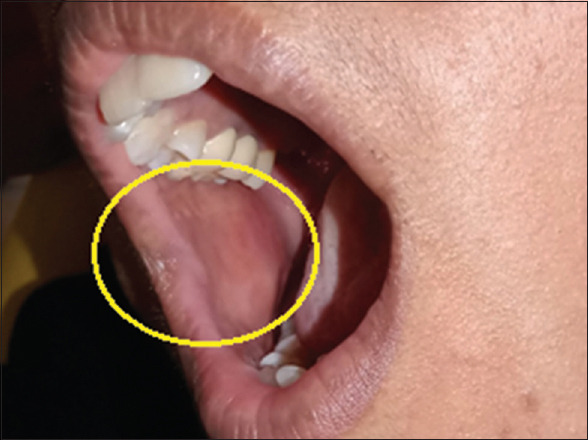
Intra oral
Figure 3.

Ultrasound image
Figure 4.

Macroscopic features showing multiple bits of soft tissue specimen
On microscopic examination, the H&E-stained sections of the soft tissue specimen revealed connective tissue areas with intersecting/interlacing and swirls of spindle shaped cells with diffusely interspersed vascular spaces of variable sizes with extravasated RBCs. The tumour cells showed an eosinophilic cytoplasm with nuclei appearing round to oval and elongated shape with tapered or blunt ends. The tumour cells seem to be surrounding blood vessels in most of the areas. Occasionally dispersed inflammatory cells, focal areas of tissue degeneration and few adipocytes were also seen. In one section, muscle bundles were seen in close proximity to the tumour with adjacent mass of adipose tissue [Figure 5]. As the tumour cells closely resemble vascular smooth muscle cells, IHC study was performed in few sections. On immunohistochemistry analysis, Vimentin showed strong positivity in tumour cells, SMA showed diffuse strong positivity of tumour cell cytoplasm, CD 34 showed positivity in endothelial cells, Desmin showed focal positivity in tumour cells, whereas S-100 and CD 68 were negative [Supplementary Figure (213.7KB, tif) ]. Based on the histopathology, IHC, clinical and radiographic findings, a final diagnosis of ‘Benign spindle cell neoplasm—Angioleiomyoma’ was made. On post operative review and follow-up no recurrence has been so far reported.
Figure 5.

(a) Histopathological features (20x), (b) Histopathological features (40x)
DISCUSSION
Benign spindle cell tumors known as leiomyomas are frequently located in anatomical regions rich in smooth muscles, including the myometrium of the uterus (referred to as uterine fibroids) and the gastrointestinal tract. This characteristic rarity extends to the oral tissues, with an incidence of 0.42% originating from the tunica media of blood vessels, hence termed vascular leiomyomas or angioleiomyomas.[11] According to the World Health Organization (2020), Leiomyomas are classified within the pericytic (perivascular) tumour group of soft tissue tumors and are categorized into two types, angioleiomyoma and leiomyoma of deep soft tissue. Angioleiomyomas, the solid form of leiomyomas, typically manifest in the subcutaneous tissue and represent the most prevalent variant of leiomyomas observed in the oral cavity.[12] Less than 0.06% of angioleiomyomas are discovered to be linked with the head and neck area. Reports indicate that oral angioleiomyomas occur with decreasing frequency on the lips, buccal mucosa, palate, gingiva and tongue. Additionally, rare cases of intraosseous tumors have also been documented.[13,14]
Minor injuries, venous congestion, and hormonal fluctuations, particularly those linked to estrogenic, have been suggested as potential causes of angioleiomyomas.[15]
Radiographs may reveal a nonspecific soft tissue mass while CT imaging reveals a clear soft tissue mass exhibiting tissue attenuation similar to that of skeletal muscle. Ultrasound examination depicts a solid, oval-shaped, well-defined mass with primarily uniform, hypoechoic texture. Colour Doppler analysis reveals significant hyper-vascularity and may indicate accompanying feeding vessels. The echogenicity and vascularity of angioleiomyoma can vary depending on its histological subtypes. The ultrasound imaging in our case showed a well-defined round to oval, hypoechoic mass.[16,17]
On macroscopic examination, angioleiomyoma presents as an oval-shaped, clearly defined, rubbery mass with a Gray-white or brown appearance. If calcification is present, the texture may be gritty/granular upon cutting. Additionally, a blood vessel wall might be visibly attached to the lesion. Histologically, the lesion consists of well-differentiated smooth muscle cells organized around vascular channels. Adipocytic metaplasia, hyalinization, and calcification may be present, while mitotic figures are typically absent. Angioleiomyoma is classified into three histological subtypes—solid, venous, and cavernous—depending on vascular structures. The solid subtype exhibits densely packed smooth muscle cells surrounding thin-walled, slit-like vascular channels. In the venous subtype, blood vessels are enveloped by thick muscle layers, along with smooth muscle bundles positioned between vessels. The cavernous subtype exhibits dilated vascular channels with walls composed of smooth muscle cells. In our case, spindle shaped cells with an eosinophilic cytoplasm and nuclei appearing round to oval and elongated shape with tapered or blunt ends seem to be surrounding blood vessels in most of the areas more in favour of solid variant. Muscle bundles were also seen in close proximity to the tumour with adjacent mass of adipose tissue. Angioleiomyoma typically exhibits positive immunohistochemical staining for Smooth Muscle Actin (SMA), Muscle-Specific Actin (MSA), and Calponin. It may variably stain positive for H-caldesmon and Desmin, while typically showing negative staining for S-100 protein and HMB45.[18] In our case immunohistochemistry showed strong positivity for Vimentin, SMA, CD 34 and Desmin but negative for CD 68 and S 100, proved that the lesion is smooth muscle in origin with vascular subtype and for ensuring a precise diagnosis.
The differential diagnosis includes keloid, mucocele, salivary gland tumour (such as canalicular adenoma arising from a minor salivary gland in the lip), lipoma, neurilemmoma, and neurofibroma (which might mimic angioleiomyoma clinically but has different IHC features), and the granular cell tumour.
In the most extensive study by Hachisuga et al.[19] comprising 562 cases of angioleiomyoma, (lip-11 cases), (mandible-2), (cheek-1), and (hard palate-1), found that the solid subtype was the most prevalent (66%), followed by the venous (23%) and cavernous (11%) subtypes. Angioleiomyoma may exhibit morphological similarities with various benign and malignant soft tissue tumors, such as angiomyolipoma, myopericytoma, and leiomyosarcoma.[19] In a review of 121 cases of oral angioleiomyomas, the lip was the most commonly cited location, occurring in 48.6% of patients, followed by the palate (21.1%), buccal mucosa, and tongue (each 9.2%), mandible (8.3%), and buccal sulcus, labial sulcus, floor of the mouth, and gingiva (each 0.9%).[5] Another review of 7748 cases of leiomyomas of all sites by Ernest Baden et al.[10] from 1884–1992, found that there is male sex prevalence (54.0%), the lips were the most common site (27.46%) followed by the tongue (18.30%), cheeks and palate (15.49%), gingiva 12 (8.45%), and mandible (5.63%). As reports suggest that angioleiomyoma of buccal mucosa is rare, hence this case is one among the rare incidence for reporting.
CONCLUSION
To summarise, angioleiomyoma represents the predominant histological subtype of oral leiomyoma, a condition that ranks among the rarest of benign neoplasms involving the oral cavity. Though histopathological investigations are a routine in diagnosis, IHC analysis plays a crucial role for final diagnosis and distinguishing this condition from other similar benign lesions.
Declaration of patient consent
The authors certify that they have obtained all appropriate patient consent forms. In the form, the patient(s) has/have given his/her/their consent for his/her/their images and other clinical information to be reported in the journal. The patients understand that their names and initials will not be published and due efforts will be made to conceal their identity, but anonymity cannot be guaranteed.
Conflicts of interest
There are no conflicts of interest.
(a) IHC showing positivity for Vimentin (20x), (b) IHC showing positivity for SMA (40x), (c) IHC showing positivity for CD34 (40x), (d) IHC showing positivity for Desmin (10x), (e) IHC showing negative for S100 (10x)
Funding Statement
Nil.
REFERENCES
- 1.Bhattacharyya I, Summerlin DJ, Cohen DM, Ellis GL, Bavitz JB, Gillham LL. Granular cell leiomyoma of the oral cavity. Oral Surg Oral Med Oral Pathol Oral Radiol Endod. 2006;102:353–9. doi: 10.1016/j.tripleo.2005.08.005. [DOI] [PubMed] [Google Scholar]
- 2.Hamid R, Chalkoo A, Tariq S, Bilal S, Wani S. Central angioleiomyoma of the mandible: A rare entity. J Cancer Res Ther. 2020;16:647–52. doi: 10.4103/jcrt.JCRT_960_17. [DOI] [PubMed] [Google Scholar]
- 3.Menditti D, Laino L, Nastri L, Caruso U, Fiore P, Baldi A. Oral angioleiomyoma: A rare pathological entity. In Vivo. 2012;26:161–3. [PubMed] [Google Scholar]
- 4.Heim S, Mandahl N, Kristoffersson U, Mitelman F, Rööser B, Rydholm A, et al. Structural chromosome aberrations in a case of angioleiomyoma. Cancer Genet Cytogenet. 1986;20:325–30. doi: 10.1016/0165-4608(86)90091-9. [DOI] [PubMed] [Google Scholar]
- 5.Brooks JK, Nikitakis NG, Goodman NJ, Levy BA. Clinicopathologic characterization of oral angioleiomyomas. Oral Surg Oral Med Oral Pathol Oral Radiol Endod. 2002;94:221–7. doi: 10.1067/moe.2002.125276. [DOI] [PubMed] [Google Scholar]
- 6.Iwamura R, Komatsu K, Kusano M, Kubo C, Inaba Y, Shiba E, et al. PDGFRB and NOTCH3 mutations are detectable in a wider range of pericytic tumors, including myopericytomas, angioleiomyomas, glomus tumors, and their combined tumors. Mod Pathol. 2023;36:100070. doi: 10.1016/j.modpat.2022.100070. [DOI] [PubMed] [Google Scholar]
- 7.Thakker T, Anehosur V, Dinesh US, Anand J, Kumar N. Gingival angioleiomyoma –A rare case report. Oral Maxillofac Surg Cases. 2019;5:100114. [Google Scholar]
- 8.Esguep A, Solar M. Oral vascular leiomyoma–report of 5 cases and review of the literature. J Oral Med. 1986;41:126–33. [PubMed] [Google Scholar]
- 9.Grossmann S de MC, Johann ACR, Castro WH, Friedman H, Gomez RS, Mesquita RA. Anterior midline nodule of the hard palate. Oral Surg Oral Med Oral Pathol Oral Radiol Endodontol. 2009;108:808–11. doi: 10.1016/j.tripleo.2009.06.025. [DOI] [PubMed] [Google Scholar]
- 10.Baden E, Doyle JL, Lederman DA. Leiomyoma of the oral cavity: A light microscopic and immunohistochemical study with review of the literature from 1884 to 1992. Eur J Cancer B Oral Oncol. 1994;30:1–7. doi: 10.1016/0964-1955(94)90043-4. [DOI] [PubMed] [Google Scholar]
- 11.Mehta PD, Desai N, Makwana K, Patel Y. Angioleiomyoma of the lower lip. Ann Maxillofac Surg. 2020;10:251. doi: 10.4103/ams.ams_275_19. [DOI] [PMC free article] [PubMed] [Google Scholar]
- 12.White DK, Selinger LR, Miller AS, Behr MM, Damm DD. Primary angioleiomyoma of the mandible. J Oral Maxillofac Surg. 1985;43:640–4. doi: 10.1016/0278-2391(85)90140-5. [DOI] [PubMed] [Google Scholar]
- 13.Goldblatt LI, Edesess RB. Central leiomyoma of the mandible: Report of a case with ultrastructural confirmation. Oral Surg Oral Med Oral Pathol. 1977;43:591–7. doi: 10.1016/0030-4220(77)90114-1. [DOI] [PubMed] [Google Scholar]
- 14.Inaba T, Adachi M, Yagisita H. A case of angioleiomyoma in the buccal space. Odontology. 2015;103:109–11. doi: 10.1007/s10266-013-0128-z. [DOI] [PubMed] [Google Scholar]
- 15.Suárez-Peñaranda JM, Pita da Veiga G, Pérez-Muñoz N, Fernández-Figueras MT. Acral calcified vascular leiomyoma: Report of 3 cases and literature review. Am J Dermatopathol. 2021;43:732–35. doi: 10.1097/DAD.0000000000001773. [DOI] [PubMed] [Google Scholar]
- 16.Kang BS, Shim HS, Kim JH, Kim YM, Bang M, Lim S, et al. Angioleiomyoma of the extremities: Findings on ultrasonography and magnetic resonance imaging. J Ultrasound Med. 2019;38:1201–8. doi: 10.1002/jum.14798. [DOI] [PubMed] [Google Scholar]
- 17.Matsuyama A, Hisaoka M, Hashimoto H. Angioleiomyoma: A clinicopathologic and immunohistochemical reappraisal with special reference to the correlation with myopericytoma. Hum Pathol. 2007;38:645–51. doi: 10.1016/j.humpath.2006.10.012. [DOI] [PubMed] [Google Scholar]
- 18.Hachisuga T, Hashimoto H, Enjoji M. Angioleiomyoma. A clinicopathologic reappraisal of 562 cases. Cancer. 1984;54:126–30. doi: 10.1002/1097-0142(19840701)54:1<126::aid-cncr2820540125>3.0.co;2-f. [DOI] [PubMed] [Google Scholar]
- 19.Koga M, Nishio J, Koga T, Koga K, Nakayama S, Yamamoto T. An update on clinicopathological, imaging, and genetic features of angioleiomyoma. Cancer Diagn Progn. 2023;3:145–50. doi: 10.21873/cdp.10193. [DOI] [PMC free article] [PubMed] [Google Scholar]
Associated Data
This section collects any data citations, data availability statements, or supplementary materials included in this article.
Supplementary Materials
(a) IHC showing positivity for Vimentin (20x), (b) IHC showing positivity for SMA (40x), (c) IHC showing positivity for CD34 (40x), (d) IHC showing positivity for Desmin (10x), (e) IHC showing negative for S100 (10x)