To the Editor:
Acute myeloid leukemia (AML) accounts for about 20% of acute leukemias in children and still has a poor prognosis compared to lymphoblastic leukemia (5-year survival rate about 68%) [1]. Over the years, knowledge of oncogenetic abnormalities has improved, allowing AML to be classified into groups with different prognosis, leading to different treatment intensity [2]. However, most genetic alterations have been described in adult cohorts. Bolouri et al. [3] and Marceau-Renaut et al. [4] have shown that the molecular landscape in children differs from that of adult AMLs and further studies are needed for a comprehensive classification of pediatric AML.
RUNX1, runt related transcription factor 1, is a transcription factor expressed in hematopoietic cells, plays a role in the early differentiation of progenitor and stem cells, and is known to be involved in hematologic diseases and leukemogenesis as a site of mutations [5].
In adult AML cohorts, RUNX1 mutations are identified in 5–8% of younger patients, associated with M0 FAB subtype, normal karyotype and correlate with poor clinical outcome [6–8]. As an unfavorable marker, adult patients with RUNX1 mutation are stratified in high risk group of treatment [9]. In contrast to RUNX1 mutations, RUNX1 deletions have rarely been studied and their impact is therefore unknown. In children, the impact of RUNX1 mutations or deletions remains unclear because of their low frequency and is not used for risk stratification and choice of treatment intensity.
438 children with de novo AML were treated in the ELAM02 trial and RUNX1 gene status was screened in 386 of them. RUNX1 abnormality was found in 8% (29 of 386) of the cases, 24 patients with mutation and 5 with deletion. Because the majority of RUNX1 mutations in AML behave as loss-of-function mutations, we decided to study both RUNX1 mutations and deletions as one group.
Main clinical, cytological and cytogenetic characteristics of children with RUNX1 mutated and deleted (RUNX1m/del) compared with RUNX1 wild type (RUNX1wt) are reported in Table 1.
Table 1.
Clinical, cytological, cytogenetic characteristics and outcome of RUNX1m/del and RUNX1wt children.
| RUNX1m/del | RUNX1wt | P value | |
|---|---|---|---|
| n = 29 (8%) | n = 357 (92%) | ||
| Clinical data, n (%) | |||
| Male gender | 14 (48%) | 196 (55%) | 0.56 |
| Age at diagnosis | |||
| ≤2 years old | 2 (7%) | 79 (22%) | 0.06 |
| Median age [min-max] | 11.2 [0.7–17.2] | 8.4 [0–18.6] | 0.07 |
| CNS involvement | 3 (10%) | 57 (16%) | 0.60 |
| White blood cells count at diagnosis | |||
| >100 G/L | 4 (14%) | 57 (16%) | 0.62 |
| Median count | 10.8 [1.7 –445] | 17.7 (0.4–575) | 0.20 |
| Risk group | |||
| Standard | 2 (7%) | 90 (25%) | |
| Intermediate | 20 (69%) | 181 (51%) | ns |
| Adverse | 6 (21%) | 84 (24%) | |
| Cytogenetics, n (%) | |||
| Normal | 10 (34%) | 90 (25%) | 0.28 |
| CBF | 2 (7%) | 90 (25%) | 0.02 |
| MLL | 0 | 78 (22%) | 0.002 |
| −7/del7q | 5 (17%) | 28 (8%) | 0.09 |
| Complex karyotype | 6 (21%) | 39 (11%) | 0.13 |
| Missing | 1 (4%) | 2 (1%) | 0.21 |
| FAB classification, n (%) | |||
| M0 | 5 (17%) | 19 (5%) | 0.03 |
| M1 | 8 (28%) | 54 (15%) | 0.11 |
| M2 | 8 (28%) | 82 (23%) | 0.65 |
| M4 | 3 (10%) | 76 (21%) | 0.23 |
| M5 | 1 (3%) | 85 (24%) | 0.009 |
| M6 | 2 (7%) | 9 (3%) | 0.20 |
| M7 | 1 (3%) | 18 (5%) | 1 |
| Unclassified + basophil | 1 (3%) | 9 (3%) | 0.55 |
| Chloroma | 0 | 5 (1%) | 1 |
| Outcome, n (%) | |||
| Complete remission | 24 (83%) | 327 (92%) | 0.17 |
| Relapse | 11 (38%) | 122 (34%) | 0.69 |
| HSCT | 9 (31%) | 99 (28%) | 0.67 |
| Death | 17 (59%) | 86 (24%) | <0.001 |
| Survival, [CI 95%] | |||
| 5-years OS | 33.6% [18.6–60.8] | 75.7% [71.3–80.4] | <0.001 |
| 5-years EFS | 32.5% [16.8–62.8] | 61.4% [56.2–67.2] | 0.003 |
Data presented as number (%), unless otherwise indicated.
CNS central nervous system, FAB French-American-British classification.
CBF Core binding factor define by inv(16) or t(8;21).
HSCT hematopoietic stem cell transplantation.
OS Overall survival, EFS Event-free survival.
There were no differences between RUNX1m/del and RUNX1wt regarding sex, age, white blood cell count, or central nervous system involvement.
RUNX1m/del AMLs were more likely to be AML-FAB M0 (5/29 (17%) vs 19/357 (5%), p value = 0.03), and exclusive with AML-FAB M5. RUNX1 mutations were associated with a normal karyotype (10/24 (42%) vs 90/357 (25%), p = 0.09) as previously described in adult studies [6, 8], exclusive with KMT2A (11q23) rearrangement, and rarely associated with Core Binding Factor (CBF) abnormalities as t(8;21)(q22;q22). Therefore, 69% of RUNX1m/del patients were classified in the intermediate risk group. The distribution between risk groups was similar between RUNX1m/del and RUNX1wt patients leading to a comparable treatment intensity.
We identified 30 RUNX1 mutations in 24 patients (6 of them cumulating 2 mutations), 16 of them in the RUNT Homology domain, and 8 mutations had been previously described in the literature by Brown et al. [10]. We found no association between the level of variant allele frequency (VAF), type (frameshift, missense, nonsense), and location of the mutation with leukemia prognosis. In addition to mutated patients, 5 patients had RUNX1 deletion (between 50 kb and 1,6 Mb; all involving the RUNT domain).
Based on the 2015 American College of Medical Genetics and Genomics and Association for Molecular Pathology guidelines [11], all RUNX1 alterations are classified as pathogenic or likely pathogenic except for 2; the first is known to be a benign variant of RUNX1 gene and the second is of unknown significance. These 2 patients are cured and were treated in the favorable group because of CBF alteration.
RUNX1 mutated patients had a higher number of co-mutations compared with the rest of the cohort (2.71 on average vs 1.43, p < 0.001), as described by Brown et al. [10]. The most common class of co-mutated genes involved control kinase signaling (50%) especially FLT3-ITD, NRAS, FLT3-TKD and KRAS or WT1. RUNX1 alterations were also associated with EZH2 and BCOR mutations, as reported by Gaidzik [8] in adults.
We found no CEBPA, NPM1, TET2, SETBP1, RAD21, CBL mutations in RUNX1 mutated patients. Except for 1 patient with RUNX1 deletion who had a co-mutation in U2AF1, RUNX1 mutated patients had no alteration in splicing factor (SF) (such as SRSF2 or SF3B1). In contrast to what was recently reviewed by Inge van der Werf et al. [12] in adult AML, the prognostic value of RUNX1 mutations in our cohort was not limited to their co-occurrence with SF mutations.
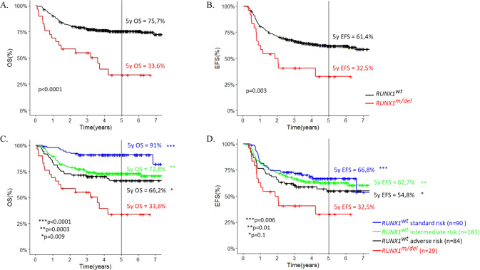
We observed a significantly worse outcome for RUNX1m/del patients compared with RUNX1wt (5-year EFS = 32.5% [95% confidence interval = 16.8–62.8] vs 61.4% [CI = 56.2–67.2]; and 5-yOS = 33.6% [CI = 18.6–60.8] vs 75.7% [CI = 71.3–80.4]). Hazard ratios for EFS and OS were 2.2 (CI = 1–4,7; p value = 0.003) and 3.3 (CI = 1.4–7.5; p < 0.0001), respectively. Comparing by risk groups, RUNX1m/del patients still had a worse outcome than patients in adverse risk group (5-y OS = 33.6% for RUNX1m/del vs 66.2% for RUNX1wt in adverse risk group). (Fig. 1).
Fig. 1. Childhood AML outcome according RUNX1 status.
A Overall survival according RUNX1 status - B Event-free survival according RUNX1 status - C Overall survival according to risk group and RUNX1m/del - D EFS according to risk group and RUNX1m/del . *Significant difference between RUNX1m/del and RUNX1wt risk subgroup.
However, RUNX1m/del patients achieved complete remission (CR) as RUNX1wt patients (83% vs 92%, p = 0.17) and had the same relapse rate (11/29 (38%) vs 122/357 (34%), p = 0.69).
Causes of death included leukemia (7/17), infection (4/17), or post-transplant toxicity (4/17). To elucidate the reason for this high toxicity and to understand how the RUNX1 alteration leads to such a poor outcome, further studies need to be performed on a larger cohort.
Three arguments led us to question whether some of these children with mutated RUNX1 might have a constitutional mutation: (1) the high toxic death rate, which could be explained by abnormal hematopoiesis exacerbating treatment toxicities(2), the high number of patients (16/24, 67%) with an increased VAF (>30%), suggesting a possible germline origin, and (3) the presence of two different RUNX1 mutations in 6 patients, one of which may be of germline origin and a second of somatic origin [13].
In our study, among these 16 patients with VAF > 30%, only 1/12 of the patients tested was confirmed to have a RUNX1 germline mutation; this patient had only one RUNX1 mutation, is still alive, and showed no treatment-related toxicity.
Among the 5 deletions, only one patient had a large germline RUNX1 deletion (1.6 Mb), already described by Preudhomme et al. [14].
Although further studies are needed to determine whether these constitutional mutations require specific treatment, the systematic search for germline mutations in complete remission may be of interest to adjust therapeutic agents.
In conclusion, our study demonstrates the prevalence, co-mutation profile, and poor survival of RUNX1-mutated or -deleted AML in a well-described pediatric cohort. The EZH2 and BCOR genes, known as chromatin modifiers, are frequent co-mutations in RUNX1m/del leukemia and may play a role in the unfavorable future of this leukemia. Considering other pediatric studies [3, 15], RUNX- mutated and -deleted AML in children should be classified into a poor risk group to benefit from optimal intensified treatment, taking into account the high mortality due to toxicity.
Supplementary information
Acknowledgements
The authors are grateful to Christophe Roumier and Olivier Nibourel (Tumor Bank, CHU Lille) and technical support of Trousseau Laboratory (National ELAM02 biobanking) for handling, conditioning, and storing patient samples. The work of all clinical research assistants is also acknowledged here. Thank you to Alain Degenne for his work on statistical analysis. The authors would like to thank all the patients, their families, the investigators, and staff of all the centers of Société Française de lutte contre les Cancers et leucémies de l’Enfant et de l’Adolescent (SFCE) for their implication in the trial.
Author contributions
GL was the principal investigator of ELAM02 study. LLD, HL, and GL performed the research. LLD and HL wrote the paper. AMR, LF, WC, PB, CP, and HL performed genetic analysis. LLD and MF performed statistical analysis. AG ensured the database management. AP, VG, FR, PS, GM, YB, AB, and GL enrolled patients in the study.
Funding
This work was supported by the French National Cancer Institute (INCA-DGOS_5797) and by a grant from the French Ministry of Health (PHRC-K 2003 no 03142). The Trousseau laboratory received funding from the Association Laurette Fugain (ALF2015/12) and La Fondation de France (RFDF9) to support molecular study and the ELAM02 national tumor Bank.
Data availability
The datasets analyzed during the current study are available from the corresponding author on reasonable request.
Competing interests
The authors declare no competing interests.
Footnotes
Publisher’s note Springer Nature remains neutral with regard to jurisdictional claims in published maps and institutional affiliations.
Supplementary information
The online version contains supplementary material available at 10.1038/s41375-023-01931-y.
References
- 1.Siegel RL, Miller KD, Fuchs HE, Jemal A. Cancer Statistics, 2021. CA Cancer J Clin. 2021;71:7–33. doi: 10.3322/caac.21654. [DOI] [PubMed] [Google Scholar]
- 2.Rubnitz JE. Current Management of Childhood Acute Myeloid Leukemia. Paediatr Drugs. 2017;19:1–10. doi: 10.1007/s40272-016-0200-6. [DOI] [PubMed] [Google Scholar]
- 3.Bolouri H, Farrar JE, Triche T, Ries RE, Lim EL, Alonzo TA, et al. The molecular landscape of pediatric acute myeloid leukemia reveals recurrent structural alterations and age-specific mutational interactions. Nat Med. 2018;24:103–12. doi: 10.1038/nm.4439. [DOI] [PMC free article] [PubMed] [Google Scholar]
- 4.Marceau-Renaut A, Duployez N, Ducourneau B, Labopin M, Petit A, Rousseau A, et al. Molecular Profiling Defines Distinct Prognostic Subgroups in Childhood AML: A Report From the French ELAM02 Study Group. HemaSphere. 2018;2:e31. doi: 10.1097/HS9.0000000000000031. [DOI] [PMC free article] [PubMed] [Google Scholar]
- 5.Sood R, Kamikubo Y, Liu P. Role of RUNX1 in hematological malignancies. Blood. 2017;129:2070–82. doi: 10.1182/blood-2016-10-687830. [DOI] [PMC free article] [PubMed] [Google Scholar]
- 6.Khan M, Cortes J, Kadia T, Naqvi K, Brandt M, Pierce S, et al. Clinical Outcomes and Co-Occurring Mutations in Patients with RUNX1-Mutated Acute Myeloid Leukemia. Int J Mol Sci. 2017;18:1618. doi: 10.3390/ijms18081618. [DOI] [PMC free article] [PubMed] [Google Scholar]
- 7.Jalili M, Yaghmaie M, Ahmadvand M, Alimoghaddam K, Mousavi SA, Vaezi M, et al. Prognostic Value of RUNX1 Mutations in AML: A Meta-Analysis. Asian Pac J Cancer Prev APJCP. 2018;19:325–9. doi: 10.22034/APJCP.2018.19.2.325. [DOI] [PMC free article] [PubMed] [Google Scholar]
- 8.Gaidzik VI, Teleanu V, Papaemmanuil E, Weber D, Paschka P, Hahn J, et al. RUNX1 mutations in acute myeloid leukemia are associated with distinct clinico-pathologic and genetic features. Leukemia. 2016;30:2160–8. doi: 10.1038/leu.2016.126. [DOI] [PubMed] [Google Scholar]
- 9.Döhner H, Estey E, Grimwade D, Amadori S, Appelbaum FR, Büchner T, et al. Diagnosis and management of AML in adults: 2017 ELN recommendations from an international expert panel. Blood. 2017;129:424–47. doi: 10.1182/blood-2016-08-733196. [DOI] [PMC free article] [PubMed] [Google Scholar]
- 10.Brown AL, Arts P, Carmichael CL, Babic M, Dobbins J, Chong CE, et al. RUNX1-mutated families show phenotype heterogeneity and a somatic mutation profile unique to germline predisposed AML. Blood Adv. 2020;4:1131–44. doi: 10.1182/bloodadvances.2019000901. [DOI] [PMC free article] [PubMed] [Google Scholar]
- 11.Luo X, Feurstein S, Mohan S, Porter CC, Jackson SA, Keel S, et al. ClinGen Myeloid Malignancy Variant Curation Expert Panel recommendations for germline RUNX1 variants. Blood Adv. 2019;3:2962–79. doi: 10.1182/bloodadvances.2019000644. [DOI] [PMC free article] [PubMed] [Google Scholar]
- 12.van der Werf I, Wojtuszkiewicz A, Meggendorfer M, Hutter S, Baer C, Heymans M, et al. Splicing factor gene mutations in acute myeloid leukemia offer additive value if incorporated in current risk classification. Blood Adv. 2021;5:3254–65. doi: 10.1182/bloodadvances.2021004556. [DOI] [PMC free article] [PubMed] [Google Scholar]
- 13.Brown AL, Hahn CN, Scott HS. Secondary leukemia in patients with germline transcription factor mutations (RUNX1, GATA2, CEBPA) Blood. 2020;136:24–35. doi: 10.1182/blood.2019000937. [DOI] [PMC free article] [PubMed] [Google Scholar]
- 14.Preudhomme C, Renneville A, Bourdon V, Philippe N, Roche-Lestienne C, Boissel N, et al. High frequency of RUNX1 biallelic alteration in acute myeloid leukemia secondary to familial platelet disorder. Blood. 2009;113:5583–7. doi: 10.1182/blood-2008-07-168260. [DOI] [PubMed] [Google Scholar]
- 15.Yamato G, Shiba N, Yoshida K, Hara Y, Shiraishi Y, Ohki K, et al. RUNX1 mutations in pediatric acute myeloid leukemia are associated with distinct genetic features and an inferior prognosis. Blood. 2018;131:2266–70. doi: 10.1182/blood-2017-11-814442. [DOI] [PubMed] [Google Scholar]
Associated Data
This section collects any data citations, data availability statements, or supplementary materials included in this article.
Supplementary Materials
Data Availability Statement
The datasets analyzed during the current study are available from the corresponding author on reasonable request.


