



Abstract
Chronic myelogenous leukemia (CML) is a myeloproliferative disease associated with a characteristic chromosomal translocation called the Philadelphia chromosome. This results in the expression of the Bcr‐Abl fusion protein, a constitutively active protein tyrosine kinase. Although there are a few treatment options with Bcr‐Abl kinase inhibitors, drug resistance is often encountered. One of the major obstacles in overcoming drug resistance in CML is the high endogenous levels of heat shock protein 70 (Hsp70). Resveratrol is a phytoalexin produced by several plants. We studied the chemotherapeutic effects and mode of action of resveratrol on K562 (CML) cells. Resveratrol induced apoptosis in K562 cells in a time‐dependent manner. This was established by increased annexin V binding, corroborated with an enhanced caspase‐3 activity and a rise in the sub‐G0/G1 population. Resveratrol treatment also caused suppression of Hsp70 both in mRNA and protein levels. The downregulation of Hsp70 by resveratrol exposure was correlated with a diminished presence of heat shock factor 1 (HSF1) in the nucleus, and the downregulation of transcriptional activity of HSF1. High endogenous levels of Hsp70 have been found to be a deterrent for sensitivity to chemotherapy. We show here that resveratrol could considerably enhance the apoptosis induction in K562 cells by 17‐allylamino‐17‐demethoxygeldanamycin, an anticancer agent that inhibits Hsp90 but augments Hsp70 levels. We conclude that resveratrol significantly downregulated Hsp70 levels through inhibition of HSF1 transcriptional activity and appreciably augmented the pro‐apoptotic effects of 17‐allylamino‐17‐demethoxygeldanamycin. (Cancer Sci 2008; 99: 1109–1116)
Abbreviations:
- 17‐AAG
17‐allylamino‐17‐demethoxygeldanamycin
- CML
chronic myelogenous leukemia
- HSE
heat shock element
- HSF1
heat shock factor 1
- Hsp
heat shock protein
- PBS
phosphate‐buffered saline
- PCR
polymerase chain reaction
- pHSE‐SEAP
vector containing heat shock element which controls the expression of secreted alkaline phosphatase
- pTAL‐SEAP
vector containing TATA‐like promoter expressing secreted alkaline phosphatase
- PS
phosphatidylserine
- TK
tyrosine kinase
- WBC
white blood cells
CML is characterized by the balanced reciprocal translocation between chromosomes 9 and 22 t (9:22) q (34:11) giving rise to the Philadelphia chromosome.( 1 ) As a consequence of this translocation, a chimerical Bcr‐Abl fusion protein is expressed. This protein has constitutively active non‐receptor TK activity and plays a pivotal role in the pathogenesis of the disease.( 1 ) Bcr‐Abl TK activity modulates a wide range of cellular activities and signaling pathways that confer resistance to chemotherapeutic drugs such as Ara‐C and etoposide.( 2 , 3 ) For example, Bcr‐Abl TK activity leads to phosphorylation and activation of PI‐3kinase,( 4 ) p38 mitogen‐activated protein kinase,( 5 ) and c‐jun N‐terminal kinase.( 6 ) Furthermore, Bcr‐Abl regulates the activation of transcription factors like nuclear factor κB( 7 ) and Stat5( 8 ) that play important roles in the genesis of leukemia. The universal presence of Bcr‐Abl fusion protein in CML patients and its diverse range of substrates made it an obvious target for drug development. This has led to the development of specific small molecule kinase inhibitors against Bcr‐Abl. Imatinib mesylate (STI571) is one such TK inhibitor of Bcr‐Abl and cAbl that was approved for treatment for all stages of the disease.( 9 , 10 ) Unfortunately, resistance to imatinib mesylate also occurs,( 11 , 12 , 13 ) and an increasing number of recent reports has documented a lack of patient response to imatinib. This might be due to point mutations resulting in impaired imatinib binding at the kinase activity site of Bcr‐abl.( 11 , 12 , 13 ) Therefore, the search for other novel targets and new strategies for the management of CML continues. Numerous natural products are under investigation for their clinical efficacy in prevention and treatment of a wide array of diseases including cancer.( 14 , 15 ) Among these, trans‐resveratrol (trans‐3,4′,5‐tri hydroxyl stilbenes), found in considerable amounts in grapes, red wine, peas, nuts, and mullberries, has attracted attention due to its many beneficial effects.( 16 ) Different studies have established the beneficial effects of resveratrol in cancer therapy, showing inhibition of proliferation and induction of apoptosis in various tumor cell lines (of different origin). Resveratrol was found to have pro‐apoptotic effects on cancer cells but displayed minimal toxicity to human peripheral blood lymphocytes, indicating its lack of toxicity for normal cells.( 17 ) Efficacy of resveratrol has been shown in rodent models of carcinogenesis.( 18 ) Interestingly, resveratrol also acts as a sensitizer in drug‐induced apoptosis.( 19 ) In spite of these findings, the key molecular targets and the precise mechanisms by which resveratrol exerts its anticancer effect remain largely unknown.
The heat shock proteins or stress proteins Hsp70 and Hsp90 have been identified as contributors to oncogenesis, suggested by the fact that overexpression of Hsp70 and Hsp90 leads to the tumor‐promoting potential of cells.( 20 ) The expression of these proteins is under the tight regulation of transcription factor HSF1. HSF1 becomes phosphorylated due to induction by heat, other forms of stress, or any other imbalance in cellular homeostasis, and phopho‐HSF1 trimerizes and translocates to the nucleus to switch on the expression of Hsp70 and Hsp90.( 21 , 22 ) The cytoprotective role of the Hsps is attributed to their powerful chaperoning activity, their ability to modulate the signal transduction pathways, and their role in proteasome‐mediated degradation of the apoptotic and survival proteins.( 22 ) Hsp90 inhibition is thought to be a powerful tool in cancer therapy by virtue of its involvement in chaperoning dozens of proteins known to be associated with oncogenesis.( 23 ) The Hsp90 inhibitor geldanamycin is reported to selectively sensitize Bcr‐Abl‐expressing leukemia cells to cytotoxic chemotherapy.( 24 ) Unfortunately, Hsp90 inhibition induces Hsp70 expression at the mRNA and protein level.( 25 ) K562 cells also have high endogenous levels of Hsp70. Also, anti‐apoptotic effects of Bcr‐Abl are found to be partially mediated by Hsp70 upregulation.( 25 ) Current reports also link the increased expression of Hsp70 in Ph1+ leukemia cells with imatinib resistance.( 26 ) Even a weak overexpression of Hsp70 is enough to make the CML cells insensitive to treatment with imatinib as well as second generation TK inhibitor nilotinib.( 26 ) This is a major drawback in successfully treating CML patients, especially those in the advanced stages of disease. Abrogation of Hsp70 induction by small interfering RNA has been shown to increase antileukemia activity of Hsp90 inhibitor geldanamycin.( 25 ) However, at present the small interfering RNA approach as a therapy can not be accomplished because the extent of inhibition can not be modulated. Therefore, the search for such a small molecule is warranted that would directly compromise without completely inhibiting the chaperoning activities of Hsp70. In our present study we put forward resveratrol as such a small molecule that induces apoptosis in Ph1+ leukemia cells (K562) by targeting Hsp70, one of the chief players involved in the pathogenesis of CML. Furthermore, we elucidate some causative mechanisms by which resveratrol influences the expression of Hsp70.
Materials and Methods
Materials. Resveratrol and fluorogenic caspase substrate acetyl‐Asp‐Glu‐Val‐Asp‐aminomethylcoumarin were obtained from Calbiochem, UK. Monoclonal mouse anti‐actin and mouse anti‐Hsp70 antibodies were purchased from BD Pharmingen. Rabbit anti‐HSF1 was obtained from Abcam and secondary antibodies were obtained from Calbiochem. pHSE‐SEAP and pTAL‐SEAP vectors were from Clontech. Caspase‐3 inhibitor acetyl‐asp‐glu‐val‐asp‐CHO (aldehyde) and general caspase inhibitor Z‐val‐ala‐asp‐fluoromethylketone were purchased from BD Pharmingen.
Cell culture and drug treatment conditions. Human chronic myeloid (K562, KU812, and KCL 22) leukemia cells were maintained in exponential growth in RPMI‐1640 medium supplemented with 10% heat‐inactivated fetal bovine serum, 100 units/mL penicillin and 100 µg/mL streptomycin in a humidified atmosphere of 5% CO2 at 37°C. Resveratrol was dissolved in dimethylsulfoxide at a concentration of 40 mM and added to cells at the indicated concentrations.
Flow cytometric analysis. K562 cells were treated with resveratrol (40 µM) for varying time intervals. Cells were harvested and fixed with 70% chilled ethanol and kept at –20°C for 24 h. These cells were washed with PBS twice and suspended in PBS containing 20 µg RNase and kept for 2 h. These cells were then treated with propidium iodide (100 µg/mL) and kept in the dark for 15 min. The cell cycle of these cells was analyzed in a Becton Dickinson flow cytometer (FACSCalibur).
Determination of caspase‐3 activity. Fifty micrograms of protein was incubated with 10 µg acetyl‐Asp‐Glu‐Val‐Asp‐aminomethylcoumarin, the fluorogenic caspase‐3 substrate, in 1 mL assay buffer (20 mM HEPES [pH 7.5], 10% glycerol, 2 mM dithiothreitol) for 1 h and the reaction was stopped by chilling. Fluorescence was measured at 380 nm excitation and 420–460 nm emission by a spectrofluorimeter.
Western blot analysis. Protein extracts (20–50 µg) were separated by 10% sodium dodecyl sulfate–polyacrylamide gel electrophoresis and were transferred onto polyvinylidene difluoride membranes. The membranes were blocked for 2 h at room temperature with 5% bovine serum albumin and incubated overnight at 4°C with respective primary antibodies. Levels of β‐actin were confirmed to ensure equal loading of the samples. The membranes were incubated with 1:5000 dilution of the appropriate peroxidase‐conjugated secondary antibodies and/or alkaline phosphatase‐conjugated antibodies and developed for detection by chemiluminescence or colorimetry. Western blots were scanned by a UMAX Astra scanner and the bands were quantified using ImageJ software.
Reverse transcription–PCR. Total RNA (2.5 µg) was reverse transcribed with oligo(dT) primer using 0.2 mM dNTP mix, 25 U RNase inhibitor, and 100 U Moloney murine leukemia virus reverse transcriptase. For PCR, 1 µL cDNA from the reverse transcription reaction mixture was taken to amplify in a 25 µL reaction volume containing 10 pmol of each primer, 200 µM each dNTP, in buffer containing 1.5 mM MgCl2 and 5 U of Taq polymerase. A 10 µL portion of PCR product was electrophoresed in a 1.0% agarose gel. The following primers were used for Hsp70 (forward, 5′‐ACCCTGTCGTCCAGCACCCA3′; reverse, 5′CTGCGTCTGCTTGGTGGGGA3′) and β‐Actin (forward, 5′‐GAAGCA TTTGCGGTGGACCAT‐3′; reverse, 5′‐TCCTGTGG CATCCACCAAACT‐3′). The primer sequence of Hsp70 was taken from the sequence of the inducible form of Hsp70 (GenBank accession number NM‐005346).
Promoter assay: measurement of transcription factor‐induced gene expression. Transcription factor‐induced gene regulation was monitored by Mercury Pathway Profiling SEAP system kit from Clontech following the manufacturer's instructions. Cells were transfected by corresponding plasmids as described in the manuals. Mercury vectors are engineered in such a way that they contain a specific cis‐acting DNA binding sequence and a sensitive reporter gene. That is, if any transcription factor binds to the cis‐acting DNA binding domain, secreatory alkaline phosphatase will be expressed. This secreted alkaline phosphatase was measured flurometrically by using its substrate, 4‐methyl umbeliferryl phosphate (10 µM). The transfections were induced by calcium phosphate and the vectors pHSE‐SEAP and pTAL‐SEAP were used.
Confocal microscopy. Resveratrol treated cells and control samples were washed several times in PBS and finally suspended in 200 µL of 1X annnexin V binding buffer containing annexin V–fluorescein‐isothiocyanate and incubated at room temperature for 15 min in the dark. Then the samples were washed three times and finally suspended in annexinV binding buffer. A drop of the final suspension was mounted on a glass slide covered with a coverslip and scanned with a confocal laser microscope (Zeiss).
Electrophoretic mobility shift assay. Protein was collected using the NE‐PER Nuclear and Cytoplasmic Extraction Reagents Kit (Pierce Biotechnology Inc, Rockford, IL, USA). The cytoplasmic fraction and the nuclear fraction were subjected to Western blot analysis for HSF1 quantification. The nuclear fraction thus isolated was further used for electrophoretic mobility shift assay for HSF1 and HSE binding.
Oligonucleotides bearing the sequence of the HSE (5′‐GAT CCT CGA AGG TTC GAG GAT CCT CGA AGG TTC GAG‐3′ and 5′‐GAT CCT CGA ACC TTC GAG GAT CCT CGA ACC TTC GAG‐3′)( 27 ) were end‐labeled with [γ‐32P] dATP by incubating annealed oligonucleotides with 5 × reaction buffer and 10 U T4 polynucleotide kinase (Invitrogen). Then the labeled oligos were allowed to anneal at room temperature for 30 min. An equal amount of protein (20 µg) from each sample was used in 25 µL binding reactions, which consisted of 1 µg poly dI‐dC, 5 × binding buffer (20 mM Tris [pH 7.5], 100 mM NaCl, 2 mM ethylenediaminetetraacetic acid, 10% glycerol, and 10 mg/mL bovine serum albumin), and the labeled probe. Protein lysate volumes were made to equal 5 µL by addition of buffer (20 mM HEPES [pH 7.8], 25% glycerol, 420 mM NaCl, 1.5 mM MgCl2, 0.2 mM ethylenediaminetetraacetic acid, and 0.5 mM phenylmethylsulphonyl fluoride). To determine specificity of DNA binding, unlabeled competitor DNA was added to the binding reactions and allowed to incubate for 10 min at room temperature. Following this incubation, labeled probe was added and samples were further incubated for 20 min at room temperature. Samples were then electrophoresed on a 5% non‐denaturing polyacrylamide gel in 0.5 × TBE for 1.5 h at 120 V and visualized by autoradiography.
Statistical analysis. Student's t‐test was used to calculate statistical significance of the data.
Results
Effect of resveratrol on Hsp70 protein and mRNA levels in K562 cells. We found that resveratrol downregulates the levels of Hsp70 both at the mRNA and protein level (Fig. 1). This downregulation was found to be dose‐dependent (Fig. 1) and time‐dependent (Fig. 2). We observed the effects of 20, 40, and 100 µM resveratrol on the Hsp70 levels of K562 cells and found that 20 µM resveratrol caused less than 20% reduction in Hsp70 protein levels, but 40 µM and 100 µM markedly reduced Hsp70 protein levels (50–80%) after a period of 48–72 h. One hundred micromols of resveratrol caused a drastic decline of 90% in the mRNA level of Hsp70 and a steep 80% drop in Hsp70 protein after 48 h. As Hsp70 is also an important cellular chaperone, we chose a dose of 40 µM resveratrol, which caused a considerable but a less severe drop in Hsp70 levels for all further experiments. After the initial 24 h of exposure to resveratrol, a modest drop (approximately 20%) was observed in Hsp70 protein levels. Resveratrol exposure for 48 h produced a significant drop (P < 0.05) in Hsp70 protein levels (Fig. 2a) which declined further on prolongation of incubation to 72 h (>60% decrease from control levels; P < 0.05). Hsp70 mRNA levels declined significantly (P < 0.01; 61% decline) on resveratrol exposure for 48 h after a moderate but significant decline (P < 0.05; 39% decrease) at 24 h, and at 72 h the decline was maintained (drop >60%; P < 0.001). (Fig. 2b). Similarly, resveratrol diminished Hsp70 protein levels in another CML cell line, KCL22, with high endogenous Hsp70 levels. In contrast, resveratrol did not have a lowering effect on already lower levels of Hsp70 in KU812 basophilic CML cells (Fig. 2c).
Figure 1.
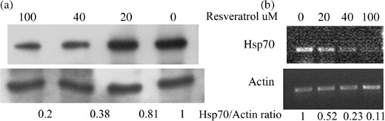
Dose–response of resveratrol‐induced inhibition of heat shock protein 70 (Hsp70) levels in K562 chronic myelogenous leukemia cells. (a) The upper panel depicts Hsp70 protein levels after treatment for 48 h with various doses of resveratrol. The lower panel shows loading control β‐actin levels. The numbers at the bottom represents Hsp70/Actin protein band densities (normalized to control). The blots are representative of three separate experiments. (b) The upper panel depicts Hsp70 mRNA levels after treatment for 48 h with various doses of resveratrol. The lower panel shows loading control β‐actin levels. The numbers at the bottom represents Hsp70/actin mRNA band densities (normalized to control). The blots are representative of two separate experiments.
Figure 2.
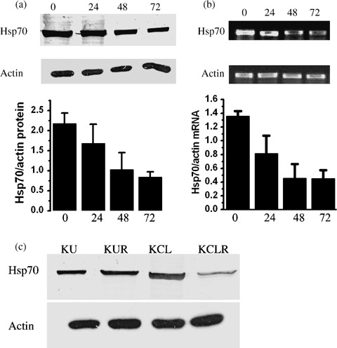
Effect of resveratrol on heat shock protein 70 (Hsp70) protein and mRNA levels in K562 chronic myelogenous leukemia cells. (a) Hsp70 protein levels. Upper panel depicts a Western blot showing Hsp70 protein levels in K562 cells after resveratrol exposure for 24, 48, and 72 h. The Hsp protein is recognized by an antibody against human Hsp70. The middle panel shows a β‐actin immunoblot to indicate equal protein loading. The Western blots are representative of three separate experiments. The lower panel displays the ratio (mean ± SD) of the Hsp70 protein/actin protein band densities from two experiments. (b) Hsp70 mRNA levels. Upper panel depicts mRNA levels of Hsp70 in K562 cells after resveratrol exposure for 24, 48, and 72 h. The middle panel shows β‐actin mRNA to indicate equal loading. The gels are representative of two separate experiments. The lower panel displays the ratio (mean ± SD) of the Hsp70 mRNA/actin mRNA band densities from two experiments. (c) Hsp70 protein levels in KU812 and KCL22 human chronic myeloid leukemia cells. Upper panel depicts Hsp70 levels and lower panel shows β‐actin levels indicatiing equal loading. KCL, untreated KCL22 cells; KCLR, KCL22 cells after 72 h of treatment with 40 µM resveratrol; KU, untreated KU812 cells; KUR, KU812 cells after 72 h of treatment with 40 µM resveratrol.
Resveratrol induces apoptosis in K562 cells. First we tried to understand whether resveratrol possessed significant apoptotic properties on Bcr‐Abl Ph1+ K562 cells. Resveratrol was found to induce apoptosis in K562 cells in a time‐dependent manner. Cell cycle analysis with 40 µM resveratrol (Fig. 3a–d) showed that the percentage of cells at sub‐G0/G1 (fragmented DNA) rose significantly (approximately 4–7‐fold higher than the control; P < 0.01) at 48 h of resveratrol exposure and remained similarly high at 72 h of exposure (P < 0.05 from control). Resveratrol exposure also caused a 2.08 ± 0.53‐fold increase in caspase‐3 activity (Fig. 3e) at 24 h that climbed to approximately 3.2 ± 0.73‐fold by 48 h and further to 4.31 ± 0.01‐fold by 72 h (P < 0.05). The increase in caspase activity was not responsible for the decrease in Hsp70 levels. As Fig. 3f shows, the caspase‐3 inhibitor and general caspase inhibitor failed to block the decline in Hsp70 levels brought about by resveratrol. During apoptosis, PS translocates from the cytoplasmic face of the plasma membrane to the cell surface. Annexin V has a strong, Ca2+‐dependent affinity for PS and therefore is used as a probe for detecting apoptosis. K562 cells treated with resveratrol showed a considerable translocation (Fig. 3g) of PS to the cell surface over the control untreated cells, as detected by confocal imaging.
Figure 3.

Apoptosis induced by 40 µM resveratrol in K562 chronic myelogenous leukemia cells. (a–c) Cell cycle analysis of K562 cells by flow cytometry. (a) Cell cycle analysis of control (0 h) K562 cells, cells treated with resveratrol for 48 h (b), and cells treated with resveratrol for 72 h (c). M1, sub‐G0/G1; M2, G1; M3, S; M4, G2 phase of the cell cycle. (d) Percentage of cells in sub‐G0/G1 (fragmented DNA), as determined by flow cytometry, after 0 h, 48 h, and 72 h of resveratrol treatment. Data are presented as mean ± SD from two or three separate experiments. (e) Caspase‐3 activity after K562 cells were exposed to resveratrol, presented as the fold increase over control levels (mean ± SD) from two separate experiments. (f) Western blot analysis of heat shock protein 70 (Hsp70). Upper panel, Hsp70; lower panel, β‐actin indicating equal loading. K‐ctl, untreated K562 cells; K+C3I+R, K562 cells pretreated for 1 h with caspase‐3 inhibitor and further treated with both inhibitor and 40 µM resveratrol for 72 h; K+PCI+R, K562 cells pretreated for 1 h with general caspase inhibitor and further treated with both inhibitor and 40 µM resveratrol for 72 h; K+R, K562 cells treated for 72 h with 40 µM resveratrol. (g) Confocal micrographs of annexin V binding to K562 cells after exposure to resveratrol for 72 h. 1, Control annexinV–fluorescein‐isothiocyanate (FITC); 2, differential interference contrast of the same field as 1; 3, 72 h resveratrol treated annexinV–FITC; 4, differential interference contrast of the same field as 3.
Resveratrol prevents HSF1 transcriptional activity and nuclear localization of HSF1. Hsp70 synthesis is controlled by the transcription factor HSF1 which binds to the HSE controlling Hsp70 gene expression.( 21 ) As resveratrol inhibits Hsp70 at the mRNA level, we next investigated if resveratrol has any critical impact on HSF1. To explore the mechanism underlying the reduction of Hsp70, we measured the levels of cytoplasmic and nuclear HSF1 by Western blot analysis (Fig. 4a,b). It was found that resveratrol promoted (Fig. 4a) a considerable augmentation (approximately 7‐fold) of HSF1 in the cytoplasmic fraction at 36 h of treatment in a time‐dependent manner. In contrast, resveratrol prominently decreased the levels of nuclear HSF1 (Fig. 4b). In accordance with this finding, it was also found that resveratrol clearly decreased (approximately 50%) binding of HSF1 from the nuclear extracts of K562 cells to the HSE DNA sequence (Fig. 4c). The promoter assay for HSE (Fig. 4d) indicated that HSF transcriptional activity is significantly inhibited by resveratrol (P < 0.01).
Figure 4.
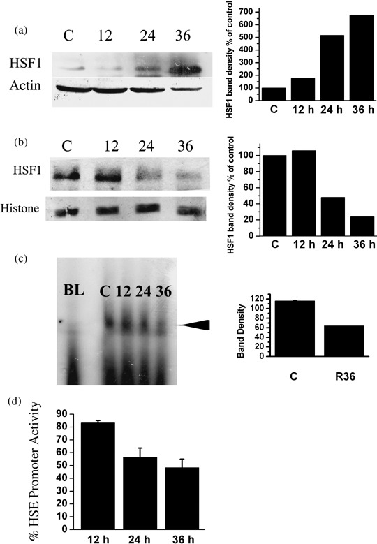
Heat shock factor 1 (HSF1) cellular localization, HSF1 binding to heat shock element (HSE), and HSE promoter activity in K562 chronic myelogenous leukemia cells after treatment with resveratrol. (a) Upper left panel, cytoplasmic localization of HSF1 at different time points after resveratrol treatment. Lower left panel, β‐actin immunoblot to indicate protein loading. Right‐hand panel, cytoplasmic HSF1 band densities of control and resveratrol treated K562 cells expressed as percentage of control. Control has been taken as 100%. The blot is representative of two separate experiments. (b) Upper left panel, nuclear localization of HSF1 at different time points after resveratrol treatment. Lower left panel, histone 2B immunoblot to indicate equal protein loading. Right‐hand panel, nuclear HSF1 band densities of control and resveratrol treated K562 cells expressed as percentage of control. Control has been taken as 100%. The blot is representative of two separate experiments. (c) Left panel, electrophoretic mobility shift assay. Arrow indicates HSF1–HSE binding. 12, nuclear extract of K562 cells treated with resveratrol for 12 h; 24, nuclear extract of K562 cells treated with resveratrol for 24 h; 36, nuclear extract of K562 cells treated with resveratrol for 36 h; BL, blank without nuclear extract; C, control. Right‐hand panel, HSF1–HSE binding band densities of control and K562 cells treated with resveratrol for 36 h (R36). Representative of two separate experiments. (d) HSE promoter activity. Data are presented as mean ± SD of percentage of control HSE promoter activity from two or three experiments. The secreted alkaline phosphatase assay fluorescence value for untreated control K562 cells were taken as 100%. Details of the experimental procedure are given in the Materials and Methods section.
Resveratrol and Hsp90 inhibitor 17‐AAG mutually potentiate their anti‐apoptotic effects in K562 cells. Fig. 5 shows that resveratrol in combination with moderate concentrations of 1, 2.5, and 5 µM 17‐AAG induced apoptosis in K562 cells. Fig. 5b–d shows that 17‐AAG alone at concentrations of 1–5 µM caused a moderate amount of apoptosis, as indicated by the presence of a significantly (P < 0.01) higher percentage of cells in sub‐G0/G1. Fig. 5f–h shows that, in combination with resveratrol, 17‐AAG treatment promoted a higher amount of DNA fragmentation (P < 0.01 for 1 µM and P < 0.05 for 2.5 and 5 µM 17‐AAG; Fig. 5i). 17‐AAG treatment, along with resveratrol, also enhanced the apoptotic effect of resveratrol in K562 cells (P < 0.01). Fig. 6a–c shows the effect of 17‐AAG treatment on Hsp70 levels of K562 cells. Hsp70 protein and mRNA levels were brought down by concomitant use of resveratrol and 17‐AAG when compared with cells treated only with 17‐AAG (Fig. 6a–c). Hsp70 mRNA levels were noticeably reduced by resveratrol and 1 µM 17‐AAG combination (Fig. 6a and 33% drop from control levels) and Hsp70 protein levels were also similarly reduced from control levels (41 ± 1.4% decline; data not shown). However, there was no boost in Hsp70 mRNA levels (Fig. 6a) and only a slight increase in Hsp70 protein levels by 1 µM 17‐AAG treatment alone (18 ± 8.5% increase; data not shown). In contrast, on addition of 2.5 µM 17‐AAG alone, the Hsp70 protein and mRNA levels (Fig. 6b,c) were markedly increased (2‐fold and 3‐fold, respectively). When resveratrol was combined with 2.5 µM 17‐AAG, both Hsp70 mRNA and protein levels diminished from the 17‐AAG‐induced enhanced levels (Fig. 6b,c). However, the drop in mRNA and protein levels of Hsp70 caused by the combined treatment of 17‐AAG (2.5 µM) and resveratrol was not sufficient enough to bring the values below control levels.
Figure 5.
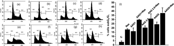
Combined activity of anticancer drug 17‐allylamino‐17‐demethoxygeldanamycin (17‐AAG) and resveratrol in K562 chronic myelogenous leukemia cells. (a–d) Cell cycle analysis by flow cytometry of control and 17‐AAG treated K562 cells. (a) Control untreated K562 cells; (b) 1 µM 17‐AAG; (c) 2.5 µM 17‐AAG; (d) 5 µM 17‐AAG. (e–h) Cell cycle analysis by flow cytometry of K562 cells treated for 48 h with 40 µM resveratrol only (e) and with both resveratrol (40 µM) and 17‐AAG at concentrations of 1 µM (f), 2.5 µM (g), or 5 µM (h). M1, sub‐G0/G1; M2, G1; M3, S; M4, G2 phase of the cell cycle. (i) Percentage of sub‐G0/G1 cells in K562 cells treated with 17‐AAG and resveratrol. Data are presented as the mean ± SD of three or four separate experiments. The cells were subjected to the treatments detailed above for a period of 48 h.
Figure 6.

Heat shock protein 70 (Hsp70) status of K562 chronic myelogenous leukemia cells treated with 17‐allylamino‐17‐demethoxygeldanamycin (17‐AAG) and resveratrol. (a) Upper panel, Hsp70 mRNA levels in K562 cells treated with 1 µM 17‐AAG and 40 µM resveratrol; lower panel, β‐actin levels in K562 cells treated with 1 µM 17‐AAG and 40 µM resveratrol. C, control; C+Res, K562 cells treated with resveratrol; AAG1, 1 µM 17‐AAG; AAG1+Res, resveratrol and 1 µM 17‐AAG. Ratio of Hsp70/β‐actin mRNA (normalized to control) is presented as the mean of data from two separate experiments. (b) Upper panel, Hsp70 protein levels in K562 cells treated with 2.5 µM 17‐AAG and 40 µM resveratrol; lower panel, β‐actin levels in cells treated with 2.5 µM 17‐AAG and 40 µM resveratrol. C, control; AAG2.5, 2.5 µM 17‐AAG; AAG2.5+Res, resveratrol and 2.5 µM 17‐AAG. Ratio of Hsp70/β‐actin protein (normalized to control) is presented at the bottom. The scan is representative of two separate experiments. (c) Upper panel, Hsp70 mRNA levels in 2.5 µM 17‐AAG and 40 µM resveratrol treated K562 cells; lower panel, β‐actin mRNA levels in 2.5 µM 17‐AAG and 40 µM resveratrol treated K562 cells. C, control; AAG 2.5, 2.5 µM 17‐AAG; AAG2.5+Res, resveratrol and 2.5 µM 17‐AAG. Ratio of Hsp70/β‐actin mRNA (normalized to control) is presented at the bottom. The cells were subjected to the treatments mentioned above for a period of 48 h. The scan is representative of two separate experiments.
Effect of resveratrol on non‐adherent WBC from normal human blood. Non‐adherent WBC were collected from normal volunteers after obtaining informed consent. The Hsp70 protein levels in these cells were found to be extremely low in comparison with K562 cells (Fig. 7, upper panel). Furthermore, the levels of sub‐G0/G1 fragmented DNA observed in these cells with or without exposure to resveratrol were similar (Fig. 7, lower panel) indicating that resveratrol (40 µM) exposure for 48 h did not cause any change in apoptosis of normal non‐adherent WBC. This is in agreement with previous findings by Clement et al.( 17 ) that resveratrol shows minimal toxicity in normal peripheral blood lymphocytes.
Figure 7.
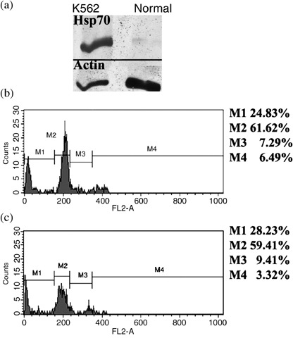
Status of heat shock protein 70 (Hsp70) levels in normal non‐adherent white blood cells (WBC) and the effect of resveratrol on sub‐G0/G1 DNA in these cells. (a) Western blot of Hsp70 protein in K562 chronic myelogenous leukemia cells and normal non‐adherent WBC. Upper panel, Hsp 70 protein levels; lower panel, β‐actin levels. (b,c) Cell cycle analysis by flow cytometry of normal non‐adherent WBC with (c) or without (b) 40 µM resveratrol exposure. M1, sub‐G0/G1; M2, G1; M3, S; M4, G2 phase of the cell cycle.
Discussion
Resveratrol is a phytoalexin with potent chemopreventive and chemotherapeutic properties against a wide array of diseases.( 28 ) In this study we tried to establish the pro‐apoptotic role of resveratrol in CML cells and to elucidate the mechanisms underlying this action. Although there are reports of induction of apoptosis by resveratrol in other forms of leukemia and cancer models,( 29 ) its effects on the promotion of apoptosis in CML have not been extensively studied. Our results show that resveratrol can induce apoptosis in K562 cells in a time‐dependent manner. This apoptosis induction involves PS externalization, the activation of caspase‐3, and increased percentage of cell population with fragmented DNA (sub‐G0/G1) (Fig. 3). All these data put together confirm the ability of resveratrol to induce apoptosis in K562 cells.
The anti‐apoptotic chaperone Hsp70 is found to be highly expressed in K562 cells and it may present a major challenge in inducing apoptosis in K562 cells. An important role for Hsp70 has been described, in its inhibition of apoptosis in acute leukemia and in imatinib resistance in CML.( 26 )Thus, the challenge to induce apoptosis in K562 cells is to overcome the highly overexpressed levels of Hsp70.
We found that resveratrol possesses the property of downregulating Hsp70 levels both at the mRNA and protein levels (Fig. 1) in K562 cells. This downregulation was found to be time and dose dependent in nature. As Hsp70 is a chaperone and is required for providing a cytoprotective role in stress conditions, the complete abrogation of Hsp70 might be lethal to normal cells. Our results suggest that a concentration of 40 µM resveratrol was sufficient in inducing apoptosis in K562 cells without completely abrogating the Hsp70 status. Therefore, inhibition of Hsp70 levels with resveratrol provides a good compromise between maintenance of normal chaperone activity and induction of apoptosis.
As the two main chaperones, Hsp70 and Hsp90, are associated with the anti‐apoptotic property of cells, they also play a crucial role in promoting tumorigenesis and imparting greater survival of cancerous cells under cytotoxic stress.( 30 ) STA‐4783, a novel small molecule drug candidate, induces a rapid and significant oxidative stress response, leading to apoptosis in many human cancer cells.( 31 ) As a protective response to substantial oxidative stress, the expression of Hsps, metallothioneins, and antioxidant genes are enhanced by STA‐4783. But this protective response fails because the sustained production of reactive oxygen species overpowers the protective response and drives the cells to apoptotic death.( 31 ) Therefore, in all likelihood, the heat shock response induced by STA‐4783 is a minor effect completely overwhelmed by the major effect of the drug, which is sustained production of reactive oxygen species in the cancer cells. Hence, the anticancer effect of STA‐4783 might not involve the increase in heat shock proteins. Much of the focus of targeted therapy for cancer has been the designing of Hsp90 inhibitors, namely geldanamycin, 17‐AAG, and spheredin.( 32 ) But the efficacy of these drugs is rendered low as they do not inhibit Hsp70, and inhibition of Hsp90 might lead to HSF1‐mediated higher expression of Hsp70.( 25 ) 17‐AAG is known to decrease Bcr‐Abl levels,( 33 ) and Bcr‐Abl is associated with increased expression of Hsp70.( 25 ) However, 17‐AAG is also known to induce Hsp70 expression.( 25 ) In this study, 1 µM 17‐AAG did not appreciably increase Hsp70 levels, but a combination of 17‐AAG (1 µM) and resveratrol decreased Hsp70 levels (30–40% decrease from control levels). In contrast, 2.5 µM 17‐AAG augmented both protein and mRNA levels of Hsp70 (Fig. 6b,c) in agreement with previous reports.( 25 ) This was brought down by combining 17‐AAG (2.5 µM) with resveratrol, but to levels still higher than those of the control. In our study, 17‐AAG alone did not diminish Hsp70 levels, rather, 17‐AAG treatment resulted in a noticeable increase in Hsp70 levels. The addition of resveratrol with 17‐AAG significantly increased the apoptotic effects of 17‐AAG and diminished Hsp70 levels. Therefore, the increase in DNA fragmentation observed by the simultaneous application of resveratrol and 17‐AAG over treatment by 17‐AAG alone could be connected to the status of Hsp70. In essence, resveratrol treatment along with 17‐AAG produced a higher amount of apoptosis in K562 cells compared to 17‐AAG alone as the former abrogated the levels of Hsp70, whereas Hsp90 was inhibited by 17‐AAG. As described in recent published reports,( 25 , 33 ) 17‐AAG could have two entirely opposite effects on Hsp70. 17‐AAG, through its lowering effect on Bcr‐Abl,( 33 ) might bring down Hsp70 levels as Bcr‐Abl is known to enhance the expression of Hsp70.( 25 ) In total contrast, 17‐AAG‐induced inhibition of Hsp90 can trigger Hsp70 increase by enhancing the availability of HSF1 required for Hsp70 transcription.( 25 ) In the present study it seems likely that 17‐AAG treatment of K562 cells might have followed the second possibility. Hsp90 from normal cells bind 17‐AAG with a 100‐fold lower affinity than Hsp90 derived from cancer cells.( 34 ) This high affinity of cancer cell‐derived Hsp 90 is explained by the presence of activated Hsp90 complexes in cancer cells. Also, Hsp90 client proteins in normal cells are less sensitive to 17‐AAG than similar proteins expressed in cancer cells.( 35 ) Therefore, cytotoxicity of 17‐AAG to normal cells is not foreseen.
Once the inhibitory effects of resveratrol on Hsp70 levels were confirmed, the need to uncover the underlying mechanisms became urgent. Resveratrol clearly blocked translocation of HSF1 to the nucleus, resulting in a predominantly cytoplasmic localization of HSF1 (Fig. 4). As HSF1 requires phosphorylation at specific amino acid residues for it to trimerize and translocate to the nucleus, and subsequently to act as the active transcription factor for the Hsp70 gene, resveratrol might block this phosphorylation by interacting with the kinases involved in the process.
The efficacy of resveratrol as a chemopreventive and sensitizer for anticancer drug therapy has been stressed by many recent studies.( 19 , 36 ) Apart from directly targeting Hsp70, resveratrol could exert its pro‐apoptotic effects through downregulation of inhibitors of apoptosis like survivin. The downregulation of inhibitors of apoptosis may be direct or through inhibition of the chaperone function of Hsp70. Recently, the option of using natural products in the treatment of hematological malignancies received encouragement when a report came out that the tea polyphenol epigallocatechin gallate could produce favorable responses in chronic lymphoid leukemia and follicular lymphoma.( 37 ) For the first time we report that resveratrol could downregulate the high endogenous Hsp70 levels in K562 cells. Recent studies have shown( 26 ) that the anti‐apoptotic effects of Bcr‐Abl in leukemia are partially mediated by increased expression of Hsp70. So identification of natural compounds that could downregulate Hsp70 can provide an option for sensitizing cancerous cells to conventional drugs and radiation therapy. Therefore, resveratrol might offer an option for adjunct therapy alongside inhibitors of Bcr‐Abl and Hsp90. Our study also established that resveratrol downregulated Hsp70 through inhibition of HSF1 transcriptional activity, mediated by a blockage of nuclear translocation of HSF1.
Acknowledgments
We are grateful to Swasti Ray Chaudhuri of the Structural Genomics section of Saha Institute of Nuclear Physics for his help with the confocal microscopy. We thank Dr Santu Bandyopadhayay of the Indian Institute of Chemical Biology, Kolkata for giving us the KU812 and KCL22 cell lines.
References
- 1. Deininger MWN, Goldman JM, Melo JV. The molecular biology of chronic myeloid leukemia. Blood 2000; 96: 3343–56. [PubMed] [Google Scholar]
- 2. Bedi A, Barber JP, Bedi GC et al . BCR‐ABL‐mediated inhibition of apoptosis with delay of G2/M transition after DNA damage: a mechanism of resistance to multiple anticancer agents. Blood 1995; 86: 1148–58. [PubMed] [Google Scholar]
- 3. Amarante‐Mendes GP, Kim CN, Liu L et al . Bcr‐Abl exerts its antiapoptotic effect against diverse apoptotic stimuli through blockage of mitochondrial release of cytochrome C, activation of caspase‐3. Blood 1998; 91: 1700–5. [PubMed] [Google Scholar]
- 4. Kharas MG, Fruman DA. ABL oncogenes and phosphoinositide 3‐kinase: mechanism of activation and downstream effectors. Cancer Res 2005; 65: 2047–53. [DOI] [PubMed] [Google Scholar]
- 5. Sánchez‐Arévalo Lobo VJ, Aceves Luquero CI, Álvarez‐Vallina L et al . Modulation of the p38 MAPK (mitogen‐activated protein kinase) pathway through Bcr/Abl: implications in the cellular response to Ara‐C. Biochem J 2005; 387: 231–8. [DOI] [PMC free article] [PubMed] [Google Scholar]
- 6. Raitano AB, Halpern JR, Hambuch TM, Sawyers CL. The Bcr‐abl leukemia oncogene activates jun kinase and requires jun for transformation. Proc Natl Acad Sci USA 1995; 92: 1746–50. [DOI] [PMC free article] [PubMed] [Google Scholar]
- 7. Kirchner D, Duyster J, Ottmann O, Schmid RM, Bergmann L, Munzert G. Mechanisms of Bcr‐Abl‐mediated NF‐κB/Rel activation. Exp Hematol 2003; 31: 504–11. [DOI] [PubMed] [Google Scholar]
- 8. Benekli M, Baer M, Baumann RH, Wetzler M. Signal transducer and activator of transcription proteins in leukemias. Blood 2003; 101: 2940–54. [DOI] [PubMed] [Google Scholar]
- 9. Sawyers CL, Hochhaus A, Feldman E et al . Imatinib induces hematologic and cytogenetic responses in patients with chronic myelogenous leukemia in myeloid blast crisis: results of a phase II study. Blood 2002; 99: 3530–9. [DOI] [PubMed] [Google Scholar]
- 10. Schindler T, Bornmann W, Pellicena P, Miller WT, Clarkson B, Kuriyan J. Structural mechanism for STI‐571 inhibition of abelson tyrosine kinase. Science 2000; 15: 1938–42. [DOI] [PubMed] [Google Scholar]
- 11. Walz C, Sattler M, Ellwood K. et al . Novel targeted therapies to overcome imatinib mesylate resistance in chronic myeloid leukemia (CML). Crit Rev Oncol Hematol 2006; 57: 145–64. [DOI] [PubMed] [Google Scholar]
- 12. Gorre ME, Mohammed M, Ellwood K et al . Clinical resistance to STI‐571 cancer therapy caused by BCR‐ABL gene mutation or amplification. Science 2001; 3: 876–80. [DOI] [PubMed] [Google Scholar]
- 13. Shah NP, Nicoll JM, Nagar B et al . Multiple BCR‐ABL kinase domain mutations confer polyclonal resistance to the tyrosine kinase inhibitor imatinib (STI571) in chronic phase and blast crisis chronic myeloid leukemia. Cancer Cell 2002; 2: 117–25. [DOI] [PubMed] [Google Scholar]
- 14. Goh SS, Woodman OL, Pepe S, Cao AH, Qin C, Ritchie RH. The red wine antioxidant resveratrol prevents cardiomyocyte injury following ischaemia‐reperfusion via multiple sites and mechanisms. Antioxid Redox Signal 2007; 9: 101–13. [DOI] [PubMed] [Google Scholar]
- 15. Winkelmann I, Diehl D, Osterle D et al . The suppression of aberrant crypt multiplicity in colonic tissue of 1,2‐dimethylhydrazine‐treated C57BL/6J mice by dietary flavone is associated with an increased expression of Krebs cycle enzymes. Carcinogenesis 2007; 28: 1446–54. [DOI] [PubMed] [Google Scholar]
- 16. Pervaiz S. Chemotherapeutic potential of the chemopreventive phytoalexin resveratrol. Drug Resist Updat 2004; 7: 333–44. [DOI] [PubMed] [Google Scholar]
- 17. Clement MV, Hirpara JL, Chawdhury SH, Pervaiz S. Chemopreventive agent resveratrol, a natural product derived from grapes, triggers CD95 signaling‐dependent apoptosis in human tumor cells. Blood 1998; 92: 996–1002. [PubMed] [Google Scholar]
- 18. Ziegler CC, McEntee MF, Hansen‐Petrik M et al . In vivo effects of trans‐resveratrol on intestinal tumorigenesis. FASEB J 2001; 15: A617. [Google Scholar]
- 19. Fulda S, Debatin KM. Sensitization for anticancer drug‐induced apoptosis by the chemopreventive agent resveratrol. Oncogene 2004; 23: 6702–11. [DOI] [PubMed] [Google Scholar]
- 20. Caroline J, Morimoto RI. Role of the heat shock response and molecular chaperones in oncogenesis and cell death. J Natl Cancer Inst 2000; 92: 1564–72. [DOI] [PubMed] [Google Scholar]
- 21. Lila P, Nykanen P, Sistonen L. Roles of the heat shock transcription factors in regulation of the heat shock response and beyond. FASEB J 2001; 15: 1118–31. [DOI] [PubMed] [Google Scholar]
- 22. Sreedhar AS, Csermely P. Heat shock proteins in the regulation of apoptosis: new strategies in tumor therapy: a comprehensive review. Pharmacol Ther 2004; 101: 227–57. [DOI] [PubMed] [Google Scholar]
- 23. Beere HM. The stress of dying: the role of heat shock proteins in the regulation of apoptosis. J Cell Sci 2004; 117: 2641–51. [DOI] [PubMed] [Google Scholar]
- 24. Blagoskonny MV, Fojo T, Bhall KN et al . The Hsp90 inhibitor geldanamyein selectively sensitizes Bcr‐Abl expressing leukemia cells to cytotoxic chemotherapy. Leukemia 2001; 15: 1537–43. [DOI] [PubMed] [Google Scholar]
- 25. Guo F, Rocha K, Bali P et al . Abrogation of heat shock protein 70 induction as a strategy to increase antileukemia activity of heat shock protein 90 inhibitor 17‐allylamino‐demethoxy geldanamycin. Cancer Res 2005; 65: 10536–44. [DOI] [PubMed] [Google Scholar]
- 26. Pocaly M, Lagarde V, Etienne G et al . Overexpression of the heat‐shock protein 70 is associated to imatinib resistance in chronic myeloid leukemia. Leukemia 2007; 21: 93–101. [DOI] [PubMed] [Google Scholar]
- 27. Seo HR, Chung DY, Lee YJ, Lee DH, Kim JI, Bae S et al . Heat shock protein 25 or inducible heat shock protein 70 activates heat shock factor 1: dephosphorylation on serine 307 through inhibition Of Erk1/2 phosphorylation. J Biol Chem 2006; 281: 17220–7. [DOI] [PubMed] [Google Scholar]
- 28. Pervaiz S. Resveratrol: from grapevines to mammalian biology. FASEB J 2003; 17: 1975–85. [DOI] [PubMed] [Google Scholar]
- 29. Cal C, Garban H, Jazirehi A, Yeh C, Mizutani Y, Bonavida B. Resveratrol and cancer: chemoprevention, apoptosis, and chemo‐immunosensitizing activities. Curr Med Chem Anticancer Agents 2003; 3: 77–93. [DOI] [PubMed] [Google Scholar]
- 30. Calderwood SK, Khaleque MA, Sawyer DB, Daniel RC. Heat shock proteins in cancer: chaperones of tumorigenesis. Trends Biochem Sci 2006; 31: 164–72. [DOI] [PubMed] [Google Scholar]
- 31. Foley KP, Mihalek R, Korbut T et al . Concurrent chemoradiation therapy with the oxidative stress inducer STA‐4783 in mouse xenograft tumor models. Int J Rad Oncol Biol Phys 2007; 69: S593–4. [Google Scholar]
- 32. Powers MV, Workman P. Inhibitors of the heat shock response: biology and pharmacology. FEBS Lett 2007; 581: 3758–69. [DOI] [PubMed] [Google Scholar]
- 33. Whitney JME, Harvey AJ, Crompton MR. RNA interference of Bcr‐Abl increases chronic myeloid leukemia cell killing by 17‐allylamino‐17‐demethoxygeldanamycin. Leukemia Res 2006; 30: 553–60. [DOI] [PubMed] [Google Scholar]
- 34. Kamal A, Thao L, Sensintaffar J et al . A high‐affinity conformation of Hsp90 confers tumour selectivity on Hsp90 inhibitors. Nature 2003; 425: 407–10. [DOI] [PubMed] [Google Scholar]
- 35. Da Rocha Dias S, Friedlos F, Light Y, Springer C, Workman P, Marais R. Activated B‐RAF is an Hsp90 client protein that is targeted by the anticancer drug 17‐allylamino‐17‐demethoxygeldanamycin. Cancer Res 2005; 65: 10686–91. [DOI] [PubMed] [Google Scholar]
- 36. Park EJ, Pezzuto JM. Botanicals in cancer chemoprevention. Cancer Metastasis Rev 2002; 21: 231–55. [DOI] [PubMed] [Google Scholar]
- 37. Shanafelt T, Lee Y, Call TG et al . Clinical effects of oral green tea extracts in four patients with low grade B‐cell malignancies. Leukemia Res 2006; 30: 707–12. [DOI] [PubMed] [Google Scholar]