

A 29-year-old patient, weighing 60 kg, was referred for [18 F]FDG PET/CT imaging at 18 weeks of pregnancy, a critical stage for fetal development with increased sensitivity to radiation [1]. This scan was part of the eligibility screening for external beam radiotherapy treatment for Hodgkin lymphoma IVA.
Utilizing the ultra-high-sensitivity mode of the Biograph Vision Quadra system (Siemens Healthineers) [2], we performed a 15-minute head-to-mid-femur scan (60 min post-injection). This was reconstructed into shorter scan durations to determine an acceptable duration using Likert scores and feedback from three nuclear medicine physicians. To minimize fetal exposure, only 20 MBq of [18 F]FDG was administered, and a tin filter was used to reduce CT dose (Sn140, 17 mAs, 1.7 pitch, CTDIvol = 0.4 mGy).
The effective fetal dose was quantified at 0.13 mSv for internal (PET) and 0.58 mSv for external radiation (CT) [3, 4], approximately 140 times below thresholds known to affect cognitive development and recommended levels for minimizing fetal exposure [5].
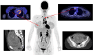
The 5-minute scan was identified as the shortest duration with excellent clinical image quality. Pathologic uptake in enlarged supraclavicular, mediastinal, and intra-abdominal lymph nodes was clearly visible, along with small pulmonary lesions in the right lung (4–14 mm). Pregnancy-related changes, such as increased mammary uptake and increased activity in the ovaries and uterus, were evident, along with traces of fetal [18 F]FDG uptake.
This case demonstrates the effectiveness of LAFOV PET/CT imaging in pregnant patients, minimizing fetal radiation exposure while maintaining high-quality maternal imaging. Other low-dose applications include patient groups with vulnerable genomes, children, and regular follow-ups.
Author contributions
All authors contributed to the study conception and design. Material preparation, data collection and analysis were performed by J.H. van Snick, O.V. Ivashchenko and K.P. Koopmans. The first draft of the manuscript was written by J.H. van Snick and O.V. Ivashchenko and all authors commented on previous versions of the manuscript. All authors read and approved the final manuscript.
Funding
The authors declare that no funds, grants, or other support were received during the preparation of this manuscript.
Data availability
The datasets generated during and/or analyzed during the current study are available from the corresponding author on reasonable request.
Declarations
Ethics approval
This case study was performed in line with the principles of the Declaration of Helsinki.
Consent to participate
Written informed consent was obtained from the patient.
Consent to publishing
The authors affirm that human research participants provided informed consent for publication of the images in figure.
Competing interests
The authors have no relevant financial or non-financial interests to disclose. The UMCG and Siemens have a partnership for building the future of health (PUSH). Siemens was not involved in the imaging process nor in the writing phase of this case.
Footnotes
Publisher’s Note
Springer Nature remains neutral with regard to jurisdictional claims in published maps and institutional affiliations.
References
- 1.Zanotti-Fregonara P et al. New fetal dose estimates from 18F-FDG administered during pregnancy: standardization of dose calculations and estimations with Voxel-based anthropomorphic phantoms. JNM. 2016; https://jnm.snmjournals.org/content/57/11/1760.long [DOI] [PubMed]
- 2.Mingles C, et al. Impact of the new ultra-high-sensitivity mode in a long axial field-of-view PET/CT. Ann Nucl Med. 2023;37:310–5. [DOI] [PMC free article] [PubMed] [Google Scholar]
- 3.Sensakovic WF, et al. Fetal dosimetry at CT: a primer. Radiographics. 2020;40(4):1061–70. [DOI] [PubMed] [Google Scholar]
- 4.Boone JM et al. Size-Specific Dose Estimation (SSDE) in Pediatric and Adult Body CT Examination. AAPM Report 204. ISBN 978-1-936366-08-8.
- 5.ICRP. 2000. Pregnancy and Medical Radiation. ICRP Publication 84. Ann. ICRP 30 (1). [DOI] [PubMed]
Associated Data
This section collects any data citations, data availability statements, or supplementary materials included in this article.
Data Availability Statement
The datasets generated during and/or analyzed during the current study are available from the corresponding author on reasonable request.