



ABSTRACT
Background:
Acute pancreatitis (AP) is an inflammatory disease of the pancreas with varying severity. The mortality rate varies from 20% to 40% among severe acute pancreatitis (SAP). Interleukin-6 (IL-6) is a pro- and anti-inflammatory cytokine that involves various infections, inflammations, and systemic disorders. Injury to acinar cells leads to necrosis, releasing proinflammatory cytokines, including IL-6, which peaks earlier. The lack of extensive data regarding the association of IL-6 with AP influences us to do this meta-analysis for early detection and treatment of AP to prevent multiorgan failure.
Methods:
We searched the PubMed, Cochrane Library, and Google Scholar databases for relevant articles published from inception to June 2024. We examined the positive and negative likelihood ratios, diagnostic odds ratios, and pooled sensitivity and specificity. We used the QUADAS-2 tool to evaluate the risk of bias.
Results:
This meta-analysis included 13 studies involving 1386 patients with AP, of which 343 had SAP and 1043 had mild and moderately severe AP. The positive and negative likelihood ratios were 3.5 (95% CI 2.6 to 4.5) and 0.25 (95% CI 0.16 to 0.40). The diagnostic odds ratio of IL-6 to diagnose SAP is 14 (95% CI: 7 to 27), and the summary receiver operating characteristic curve is 0.85 (95% CI: 0.82–0.88).
Conclusion:
Based on the results of this meta-analysis, serum IL-6 is a promising biomarker for diagnosing SAP in the early stage. However, a larger-scale study involving a more extensive population is necessary due to the considerable variation between the studies.
Keywords: Acute pancreatitis, diagnostic accuracy, IL-6, sensitivity, specificity
Introduction
Acute pancreatitis (AP) is an inflammatory condition of the pancreas that can vary in severity, ranging from mild to severe. It can result in multiorgan failure and a high fatality rate. Approximately 80% of individuals experience a mild to moderate self-limiting condition, whereas the other 20% develop severe AP with a significant risk of mortality. Patients who develop severe AP exhibit necrosis of the pancreatic or peripancreatic tissue or organ failure or both. This condition has a substantial mortality rate, which varies from 20% to 40%.[1,2] The most common cause of acute pancreatitis is a biliary cause like gallstone, followed by alcohol. Other less frequent causes are duct obstruction, metabolic disorders, smoking, trauma, and drugs like estrogen and thiazides. Acute pancreatitis increases with age and is more prevalent among males than among females.[3] The severity of AP is evaluated using clinical, biochemical, and radiological examination. Ranson’s criteria (11 criteria), the Glasgow score (8 criteria),[4,5] the Acute Physiology and Chronic Health Evaluation (APACHE II) score (14 criteria)[6] and Atlanta Classification[7] are some of the methods used to determine the severity of acute pancreatitis. Early diagnosis and treatment can reduce the complications associated with acute pancreatitis.
Interleukin-6 (IL-6) is a cytokine of a quadruple-helix structure with 184 amino acids. It is synthesized by a wide range of cells that express its effects on multiple cell types via unique receptors and causes biological activities.[8,9] IL-6 is a cytokine involved in various pathophysiological processes, including infections, inflammation, and disorders affecting the nervous system, blood vessels, and cancer.[10] IL-6 levels in the blood are very low in normal, healthy people, but they rise very quickly during the early stages of infection and inflammation. It had pro- and anti-inflammatory properties.[11] During the early phase of inflammation, IL-6 is produced at the site of inflammation. Most immune cells produce IL-6, which regulates and coordinates the immune response.[12]
The primary cause of acute pancreatitis is injury to the acinar cells. Necrotic acinar cells release damage-associated molecular patterns (DAMPs) and proinflammatory cytokines, including IL-6 and other cytokines and anti-inflammatory cytokines. IL-6 is a cytokine with both proinflammatory and anti-inflammatory properties. It is one of the first cytokines involved in the inflammation associated with AP.[13,14] Treatment with anti-IL-6 antibodies effectively neutralizes IL-6, inhibiting STAT-3 activation in pancreatic acinar cells. This results in a reduction in the severity of acute pancreatitis.[15]
There are very few meta-analyses regarding the association between IL-6 and acute pancreatitis. Identifying AP in the early stages to protect systemic inflammation and the functions of many significant organs among adults and children is of utmost importance. This prompts us to do a meta-analysis on the association of IL-6 in AP among a larger population and draw a conclusion.
This meta-analysis aims to assess the diagnostic precision of IL-6 in evaluating the severity of AP. By analyzing data from various studies, we will explore the sensitivity, specificity, and diagnostic accuracy in the clinical assessment of AP. This meta-analysis will significantly impact the detection and progression of AP. Serum IL-6 can be considered a routine biochemical marker for diagnosing AP and helps in early treatment to protect from multiple organ failure.
Methods
Our meta-analysis was conducted according to the protocol created and filed in the PROSPERO database (CRD42024553502). To define our research question, we used the PICO tool as follows:
Patients/population: The patient is suffering from AP.
Index test: Serum Interleukin-6.
Comparison: Severe Acute Pancreatitis.
Outcome: Diagnostic accuracy of serum IL-6 determined by pooled sensitivity and specificity.
Two independent reviewers thoroughly searched the PubMed, Cochrane Library, and Google Scholar databases for relevant research articles published between inception and June 2024. The search keywords and filters included (((((Interleukin-6) OR (IL-6)) AND (Acute Pancreatitis)) AND (Sensitivity)) AND (Specificity)) AND (diagnostic) with additional filters Human and English.
Inclusion criteria: We included studies if (a) patients were diagnosed with AP, (b) serum IL-6 was selected for distinguishing severe AP from mild and moderate AP, (c) studies published as full-text publications, (d) studies published as original articles in English, and (e) studies should have sufficient data to form 2 × 2 tables for estimating sensitivity and specificity.
Exclusion criteria: (a) preclinical studies, (b) reviews and abstracts without the availability of full-text research papers, (c) case reports, (d) case studies, (e) guidelines, (f) letter to editor, (g) conference abstracts, (h) studies which do not report data for pooled analysis, and (i) studies from which we cannot extract 2 × 2 data (true positive, true negative, false positive, and false negative).
Data extraction
Two researchers independently extracted all the data; the results were then cross-checked, and any discrepancies were resolved by consulting a third investigator. The following data were extracted from the studies: first author’s name; publication year; study design; study country; number of patients with SAP, MSAP, and MAP; mean/median age; IL-6 level; cutoff value of serum IL-6; and sensitivity and specificity of serum IL-6.
Methodology for quality assessment
Quality assessment of the included studies was conducted independently by two researchers using the Quality Assessment of Diagnostic Accuracy Studies (QUADAS-2) tool. The two researchers and a third researcher discussed and concluded in the event of a discrepancy. To assess quality, the QUADAS-2 implements a four-domain methodology: patient selection, index test, reference standard, and flow and timing. The risk of bias was classified into three categories: high, low, and unclear.
Statistical analysis
The random-effects model (I2 > 50%) was employed to calculate the aggregate effect size for diagnostic accuracy regarding sensitivity and specificity with a 95% confidence interval (CI). We constructed summary receiver operating characteristic curves (SROCs) to determine the discriminatory accuracy of serum IL-6 in assessing the severity of AP. We used I² statistics to determine the heterogeneity of the studies. We evaluated publication bias using Deek’s funnel plot asymmetry test. We used STATA version 13 software to conduct the statistical analyses. We used Review Manager Version 5.4 (The Nordic Cochrane Center, The Cochrane Collaboration, Copenhagen) to assess the risk of bias.
Results
Search result
A total of 427 studies were identified from the PubMed, Cochrane Library, and Google Scholar databases. After removing duplicate and irrelevant articles, we had 111 studies left for full-text review and data extraction. We excluded 29 studies for not having full-text articles, 16 for only having serum IL-6 levels with insufficient data, 22 for involving other conditions along with AP, and 31 for insufficient data. Finally, we included 13 studies in the systematic review and meta-analysis. Details are provided in the PRISMA chart [Figure 1].
Figure 1.

Study flow diagram (PRISMA) representing the study selection and inclusion
Characteristics of included study
Tables 1 and 2 give a summary of the included study. Ten studies used Revised Atlanta Classification, and three used APACHE II, Stimac D used APACHE II, Ranson-3, and Glasgow, and Heath DI used clinical and radiological data to assess the severity of AP. Cho IR used chemiluminescent immunoassay, Kumar RB did not mention, Kolber W used electrochemiluminescence immunoassay, and the rest used enzyme-linked immunosorbent assay for assessing serum IL-6 level. The total number of participants in 13 studies was 1386, of which 343 had severe AP and the rest had mild and moderate AP. The total mortality due to SAP was 21.87%. The biliary cause of AP was 44.73%, alcohol, 23.73%; and other, 31.52%. AP among males was 55.77%, and that among females was 44.22%.
Table 1.
Characteristics of included study
| Author | Year | Acute Pancreatitis case | SAP | MAP + MSAP | Total Mortality | MAP + MSAP Mortality | SAP Mortality | MALE | Female | Biliary | Alcohol | Other Causes |
|---|---|---|---|---|---|---|---|---|---|---|---|---|
| Cho IR[16] | 2023 | 103 | 8 | 95 | 2 | 0 | 2 | 70 | 33 | 33 | 39 | 31 |
| Kumar RB[17] | 2022 | 62 | 22 | 40 | 4 | 0 | 4 | 34 | 28 | 42 | 11 | 9 |
| Li J[18] | 2022 | 67 | 35 | 32 | 6 | 0 | 6 | 20 | 47 | 25 | 10 | 32 |
| Sternby H[19] | 2021 | 115 | 11 | 104 | 5 | 0 | 5 | 57 | 58 | 59 | 24 | 32 |
| Shen D[20] | 2021 | 143 | 26 | 117 | 5 | 0 | 5 | 94 | 49 | 46 | 8 | 89 |
| Jain S[21] | 2018 | 115 | 37 | 78 | 20 | 0 | 20 | 65 | 50 | 50 | 26 | 39 |
| Sternby H[22] | 2017 | 175 | 51 | 124 | 7 | 0 | 7 | 85 | 90 | 96 | 36 | 43 |
| Kolber W[23] | 2017 | 95 | 8 | 87 | 4 | 0 | 4 | 65 | 30 | 27 | 29 | 39 |
| Sternby H[24] | 2016 | 232 | 39 | 193 | 5 | 0 | 5 | 121 | 111 | 131 | 39 | 62 |
| Dambrauskas Z[25] | 2010 | 108 | 48 | 60 | 14 | 0 | 14 | 56 | 52 | 27 | 46 | 35 |
| Sathyanarayan G[26] | 2007 | 30 | 13 | 17 | 3 | 0 | 3 | 22 | 8 | 14 | 10 | 6 |
| Stimac D[27] | 2006 | 117 | 35 | 82 | 65 | 52 | 60 | 39 | 18 | |||
| Heath DI[28] | 1993 | 24 | 10 | 14 | 19 | 5 | 10 | 12 | 2 |
SAP - Severe Acute pancreatitis, MSAP - Severe Moderate Acute pancreatitis, MAP - Mild Acute pancreatitis, IL-6 - Interleukin - 6
Table 2.
Characteristics of included study
| Author | Year | Country region | Study type | Severity of pancreatitis assessed by criteria | Acute pancreatitis case | SAP | MAP + MSAP | Acute pancreatitis age (Mean±SD)/(Median (IQR) | IL-6 Test method | Cut off (pg/ml) | Sensitivity | Specificity |
|---|---|---|---|---|---|---|---|---|---|---|---|---|
| Cho IR[16] | 2023 | Korea | 1 | 1 | 103 | 8 | 95 | 49.86±19.24 | 1 | 50 | 83.3 | 62.3 |
| Kumar RB[17] | 2022 | India | 2 | 1 | 62 | 22 | 40 | 39.21±12.43 | 137 | 100 | 88.37 | |
| Li J[18] | 2022 | China | 3 | 1 | 67 | 35 | 32 | 48±17 | 2 | 121.1 | 67.65 | 67.74 |
| Sternby H[19] | 2021 | Sweden | 3 | 1 | 115 | 11 | 104 | 65 (52–78) | 2 | 196.6 | 81.9 | 91.3 |
| Shen D[20] | 2021 | China | 3 | 1 | 143 | 26 | 117 | 48.24 14.27 | 2 | 46.8 | 69.2 | 83.8 |
| Jain S[21] | 2018 | India | 3 | 1 | 115 | 37 | 78 | 39.4±14.6 | 2 | 160 | 86 | 82 |
| Sternby H[22] | 2017 | Sweden | 3 | 1 | 175 | 51 | 124 | 66 (19-97) | 2 | 23.6 | 89 | 54 |
| Kolber W[23] | 2017 | Poland | 4 | 1 | 95 | 8 | 87 | 48±16.5 | 3 | 211 | 57 | 82 |
| Sternby H[24] | 2016 | Sweden | 3 | 1 | 232 | 39 | 193 | 2 | 72 | 40 | 78 | |
| Dambrauskas Z[25] | 2010 | Lithuania | 4 | 2 | 108 | 48 | 60 | 53.2±16 | 2 | 150 | 83.63 | 77.18 |
| Sathyanarayan G[26] | 2007 | India | 4 | 1,2 | 30 | 13 | 17 | 40.27±13.89 | 2 | 122 | 81.8 | 77.7 |
| Stimac D[27] | 2006 | Croatia | 4 | 2, 3, 4 | 117 | 35 | 82 | 2 | 118 | 68.7 | 69.9 | |
| Heath DI[28] | 1993 | London | 4 | 5 | 24 | 10 | 14 | 48 (22-73) | 4 | 130 | 100 | 71 |
| Revised Atlanta Classification - 1, APACHE II - 2, Ranson -3, Glasgow -4, Clinical and radiological -5 retrospective study - 1, observational analytical study - 2, cohort study - 3, prospective observational study – 4 Chemiluminescent Immunoassay - 1, Enzyme-Linked Immunosorbent Assay - 2, Electrochemiluminescence Immunoassay – 3, 7TD1 cell line – 4 | ||||||||||||
SAP - Severe Acute pancreatitis, MSAP - Severe Moderate Acute pancreatitis, MAP - Mild Acute pancreatitis, IL-6 - Interleukin - 6, SD - standard deviation
Methodological quality
QUADAS-2 assessment results [Figures 2 and 3] reveal a mixed quality of studies, with two studies exhibiting an unclear risk of bias in patient selection. Two studies present unclear risks in index tests, indicating a need for a cautious interpretation of their findings. However, most studies demonstrate a low risk of bias across other domains and show low applicability concerns, suggesting that their findings are generally reliable and relevant to the population, index test, and reference standard of interest.
Figure 2.

Summary risk of bias for each domain
Figure 3.
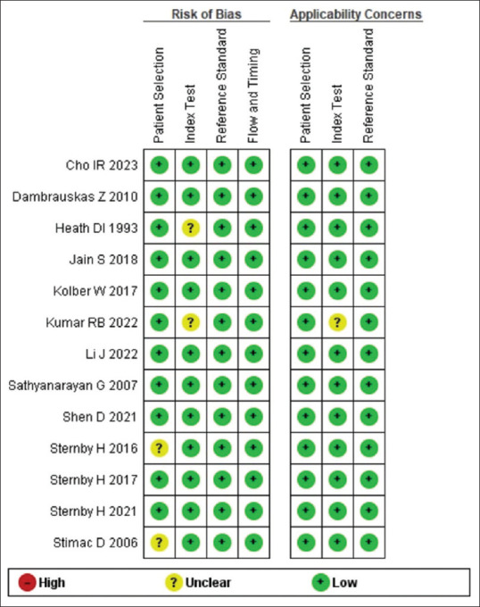
Risk of bias for individual studies. Risk of bias and applicability concerns summary: review authors’ judgements about each domain for each included study
Table 2 displays the sensitivities and specificities of IL-6 for the early diagnosis of SAP in the included studies. With a pooled sensitivity of 0.81 (95% CI 0.70 to 0.88), a pooled specificity of 0.77 (95% CI 0.70 to 0.82), and a diagnostic odds ratio (DOR) of 14 (95% CI 7 to 27), serum IL-6 is a promising biomarker in early diagnosis of SAP [Figure 4]. It effectively distinguishes patients with severe, mild, and moderate AP. SROC AUC is 0.85 (95% CI 0.82–0.88) [Figure 5]. Based on the Fagan nomogram, the serum IL-6 biomarker had a pretest probability of 20% for diagnosing SAP. The post-test probability increased to 47% with a positive likelihood ratio (PLR) of 3.5 (95% CI 2.6 to 4.6) and a negative likelihood ratio (NLR) of 0.25 (95% CI 0.16 to 0.40) [Figure 6]. Heterogeneity among the studies regarding pooled sensitivity I2 = 78.27 (95% CI 66.89 – 89.65) and pooled specificity I2 = 84.29 (95% CI 76.76 – 91.82). Table 3 provides a summary of these results.
Figure 4.

Forest plot of pooled sensitivity and specificity for diagnosing SAP
Figure 5.
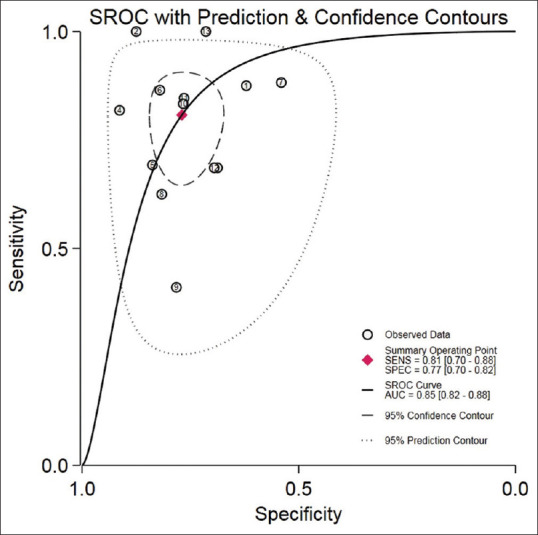
Summary ROC curve with confidence and prediction contours showing discriminatory power of IL-6 in the diagnosis of SAP. The X-axis represents the specificity of IL-6 for its discriminating accuracy in diagnosing SAP. The Y-axis represents the sensitivity of IL-6 for its discriminating accuracy in diagnosing SAP. ROC, receiver operating characteristic
Figure 6.

Fagan nomogram shows IL-6’s pretest and post-test probability in diagnosing SAP, IL-6, Interleukin-6; SAP - Sever Acute Pancreatitis
Table 3.
Diagnostic accuracy of serum IL-6 for SAP
| Parameter | n | Pooled Sensitivity (95% CI) | Pooled Specificity (95% CI) | PLR (95% CI) | NLR (95% CI) | DOR (95% CI) | SROC AUC |
|---|---|---|---|---|---|---|---|
| IL-6 | 13 | 0.81 [0.70-0.88] | 0.77 [0.70-0.82] | 3.5 [2.6-4.6] | 0.25 [0.16-0.40] | 14 [7-27] | 0.85 [0.82–0.88] |
IL-6, Interleukin-6; n, number; CI, confidence interval; PLR, positive likelihood ratio; NLR, negative likelihood ratio; DOR, diagnostic odds ratio; SROC AUC, summary receiver operating characteristics area under curve.
Publication bias
Deek’s funnel plot [Figure 7] was employed to evaluate the risk of publication bias. Publication bias will be significant only if P = <0.05. Our research revealed publication bias (P = 0.2), suggesting that the proportion of studies between positive and negative research studies published in the literature is disproportionate.
Figure 7.
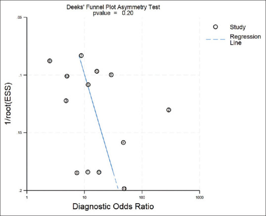
Deek’s funnel plot for assessment of risk of publication bias
Discussion
AP is an inflammatory condition of the pancreas that can vary in severity, ranging from mild to severe. It is a condition marked by inflammation of the pancreas, significantly impacting both mortality and morbidity rates. Several factors may contribute to AP, including biliary stones, alcohol use, duct blockage, smoking, trauma, and drug usage.[3] Injured acinar cells of the pancreas release several proinflammatory and anti-inflammatory cytokines. IL-6 is among the first to release cytokines from the injured cell. The amount of IL-6 released depends on the extent of injury to the cell. IL-6 is released into the serum, not stored anywhere, and quickly excreted or destroyed by anti-inflammatory antibodies. Therefore, it serves as a promising biomarker for AP.
After data analysis of 181 studies and 29 biomarkers, Van den Berg F et al.[29] concluded that IL-6 is superior in predicting MSAP/SAP early and can aid clinical decision-making. According to Rao S et al.,[30] IL-6 is the most effective inflammatory cytokine when measured within 24–36 hours of the onset of pancreatitis. The early increase and decrease of IL-6 in pancreatitis indicate that IL-6 could be a useful early indicator for the condition.[31] Staubli S et al.[32] stated that patients with high levels of IL-6 upon admission are 13.5 times more likely to develop severe pancreatitis. Meher S et al.[33] have indicated that IL-6 is effective in assessing the severity of pancreatitis According to Aoun E et al.,[34] IL-6 and IL-8 give similar results when predicting SAP. Mititelu A. et al.[35] compared data from studies on IL-6 and CRP. They found that assessing both IL-6 and CRP together is informative. However, they concluded that IL-6 should not replace CRP but instead serve as an early or complementary marker. Han Z et al.[36] studied the role of IL-6 in multiple diseases, including pancreatitis. They discovered that IL-6 had a high level of accuracy in diagnosing these conditions, suggesting its potential as a reliable diagnostic biomarker in the near future.
This meta-analysis aimed to comprehensively summarize the research on using serum IL-6 as a biomarker for diagnosing SAP. The lack of consistency and notable variations in interpreting different criterion scores emphasize the necessity for easily accessible and trustworthy blood-based biomarkers with high sensitivity and specificity. Finding reliable and efficient biomarkers can help diagnose and treat the condition quickly.
The results of our meta-analysis, which included 13 studies, show that serum IL-6 has a high level of accuracy in diagnosing SAP, with a pooled sensitivity of 0.81 (95% CI 0.70 to 0.88) and a specificity of 0.77 (95% CI 0.70 to 0.82). These data suggest that the serum IL-6 biomarker may accurately diagnose 81% of patients with SAP and exclude 77% without SAP. The area under the curve (AUC) of the SROC was 0.85 (95% CI 0.82–0.88), indicating that serum IL-6 has good diagnostic accuracy for SAP. These findings suggest that serum IL-6 can be used as a diagnostic biomarker for SAP. The likelihood ratio data (PLR = 3.5, NLR = 0.25) suggest that serum IL-6 should be carefully regarded as an independent diagnostic biomarker for SAP since PLR > 10 and NLR < 0.1 indicate high diagnostic accuracy. In our study, the DOR for IL-6 to diagnose SAP is 14 (95% CI 7 to 27), which can be considered a promising diagnostic biomarker.
Limitations
We must acknowledge certain unavoidable limitations when evaluating the results of the current meta-analysis. a) The small sample size in this study necessitates using a larger sample size to confirm the findings and conclusions. b) The primary drawback of this meta-analysis is the absence of a precise IL-6 cutoff value for diagnosing SAP. c) This study had significant heterogeneity due to the broad cutoff value range from 50 to 196.6. d) This thorough assessment included only published studies, potentially excluding unpublished work and introducing publication bias.
Conclusion
Based on the results of this meta-analysis, we conclude that serum IL-6 is a promising biomarker with high sensitivity and specificity for diagnosing SAP. Serum IL-6 has a sufficient probability of accurately detecting SAP. Separate studies for adults and adolescents are necessary. This meta-analysis will significantly impact the detection and progression of AP. Serum IL-6 can be considered a routine biochemical marker for diagnosing AP and helps in early treatment to protect from multiple organ failure. However, a larger study with an extensive sample size is required to address the heterogeneity among the studies.
Authors’ contribution
Study concept and design: SK
Data search and retrieval: SK, AS and TA
Analysis and interpretation of data: AK, AS, MKN, and RK
Tables and figures: PK, AK, MKN, RK, and AS
Drafting of the manuscript: SK, DK and MKN
Supervision: PK
Writing-original draft: SK, AS, DK and MKN
Writing-review and editing: RK, AS, MKN, DK, SK and PK.
Conflicts of interest
There are no conflicts of interest.
Acknowledgements
We would like to thank RIMS Ranchi for guiding and supporting the meta-analysis.
Funding Statement
Nil.
References
- 1.Schepers NJ, Bakker OJ, Besselink MG, Ahmed Ali U, Bollen TL, Gooszen HG, et al. Impact of characteristics of organ failure and infected necrosis on mortality in necrotizing pancreatitis. Gut. 2019;68:1044–51. doi: 10.1136/gutjnl-2017-314657. [DOI] [PubMed] [Google Scholar]
- 2.Agarwal S, George J, Padhan RK, Vadiraja PK, Behera S, Hasan A, et al. Reduction in mortality in severe acute pancreatitis: A time trend analysis over 16 years. Pancreatology. 2016;16:194–9. doi: 10.1016/j.pan.2016.01.012. [DOI] [PubMed] [Google Scholar]
- 3.Roberts SE, Akbari A, Thorne K, Atkinson M, Evans PA. The incidence of acute pancreatitis: Impact of social deprivation, alcohol consumption, seasonal and demographic factors. Aliment Pharmacol Ther. 2013;38:539–48. doi: 10.1111/apt.12408. [DOI] [PMC free article] [PubMed] [Google Scholar]
- 4.Ranson JH, Rifkind KM, Roses DF, Fink SD, Eng K, Spencer FC. Prognostic signs and the role of operative management in acute pancreatitis. Surg Gynecol Obstet. 1974;139:69–81. [PubMed] [Google Scholar]
- 5.Blamey SL, Imrie CW, O'Neill J, Gilmour WH, Carter DC. Prognostic factors in acute pancreatitis. Gut. 1984;25:1340–6. doi: 10.1136/gut.25.12.1340. [DOI] [PMC free article] [PubMed] [Google Scholar]
- 6.Neoptolemos JP, Kemppainen EA, Mayer JM, Fitzpatrick JM, Raraty MG, Slavin J, et al. Early prediction of severity in acute pancreatitis by urinary trypsinogen activation peptide: A multicentre study. Lancet. 2000;355:1955–60. doi: 10.1016/s0140-6736(00)02327-8. [DOI] [PubMed] [Google Scholar]
- 7.Banks PA, Bollen TL, Dervenis C, Gooszen HG, Johnson CD, Sarr MG, et al. Classification of acute pancreatitis-2012: Revision of the Atlanta classification and definitions by international consensus. Gut. 2013;62:102–11. doi: 10.1136/gutjnl-2012-302779. [DOI] [PubMed] [Google Scholar]
- 8.Schmidt-Arras D, Rose-John S. IL-6 pathway in the liver: From physiopathology to therapy. J Hepatol. 2016;64:1403–15. doi: 10.1016/j.jhep.2016.02.004. [DOI] [PubMed] [Google Scholar]
- 9.Mihara M, Hashizume M, Yoshida H, Suzuki M, Shiina M. IL-6/IL-6 receptor system and its role in physiological and pathological conditions. Clin Sci (Lond) 2012;122:143–59. doi: 10.1042/CS20110340. [DOI] [PubMed] [Google Scholar]
- 10.Hirano T. IL-6 in inflammation, autoimmunity and cancer. Int Immunol. 2021;33:127–48. doi: 10.1093/intimm/dxaa078. [DOI] [PMC free article] [PubMed] [Google Scholar]
- 11.Hunter CA, Jones SA. IL-6 as a keystone cytokine in health and disease. Nat Immunol. 2015;16:448–57. doi: 10.1038/ni.3153. [DOI] [PubMed] [Google Scholar]
- 12.Heinrich PC, Castell J V, Andus T. Interleukin-6 and the acute phase response. Biochem J. 1990;265:621–36. doi: 10.1042/bj2650621. [DOI] [PMC free article] [PubMed] [Google Scholar]
- 13.Hoque R. Update on innate immunity and perspectives on metabolite regulation in acute pancreatitis. Curr Opin Gastroenterol. 2016;32:507–12. doi: 10.1097/MOG.0000000000000311. [DOI] [PubMed] [Google Scholar]
- 14.Wolf J, Rose-John S, Garbers C. Interleukin-6 and its receptors: A highly regulated and dynamic system. Cytokine. 2014;70:11–20. doi: 10.1016/j.cyto.2014.05.024. [DOI] [PubMed] [Google Scholar]
- 15.Chao KC, Chao KF, Chuang CC, Liu SH. Blockade of interleukin 6 accelerates acinar cell apoptosis and attenuates experimental acute pancreatitis in vivo. Br J Surg. 2006;93:332–8. doi: 10.1002/bjs.5251. [DOI] [PubMed] [Google Scholar]
- 16.Cho IR, Do MY, Han SY, Jang SI, Cho JH. Comparison of interleukin-6, C-reactive protein, procalcitonin, and the computed tomography severity index for early prediction of severity of acute pancreatitis. Gut Liver. 2023;17:629–37. doi: 10.5009/gnl220356. [DOI] [PMC free article] [PubMed] [Google Scholar]
- 17.Kumar R, Karim T, Jain A, Arora S, Katiyar V, Patel G. Role of serum interleukin-6 and C-reactive protein in early prediction of severe acute pancreatitis. J West Afr Coll Surg. 2022;12:20. doi: 10.4103/jwas.jwas_186_22. [DOI] [PMC free article] [PubMed] [Google Scholar]
- 18.Li J, Chen Z, Li L, Lai T, Peng H, Gui L, et al. Interleukin-6 is better than C-reactive protein for the prediction of infected pancreatic necrosis and mortality in patients with acute pancreatitis. Front Cell Infect Microbiol. 2022;12:933221. doi: 10.3389/fcimb.2022.933221. [DOI] [PMC free article] [PubMed] [Google Scholar]
- 19.Sternby H, Hartman H, Thorlacius H, Regnér S. The initial course of il1β, il-6, il-8, il-10, il-12, ifn-γ and tnf-α with regard to severity grade in acute pancreatitis. Biomolecules. 2021;11:591. doi: 10.3390/biom11040591. [DOI] [PMC free article] [PubMed] [Google Scholar]
- 20.Shen D, Tang C, Zhu S, Huang G. Macrophage migration inhibitory factor is an early marker of severe acute pancreatitis based on the revised Atlanta classification. BMC Gastroenterol. 2021;21:34. doi: 10.1186/s12876-020-01598-0. [DOI] [PMC free article] [PubMed] [Google Scholar]
- 21.Jain S, Midha S, Mahapatra SJ, Gupta S, Sharma MK, Nayak B, et al. Interleukin-6 significantly improves predictive value of systemic inflammatory response syndrome for predicting severe acute pancreatitis. Pancreatology. 2018;18:500–6. doi: 10.1016/j.pan.2018.05.002. [DOI] [PubMed] [Google Scholar]
- 22.Sternby H, Hartman H, Johansen D, Thorlacius H, Regnér S. IL-6 and CRP are superior in early differentiation between mild and non-mild acute pancreatitis. Pancreatology. 2017;17:550–4. doi: 10.1016/j.pan.2017.05.392. [DOI] [PubMed] [Google Scholar]
- 23.Kolber W, Dumnicka P, Maraj M, Kuśnierz-Cabala B, Ceranowicz P, Pędziwiatr M, et al. Does the automatic measurement of interleukin 6 allow for prediction of complications during the first 48 h of acute pancreatitis? Int J Mol Sci. 2018;19:1820. doi: 10.3390/ijms19061820. [DOI] [PMC free article] [PubMed] [Google Scholar]
- 24.Sternby H, Hartman H, Johansen D, Thorlacius H, Regnér S. Predictive capacity of biomarkers for severe acute pancreatitis. Eur Surg Res. 2016;56:154–63. doi: 10.1159/000444141. [DOI] [PubMed] [Google Scholar]
- 25.Dambrauskas Z, Gulbinas A, Pundzius J, Barauskas G. Value of the different prognostic systems and biological markers for predicting severity and progression of acute pancreatitis. Scand J Gastroenterol. 2010;45:959–70. doi: 10.3109/00365521003770244. [DOI] [PubMed] [Google Scholar]
- 26.Sathyanarayan G, Garg PK, Prasad HK, Tandon RK. Elevated level of interleukin-6 predicts organ failure and severe disease in patients with acute pancreatitis. Journal of Gastroenterology and Hepatology (Australia) 2007;22:550–4. doi: 10.1111/j.1440-1746.2006.04752.x. [DOI] [PubMed] [Google Scholar]
- 27.Stimac D, Fisić E, Milić S, Bilić-Zulle L, Perić R. Prognostic Values of IL-6, IL-8, and IL-10 in Acute Pancreatitis. J Clin Gastroenterol. 2006;40:209–12. doi: 10.1097/00004836-200603000-00007. [DOI] [PubMed] [Google Scholar]
- 28.Heath DI, Cruickshank A, Gudgeon M, Jehanli A, Shenkin A, Imrie CW. Role of interleukin-6 in mediating the acute phase protein response and potential as an early means of severity assessment in acute pancreatitis. Gut. 1993;34:41–5. doi: 10.1136/gut.34.1.41. [DOI] [PMC free article] [PubMed] [Google Scholar]
- 29.van den Berg FF, de Bruijn AC, van Santvoort HC, Issa Y, Boermeester MA. Early laboratory biomarkers for severity in acute pancreatitis;A systematic review and meta-analysis. Pancreatology. 2020;20:1302–11. doi: 10.1016/j.pan.2020.09.007. [DOI] [PubMed] [Google Scholar]
- 30.Rao SA, Kunte AR. Interleukin-6: An early predictive marker for severity of acute pancreatitis. Indian J Crit Care Med. 2017;21:424–8. doi: 10.4103/ijccm.IJCCM_478_16. [DOI] [PMC free article] [PubMed] [Google Scholar]
- 31.Manohar M, Verma AK, Venkateshaiah SU, Sanders NL, Mishra A. Pathogenic mechanisms of pancreatitis. World J Gastrointest Pharmacol Ther. 2017;8:10. doi: 10.4292/wjgpt.v8.i1.10. [DOI] [PMC free article] [PubMed] [Google Scholar]
- 32.Staubli SM, Oertli D, Nebiker CA. Critical Reviews in Clinical Laboratory Sciences. Vol. 52. Taylor and Francis Ltd; 2015. Laboratory markers predicting severity of acute pancreatitis; pp. 273–83. [DOI] [PubMed] [Google Scholar]
- 33.Meher S, Mishra TS, Sasmal PK, Rath S, Sharma R, Rout B, et al. Role of biomarkers in diagnosis and prognostic evaluation of acute pancreatitis. J Biomark. 2015;2015:1–13. doi: 10.1155/2015/519534. [DOI] [PMC free article] [PubMed] [Google Scholar]
- 34.Aoun E, Chen J, Reighard D, Gleeson FC, Whitcomb DC, Papachristou GI. Diagnostic accuracy of interleukin-6 and interleukin-8 in predicting severe acute pancreatitis: A meta-analysis. Pancreatology. 2009;9:777–85. doi: 10.1159/000214191. [DOI] [PubMed] [Google Scholar]
- 35.Mititelu A, Grama A, Colceriu MC, Benţa G, Popoviciu MS, Pop TL. Role of interleukin 6 in acute pancreatitis: A possible marker for disease prognosis. Int J Mol Sci. 2024;25:8283. doi: 10.3390/ijms25158283. [DOI] [PMC free article] [PubMed] [Google Scholar]
- 36.Han Z, Li J, Yi X, Zhang T, Liao D, You J, et al. Diagnostic Accuracy of Interleukin-6 in Multiple Diseases: An Umbrella Review of Meta-Analyses. Vol. 10. Heliyon: Elsevier Ltd; 2024. [DOI] [PMC free article] [PubMed] [Google Scholar]