



Abstract
Spinal cord infarction in children is a rare condition which is becoming more widely recognized. There are few reports in the pediatric literature characterizing etiology, diagnosis, treament and prognosis. The risk factors for pediatric ischemic spinal cord infarction include obstruction of blood flow associated with cardiovascular compromise or malformation, iatrogenic or traumatic vascular inujury, cerebellar herniation, thrombotic or embolic disease, infection, and vasculitis. In many children the cause of spinal cord ischemia in the absence of vertebral fracture is unknown. Imaging diagnosis of spinal cord ischemia is often difficult due to the small transverse area of the cord, cerebrospinal fluid artifact and inadequate resolution of MRI. Physical therapy is the most important treatment option. The prognosis is dependent on the level of spinal cord damage, early identification and reversal of ischemia, and follow-up with intensive physical therapy and medical support. In addition to summarizing the literature regarding spinal cord infarction in children without vertebral fracture, this review article adds two cases to the literature which highlight the difficulties and controversies in the management of this condition.
INTRODUCTION
Spinal cord infarctions in children can have devastating long-term consequences, but there are few descriptions of the etiologies, treatments, and outcomes of pediatric cord infarction in the literature. Spinal cord infarction occurs less frequently in children than cerebral infarction and can be difficult to distinguish from other myelopathies [1-4]. Cord infarction may be ischemic or hemorrhagic and the etiologies, with some exceptions, are similar to those in cerebral infarction. Here, we present two cases of ischemic spinal cord infarction in children, and provide a review of the literature, with discussion of pathogenesis, diagnosis, treatment and outcome.
METHODS
PATIENT IDENTIFICATION
Patient cases at our institution were identified by survey of members of our pediatric neurology division and by ICD-9 search of the patient database between the years 1997 and 2005, using the following codes to identify patients with spinal cord infarction: 336.1 (acute infarction of spinal cord) and 336.9 (unspecified disease of spinal cord) [5]. Patients were included if they had focal hyperintensity on T2-weighted magnetic resonance imaging (MRI) of the spinal cord consistent with infarction and had no evidence of vertebral fracture on spinal x-ray or MRI. Of 65 patients initially identified by the search, two met criteria for inclusion.
LITERATURE REVIEW
Ovid MEDLINE and Ovid OLDMEDLINE were used to search the English-language literature using keywords “spinal cord” and “infarction”, and limiting the search to “all child 0-18”. Additional reports were identified in the reference lists of relevant articles.
REPORT OF CASES
CASE 1
The patient was a previously healthy 14-year-old female who began having difficulty breathing at school immediately after experiencing a burning sensation down her neck and back. In the nurses’ office, she vomited, then rapidly became unable to move her arms and legs. She was taken to the emergency room, where she had progressive difficulty breathing. After losing her gag reflex and ability to vocalize, she was intubated. Neurological examination revealed weakness of facial muscles and absent cough. She was flaccid and areflexic in all extremities, although she did exhibit minimal withdrawal in her legs and responded to light touch throughout. MRI of the brain on admission was normal. Spinal MRI performed 48 hours after admission showed an irregular T2 hyperintensity in the lower medulla and anterior cord from C1-C7 and at T3. There was no enhancement. Diffusion-weighted imaging was not done. The patient spent the next 4 weeks in the intensive care unit. She underwent an extensive infectious disease work-up, which was negative. There was no information regarding thrombotic work-up, or history of birth control pills or shots in her medical record. Lumbar puncture was unremarkable, but she was given a presumptive diagnosis of transverse myelitis and was treated with steroids, plasmapheresis, and IVIG, without clinical response. She required tracheotomy and g-tube placement, and spent the next 6 weeks in intensive inpatient rehabilitation, with only minimal, inconsistent increase in the movement of her right arm. In the months after the event, the patient experienced severe anxiety and had difficulty sleeping. There was no history of any illness prior to the onset of the event.
MRI of the spine 6 months after the event (Figure 1) revealed linear T2 hyperintensity in the anterior cord from C2 to C5 and at T3. All intervertebral disks and vertebral bodies were normal. Her clinical course and MRI at 6 months were felt to be more consistent with spinal cord infarction in the anterior spinal artery territory than transverse myelitis. Eighteen months after the event, she was able to turn her head and talk, had minimal movement of her right hand, and decreased pinprick sensation in the C3 dermatome and below. Still incontinent of urine and stool, she remained wheelchair-bound and ventilator-dependent, with continued anxiety and difficulty sleeping.
Figure 1.
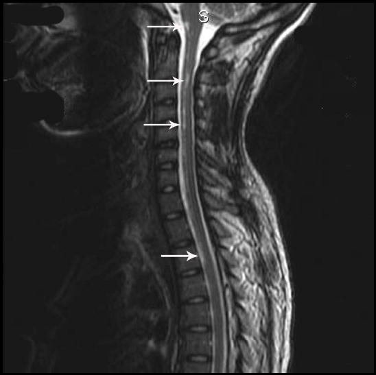
Patient 1.Digitally enhanced MRI (TR3650.0 / TE 104.0) at 6 months after the event, showing T2 hyperintensity (arrows) in the anterior medulla and along the anterior length of the cord from C2-C5 and at T3 consistent with infarction of the anterior spinal artery territory.
CASE 2
The patient was a 17-year-old male football-player with a history of back pain and palpitations. He was started on beta-blockers by his cardiologist. At home he became dizzy, fell down the stairs, and was immediately quadriparetic, with severe neck pain and numbness of his right face, left arm, and both legs. He was brought to the emergency room, where he regained movement of his upper extremities, and showed some movement of his proximal lower extremities within several hours. On exam several hours after admission, he had minimal flexion of the hips and knees. There was no voluntary movement of the feet or toes. Sensory exam revealed absence of all sensory modalities in the feet up to the shin. Reflexes were 2+ throughout the lower extremity bilaterally. Abdominal reflexes were brisk. The patient retained bowel and bladder function. Numbness of his face and arms resolved over the next few days. The initial MRI, performed on the first day of admission, was read as normal. Diffusion-weighted imaging was not done. A second MRI, one month after the event, showed a vague area of T2 hyperintensity in the thoracic cord, which was thought to be artifactual. T2-weighted axial images were not obtained. The patient continued to have lower extremity weakness and numbness with very slow improvement. He was discharged home four days later in a wheelchair with outpatient physical therapy three times weekly.
Three months after the event, MRI showed multiple Schmorl’s nodes in the mid-to-lower thoracic spine, with desiccation of intervertebral disks. Subtle T2 hyperintensity was also noted within the central region of the cord extending from T5-T9, on both axial and sagittal T2 imaging (Figure 2). Another more subtle T2 hyperintensity was seen within the upper cervical cord at the C2 level. These lesions were seen on the high resolution screens in the neuroradiology department. Retrospective viewing of previous films showed Schmorl’s nodes and similar T2 hyperintensity on T2 sagittal imaging, but no confirmation on T2 axial view was available. Thrombotic work-up revealed compound heterozygosity for the C677T and A1298C methylenetetrahydrofolate reductase (MTHFR) mutations, and the patient was started on daily aspirin and folic acid. The patient benefited from intensive physical therapy, which included two weeks at a rehabilitation hospital. Eight months following the event, the patient was able to ambulate with a cane. He continues to have decreased proprioceptive sense in his toes, and decreased vibratory sensation to the knee.
Figure 2.

- ill-defined central T2 hyperintensity is seen in axial view at mid T6-level
- enlarged sagittal image of thoracic cord demonstrating central T2 hyperintensity from T5-T8 (arrows) and Schmorl’s node (arrowhead). Infarction down to T10 can be seen on other cuts.
COMMENT
Spinal cord infarction in children is rare, and difficult to diagnose. The etiologies of spinal cord stroke in children are diverse (Table 1), and different from those commonly seen in adult patients.
Table 1.
POSSIBLE RISK FACTORS FOR SPINAL CORD ISCHEMIA IN CHILDREN WITHOUT VERTEBRAL FRACTURE
| Risk Factors | # of Patients |
|---|---|
| Cardiac | |
| Aortic dissection[40] | 1 |
| Aortic coarctation repair[27,39] | 4 |
| Hypotension | |
| Cardiac arrest[29] | 6 |
| Cardiac tamponade[30] | 1 |
| Arteriovenous malformation | |
| Vascular steal[32] | 1 |
| Mechanism unknown[31] | 5 |
| Thrombotic disorders | |
| Prothrombin variant[70,71] | 2 |
| Protein S deficiency[75] | 1 |
| Primary antiphospholipid syndrome[72] | 1 |
| Systemic lupus erythematosis[17,77] | 3 |
| Infectious disease | |
| Bacterial meningitis[53,57-67] | 15 |
| Amebic menigoencephalitis[69] | 1 |
| Viral encephalitis[68] | 1 |
| Cerebellar herniation | |
| Metabolic encephalopathy[50] | 3 |
| Minor trauma[41-48] | 18 |
| Fibrocartilaginous embolism[1-3,85-89] | 10 |
| Cancer | |
| Anterior spinal artery thrombosis[76] | 1 |
| Atlanto-axial instability | |
| Achondroplasia[22] | 1 |
| I-cell disease[20] | 1 |
| Iatrogenic | |
| Sclerotherapy of esophageal varices[56] | 1 |
| Prolonged neck flexion during surgery in sitting position[49] | 1 |
| Umbilical arterial catheters[33-36] | 8 |
| Thoracic tumor resection[37] | 2 |
| Multifactorial | |
| Achondroplasia, surgery[21] | 2 |
| Meningitis, Factor V Leiden, Chiari I malformation[74] | 1 |
| Meningitis, lumbar puncture, cardiac arrest[51-55] | 6 |
| Weight-lifting, MTHFR homozygosity[73] | 1 |
| Umbilical artery catheter, arterial switch operation[92] | 1 |
| Protein S deficiency, spine surgery, epidural catheter[71] | 1 |
| Cause unknown/unclear | |
| Not reported[93] | 2 |
| Unclear[4,91] | 6 |
| Total # | 108 |
Epidemiology
There is no comprehensive epidemiological description of ischemic spinal cord infarction in adults or children. In 1988, Sandson and Friedman reported spinal cord infarction as 1.2% of cases admitted for stroke at the Roger Williams General Hospital over a 4 year span [6]. In the adult population, spinal cord stroke is most commonly reported in the context of aortic surgery. Reported rates of peri- and post-operative spinal cord “complications”, many or most of which appear to be of vascular origin, range from 0.41% in aortic coarctation repair [7] to 12% in repair of type IIIb dissecting aortic aneurysms [8]. There are few large studies of spinal cord ischemia in children. Of 900 infants autopsied after death before 4 weeks, 21 showed evidence of hypoxic/ischemic spinal cord damage [9]. Spinal cord injury without radiographic abnormality (SCIWORA) represents a mean incidence of 34.8% of traumatic pediatric spinal cord injuries [10]. Spinal cord ischemia is one of four proposed mechanisms for SCIWORA [11]. The number of cases of SCIWORA actually attributable to spinal cord ischemia is difficult to determine because MRI was not used to define the nature of spinal cord parenchymal damage in many children studied [11].
Spinal cord infarctions are more commonly reported in adults than children: why?
Spinal cord infarction is more commonly reported in adults than in children. This might be due to age-related processes affecting perfusion of the cord. The most common causes of spinal cord infarction in adults are vascular, including atherosclerosis, abdominal aortic disease/surgery, and hypotension [6,12-17]. Compromise of blood flow to the adult cord has also been associated with cervical spondylosis [18]. With the exception of hypotension, these are diseases that take years to develop.
There are subgroups of children who are predisposed to hypotension and bony compromise of the spinal cord. Sladky and Rourke discovered spinal cord infarction in 9 of 900 asphyxiated premature neonates who underwent autopsy after death before 4 days of life [9], and Singer et al. described spinal cord infarction in an additional 3 premature neonates with paraplegia [19]. They attributed the ischemia to poor autoregulation of spinal cord blood flow during systemic hypotension secondary to prematurity. Spinal cord infarction has been associated with cervical spine changes in two children with I-cell disease after minor trauma [20] and three children with achondroplasia, two under general anesthesia [21] and one during gym class [22]. However, in general the young spinal cord is less vulnerable to infarction. The spinal cord has extensive collateral blood flow [23]. It may be easier to miss the diagnosis of spinal cord infarction in children, due to the relatively smaller volume and cross-sectional area of the pediatric spinal cord, compared to both the adult spinal cord and to the pediatric brain; the ability of radiographic imaging to detect infarctions in a small cord is limited [24].
Diagnosing spinal cord infarction
MRI allows for increased sensitivity in detecting infarction within the spinal cord. Before MRI, the diagnosis of spinal cord infarction relied upon autopsy or documented vascular occlusion by angiography. Diffusion-weighted imaging, although not commonly used on the spinal cord, now allows identification of cord ischemia within hours of the insult [25]. The limitations of spinal cord MRI include increased artifact due to cord movement and CSF flow [26]. Expectation of this artifact may lead to dismissal of actual lesions, as in patient 2. Some small infarctions can only be appreciated on high-resolution viewing monitors. Correct diagnosis of spinal cord infarction requires that the entire spine be imaged in suspicious cases, and that T2-weighted axial and sagittal views are obtained, as well as diffusion-weighted images early in the course to document ischemia. In case 2, the lesions were difficult to see in the initial MRI images on film format. When the patient was re-imaged, we included T2-weighted axial views, which revealed the infarction in more detail.
Differentiating spinal cord infarction from other myelopathy
MRI imaging and clinical presentation of spinal cord infarction can be similar to other spinal cord diseases, particularly transverse myelitis. In addition to case 1, at least six other children have been described for whom the diagnosis of ischemic spinal cord infarction was difficult to distinguish from transverse myelitis [3,4]. The MRI lesions seen in all of the these cases were in the anterior two-thirds of the spinal cord, suggesting that anterior spinal artery syndrome (ASAS) was the actual diagnosis. ASAS is characterized by the abrupt onset of symmetrical paralysis, impaired temperature and pain sensation, and loss of bowel and bladder function below the level of occlusion; vibration, light touch, and proprioceptive sensations are preserved [27]. Patient 1 clearly presented with these symptoms below the cervical level, which correlated with linear T2 hyperintensity of the anterior portion of the cervical spinal cord. The Transverse Myelitis Consortium Working Group defines inclusion and exclusion criteria for the diagnosis of acute idiopathic transverse myelitis, requiring that all inclusion criteria and no exclusion criteria be met in order to confirm diagnosis [28]. Diagnostic criteria for transverse myelitis include CSF pleocytosis or IgG index, or gadolinium enhancement representing inflammation of the spinal cord; progression of symptoms within 4 hours to 21 days following onset; and findings on MRI that do not correspond to a vascular territory. Patient 1 had unremarkable cerebrospinal fluid findings, progression of symptoms within 30 minutes, and MRI findings in the anterior spinal artery territory.
Causes of spinal cord infarction in children
As in adults, ischemic spinal cord infarction in children is caused by decreased blood flow due to hypotension; vascular injury, compression, or other impairment; or thrombosis or embolization to spinal arteries. The underlying etiologies, however, can be quite different.
Systemic hypotension leading to spinal cord infarction in children can be caused by cardiac arrest [29] or cardiac tamponade [30 ], both of which can occur in adults. Locally decreased perfusion can be caused by arteriovenous malformations. Spinal arteriovenous malformations are rare in children, but have been associated with paraplegia in 5 patients with thoracic malformations [31] and vascular steal in one patient [32].
Instrumentation during procedures or surgery, or trauma can lead to vascular injury which precipitates cord infarction. Umbilical artery catheters can cause spinal cord infarction in neonates [33-36], usually due to disruption of the artery of Ademkiewitcz, a major artery of the thoracic cord. Damage to this artery with resulting spinal cord infarction has also been reported in one child during the removal of a thoracic neuroblastoma [37]. Surgery on the aorta is a known cause of spinal cord infarction in adults[38]; children undergo aortic operations less frequently, and they are usually confined to the arch. Four children developed spinal cord infarction following repair of aortic coarctation [27,39]. Diseases of the abdominal aorta are rare in children, although there is one case of spinal cord ischemia with aortic dissection in an 18-year-old [40]. Minor trauma has also been associated with spinal cord infarction [41-48], but the exact etiology is not well understood. Pang et al. propose that in children, the relatively less elastic spinal cord is strained inside of the more flexible spinal column during hyperflexion injuries, and that this may result in reactive vasospasm of spinal cord arteries leading to ischemia [11]. The same mechanism is proposed in a case of cord infarction occurring in a child following operative removal of a pineal choriocarcinoma in the sitting position [49].
Cerebellar herniation was associated with cervical and thoracic cord infarction in three children with metabolic encephalopathy; two underwent lumbar puncture within 24 hours prior to herniation [50]. Cerebellar herniation associated with cord infarction is also suggested in children with meningitis who have undergone lumbar puncture [51-55].
There are many ways emboli and thrombi can form and impair cord perfusion. Emboli formation can be iatrogenic: embolic material to the spinal cord vasculature is reported in a pediatric patient undergoing sclerotherapy of esophageal varices [56]. Thrombotic vasculitis and/or occlusive arachnoiditis was implicated in cord infarction of many children with central nervous system infections [53,57-69]. Several thrombotic disorders have been reported in cases of spinal cord infarction [70-75]. Anterior spinal artery thrombosis is described in a child with neuroblastoma [76]. Systemic lupus erythematosus was associated with vasculitic ischemia of the spinal cord in one patient [17], and thrombosis in another [77].
Fibrocartilaginous embolism (FCE) is becoming an increasingly well characterized cause of childhood spinal cord infarction. This condition has been well characterized in animals, especially dogs [78,79], but also in cats[80,81], horses[82], swine[83], and turkeys[84]. The first human condition was described in a 15-year-old male in 1961; nucleus pulposis emboli were found within the spinal cord arteries at autopsy [85]. Patients usually experience sudden back pain with subsequent onset of rapid neurological deterioration following a “free interval” of minutes to 2 days [1]. FCE has been demonstrated, most often affecting the anterior spinal artery territory, at autopsy in 28 adults and four pediatric patients [1,86,87]. MRI has allowed FCE to become better characterized non-invasively in surviving patients [1-3,88,89]. There are reports of FCE with varying presentations and MRI findings [2,3]. In animals, FCE can be suspected when radiography and myelography demonstrate intervertebral disk changes and cord swelling at a level correlating with clinical features of spinal cord ischemia [90]. Tosi et al. propose that similar criteria may be used to diagnose FCE in humans and that MRI findings may include collapse of intervertebral disk space or Schmorl’s nodes [1]. It has been proposed that the vertical herniation of nucleus pulposis into vertebral bodies, as in the formation of Schmorl’s nodes, may allow the entry of fibrocartilaginous material into the vasculature of the spinal cord [1-3]. We believe that the pathogenesis of cord infarction in patient 2 is most likely due to FCE, because his MRI showed Schmorl’s nodes and disk changes adjacent to the area of thoracic T2 hyperintensity; presence of the MTHFR mutations may also have been contributory. This diagnosis is complicated by the immediate neurological deficit, unusual sensory distribution, and lack of neurological deterioration after onset.
For some children, the etiology of spinal cord infarction is unclear. Even after extensive work-up, the mechanism remains unclear [4,91] or is found to be multifactorial [21,51-55,71,73,74,92] with no one clear cause. The etiology of Patient 1’s spinal cord infarction is unknown. She has no history of trauma, or changes in her spine that would be consistent with FCE.
Treatment
Medical treatment of childhood spinal cord infarction has not been widely reported and it is unclear if it affects outcome. Anticoagulation with heparin and aspirin appeared to improve outcome in patients with spinal cord infarction and thrombotic disorders [70,71,73]. Patient 2 was treated with aspirin and folic acid for MTHFR heterozygosity; it is unclear if this has affected his outcome. Patient 1 and Patient 2 both underwent extensive physical therapy. Supportive care and rehabilitative physical therapy are key in the long-term treatment of children with spinal cord infarction. Both patients had improvement in their neurological function, although Patient 1’s improvement was minimal. Patient 2 continues to show significant improvement with therapy.
Outcome after spinal cord infarction
Outcomes after spinal cord infarction have been more widely reported in adults than in children. A review of outcomes in 199 patients by Cheshire et al. showed 43 (22%) cases resulted in death, 47 (24%) cases were unimproved, 70 (35%) cases showed some improvement, and 39 (19%) cases were considered markedly improved [17]. This review included some children, but results were not reported with respect to age, cause or spinal cord region involved. Two series of adults with cord infarction found that outcome was poorer in patients with more profound disability at onset [12,16]. One follow-up study of 8 patients with cord infarction included two children: one had improvement in neurological function, and the other had deterioration [93]. There are reports of varied outcomes in children with spinal cord trauma [10,11,94-98], but there is no confirmation of spinal cord infarction and many of the children had vertebral fractures. Moffett and Berkowitz reviewed outcomes in 19 children with bacterial meningitis and spinal cord infarction, finding death in 5 patients, some improvement in 12 patients, and return to normal function in 2 patients [58].
Common medical and psychosocial complications of spinal cord infarction
The complications of spinal cord infarction include ventilator-dependence and decreased bowel/bladder function, depending on the areas of spinal cord infarction. Patients are more susceptible to respiratory infections and bladder infections from indwelling catheters or urinary retention; these infections can have significant morbidity [97]. Patient 1 remains ventilator-dependent and has a chronic Foley catheter that has led to several urinary tract complications, including repeated infections and surgical removal of a bladder stone. She is followed by a pulmonologist and a urologist.
Chronic pain and spastic deformity are additional reported complications of cord infarction [16,19,93]. Patient 2 experienced chronic neuropathic pain and muscle pain associated with spasticity. He is taking medication to control this. Patient 1 receives regular botulinum injections for spasticity.
It is important to recognize and address the psychological impact of spinal cord infarction. Patient 1 has experienced anxiety, depression and insomnia as a result of her condition. Patient 2 has reported no long-term psychiatric difficulties, which may be related to his steady improvement.
CONCLUSION
The understanding of ischemic spinal cord infarction in children is an evolving area in pediatric neurology. Even as the causes become better characterized in children, our cases demonstrate the continued difficulty in diagnosis. The outcome of spinal cord infarction is difficult to change after the insult. Improvement in treatment requires more rapid and exact diagnosis, with both axial and sagittal T1, T2 and diffusion-weighted
MRI of the spinal cord to visualize both intrinsic and extrinsic lesions in suspected cases. Angiography, to assess for vascular occlusion and arteriovenous malformation, may be indicated in some cases. Thrombotic evaluation plays an important role in diagnosis, because thrombotic disorders can be medically treated. As MRI becomes more sensitive, and as new modalities of neuroimaging become available, we expect the diagnosis of spinal cord infarction to become better characterized and hopefully easier to manage. It is important to recognize and address the varied physical and psychosocial complications of spinal cord infarction in order to optimize outcome.
Acknowledgments
The authors of this paper take full responsibility for the content of this paper. We would like to thank Dr. Lawrence Walsh for contribution of one case to our paper and Ms. Nina Talib, M.Sc. for technical assistance.
Footnotes
Dr. Meredith Golomb is supported by the following grants: The National Institutes of Health NINDS grant K23 NS048024 and the Clarian Values Fund grant #VFR-171.
The authors have no conflicts of interest to report.
This paper was approved by the Institutional Review Board of the Indiana University School of Medicine (Study # 0207-55).
Publisher's Disclaimer: This is a PDF file of an unedited manuscript that has been accepted for publication. As a service to our customers we are providing this early version of the manuscript. The manuscript will undergo copyediting, typesetting, and review of the resulting proof before it is published in its final citable form. Please note that during the production process errorsmaybe discovered which could affect the content, and all legal disclaimers that apply to the journal pertain.
References
- 1.Tosi L, Rigoli G, Beltramello A. Fibrocartilaginous embolism of the spinal cord: A clinical and pathogenetic reconsideration. J Neurol Neurosurg Psychiatry. 1996;60:55–60. doi: 10.1136/jnnp.60.1.55. [DOI] [PMC free article] [PubMed] [Google Scholar]
- 2.Han JJ, Massagli TL, Jaffe KM. Fibrocartilaginous embolism - an uncommon cause of spinal cord infarction: Case report and review of the literature. Arch Phys Med Rehabil. 2004;85:153–7. doi: 10.1016/s0003-9993(03)00289-2. [DOI] [PubMed] [Google Scholar]
- 3.Davis GA, Klug GL. Acute-onset nontraumatic paraplegia in childhood: Fibrocartilaginous embolism or acute myelitis? Child's Nervous System. 2000;16:551–4. doi: 10.1007/s003810000268. [DOI] [PubMed] [Google Scholar]
- 4.Wilmshurst JM, Walker MC, Pohl KR. Rapid onset transverse myelitis in adolescence: Implications for pathogenesis and prognosis. Arch Dis Child. 1999;80:137–42. doi: 10.1136/adc.80.2.137. [DOI] [PMC free article] [PubMed] [Google Scholar]
- 5.International Classification of Diseases (icd - 9 - cm) Denver, DE: American Medical Association; 1999. [Google Scholar]
- 6.Sandson TA, Friedman JH. Spinal cord infarction: Report of 8 cases and review of the literature. Medicine (Baltimore) 1989;68:282–92. [PubMed] [Google Scholar]
- 7.Brewer LA, 3rd, Fosburg RG, Mulder GA, Verska JJ. Spinal cord complications following surgery for coarctation of the aorta. A study of 66 cases. J Thorac Cardiovasc Surg. 1972;64:368–81. [PubMed] [Google Scholar]
- 8.DeBakey ME, McCollum CH, Crawford ES, Morris GC, Jr, Howell J, Noon GP, Lawrie G. Dissection and dissecting aneurysms of the aorta: Twenty-year follow-up of five hundred twenty-seven patients treated surgically. Surgery. 1982;92:1118–34. [PubMed] [Google Scholar]
- 9.Sladky JT, Rorke LB. Perinatal hypoxic/ischemic spinal cord injury. Pediatr Pathol. 1986;6:87–101. doi: 10.3109/15513818609025927. [DOI] [PubMed] [Google Scholar]
- 10.Pang D. Spinal cord injury without radiographic abnormality in children, 2 decades later. Neurosurgery. 2004;55:1325–42. doi: 10.1227/01.neu.0000143030.85589.e6. [DOI] [PubMed] [Google Scholar]
- 11.Pang D, Wilberger JE., Jr Spinal cord injury without radiographic abnormalities in children. J Neurosurg. 1982;57:114–29. doi: 10.3171/jns.1982.57.1.0114. [DOI] [PubMed] [Google Scholar]
- 12.Salvador de la Barrera S, Barca-Buyo A, Montoto-Marques A, Ferreiro-Velasco ME, Cidoncha-Dans M, Rodriguez-Sotillo A. Spinal cord infarction: Prognosis and recovery in a series of 36 patients. Spinal Cord. 2001;39:520–5. doi: 10.1038/sj.sc.3101201. [DOI] [PubMed] [Google Scholar]
- 13.Satran R. Spinal cord infarction. Stroke. 1988;19:529–32. doi: 10.1161/01.str.19.4.529. [DOI] [PubMed] [Google Scholar]
- 14.Williams LS, Bruno A, Biller J. Spinal cord infarction. Topics in Stroke Rehabilitation. 1996;3:41–53. doi: 10.1080/10749357.1996.11754104. [DOI] [PubMed] [Google Scholar]
- 15.Foo D, Rossier AB. Anterior spinal artery syndrome and its natural history. Paraplegia. 1983;21:1–10. doi: 10.1038/sc.1983.1. [DOI] [PubMed] [Google Scholar]
- 16.Masson C, Pruvo JP, Meder JF, Cordonnier C, Touze E, De La Sayette V, Giroud M, Mas JL, Leys D. Spinal cord infarction: Clinical and magnetic resonance imaging findings and short term outcome. J Neurol Neurosurg Psychiatry. 2004;75:1431–5. doi: 10.1136/jnnp.2003.031724. [DOI] [PMC free article] [PubMed] [Google Scholar]
- 17.Cheshire WP, Santos CC, Massey EW, Howard JF., Jr Spinal cord infarction: Etiology and outcome. Neurology. 1996;47:321–30. doi: 10.1212/wnl.47.2.321. [DOI] [PubMed] [Google Scholar]
- 18.Ogino H, Tada K, Okada K, Yonenobu K, Yamamoto T, Ono K, Namiki H. Canal diameter, anteroposterior compression ratio, and spondylotic myelopathy of the cervical spine. Spine. 1983;8:1–15. doi: 10.1097/00007632-198301000-00001. [DOI] [PubMed] [Google Scholar]
- 19.Singer R, Joseph K, Gilai AN, Meyer S. Nontraumatic, acute neonatal paraplegia. J Pediatr Orthop. 1991;11:588–93. [PubMed] [Google Scholar]
- 20.Goodman ML, Pang D. Spinal cord injury in I-cell disease. Pediatr Neurosci. 1988;14:315–8. doi: 10.1159/000120411. [DOI] [PubMed] [Google Scholar]
- 21.Mayhew JF, Katz J, Miner M, Leiman BC, Hall ID. Anaesthesia for the achondroplastic dwarf. Canadian Anaesthetists' Society Journal. 1986;33:216–21. doi: 10.1007/BF03010834. [DOI] [PubMed] [Google Scholar]
- 22.Wieting JM, Krach LE. Spinal cord injury rehabilitation in a pediatric achondroplastic patient: Case report. Arch Phys Med Rehabil. 1994;75:106–8. [PubMed] [Google Scholar]
- 23.Thron AK. Vascular anatomy of the spinal cord: Neuroradiological investigations and clinical syndromes. Chapter V. New York: Springer-Verlag Wien; 1988. Postmortem angiography and microangiography of spinal cord vessels; pp. 13–64. [Google Scholar]
- 24.Bammer R, Fazekas F. Diffusion imaging of the human spinal cord and the vertebral column. Top Magn Reson Imaging. 2003;14:461–76. doi: 10.1097/00002142-200312000-00004. [DOI] [PubMed] [Google Scholar]
- 25.Kuker W, Weller M, Klose U, Krapf H, Dichgans J, Nagele T. Diffusion-weighted mri of spinal cord infarction--high resolution imaging and time course of diffusion abnormality. J Neurol. 2004;251:818–24. doi: 10.1007/s00415-004-0434-z. [DOI] [PubMed] [Google Scholar]
- 26.Pierre-Jerome C, Arslan A, Bekkelund SI. Mri of the spine and spinal cord: Imaging techniques, normal anatomy, artifacts, and pitfalls. J Manipulative Physiol Ther. 2000;23:470–5. doi: 10.1067/mmt.2000.108819. [DOI] [PubMed] [Google Scholar]
- 27.Servais LJ, Rivelli SK, Dachy BA, Christophe CD, Dan B. Anterior spinal artery syndrome after aortic surgery in a child. Pediatr Neurol. 2001;24:310–2. doi: 10.1016/s0887-8994(01)00237-5. [DOI] [PubMed] [Google Scholar]
- 28.Transverse Myelitis Consortium Working Group. Proposed diagnostic criteria and nosology of acute transverse myelitis. Neurology. 2002;59:499–505. doi: 10.1212/wnl.59.4.499. [DOI] [PubMed] [Google Scholar]
- 29.Gilles FH, Nag D. Vulnerability of the human spinal cord in transient cardiac arrest. Neurology. 1971;21:833–9. doi: 10.1212/wnl.21.8.833. [DOI] [PubMed] [Google Scholar]
- 30.Lin CC, Chen SY, Lan C, Ting-Fang Shih T, Lin MC, Lai JS. Spinal cord infarction caused by cardiac tamponade. Am J Phys Med Rehabil. 2002;81:68–71. doi: 10.1097/00002060-200201000-00013. [DOI] [PubMed] [Google Scholar]
- 31.Riche MC, Modenesi-Freitas J, Djindjian M, Merland JJ. Arteriovenous malformations (avm) of the spinal cord in children. A review of 38 cases. Neuroradiology. 1982;22:171–80. doi: 10.1007/BF00341245. [DOI] [PubMed] [Google Scholar]
- 32.Bandyopadhyay S, Sheth RD. Acute spinal cord infarction: Vascular steal in arteriovenous malformation. J Child Neurol. 1999;14:685–7. doi: 10.1177/088307389901401012. [DOI] [PubMed] [Google Scholar]
- 33.Aziz EM, Robertson AF. Paraplegia: A complication of umbilical artery catheterization. J Pediatr. 1973;82:1051–2. doi: 10.1016/s0022-3476(73)80443-3. [DOI] [PubMed] [Google Scholar]
- 34.Brown MS, Phibbs RH. Spinal cord injury in newborns from use of umbilical artery catheters: Report of two cases and a review of the literature. J Perinatol. 1988;8:105–10. [PubMed] [Google Scholar]
- 35.Haldeman S, Fowler GW, Ashwal S, Schneider S. Acute flaccid neonatal paraplegia: A case report. Neurology. 1983;33:93–5. doi: 10.1212/wnl.33.1.93. [DOI] [PubMed] [Google Scholar]
- 36.Krishnamoorthy KS, Fernandex RJ, Todres ID, De Long GR. Paraplegia associated with umbilical artery catheterization in the newborn. Pediatrics. 1976;58:443–5. [PubMed] [Google Scholar]
- 37.Boglino C, Martins AG, Ciprandi G, Sousinha M, Inserra A. Spinal cord vascular injuries following surgery of advanced thoracic neuroblastoma: An unusual catastrophic complication. Med Pediatr Oncol. 1999;32:349–52. doi: 10.1002/(sici)1096-911x(199905)32:5<349::aid-mpo7>3.0.co;2-p. [DOI] [PubMed] [Google Scholar]
- 38.Ross RT. Spinal cord infarction in diesase and surgery of the aorta. Can J Neurol Sci. 1985;12:289–95. doi: 10.1017/s0317167100035368. [DOI] [PubMed] [Google Scholar]
- 39.Castro-Vilanova MD, de Toledo M, Mateos F, Simon R. Isquemia-infarto medular. Rev Neurol. 1999;29:977–80. [PubMed] [Google Scholar]
- 40.Garland H, Greenberg J, Harriman DG. Infarction of the spinal cord. Brain. 1966;89:645–62. doi: 10.1093/brain/89.4.645. [DOI] [PubMed] [Google Scholar]
- 41.Ahmann PA, Smith SA, Schwartz JF, Clark DB. Spinal cord infarction due to minor trauma in children. Neurology. 1975;25:301–7. doi: 10.1212/wnl.25.4.301. [DOI] [PubMed] [Google Scholar]
- 42.Choi JU, Hoffman HJ, Hendrick EB, Humphreys RP, Keith WS. Traumatic infarction of the spinal cord in children. J Neurosurg. 1986;65:608–10. doi: 10.3171/jns.1986.65.5.0608. [DOI] [PubMed] [Google Scholar]
- 43.Ergun A, Oder W. Pediatric care report of spinal cord injury without radiographic abnormality (SCIWORA): Case report and literature review. Spinal Cord. 2003;41:249–53. doi: 10.1038/sj.sc.3101442. [DOI] [PubMed] [Google Scholar]
- 44.Linssen WH, Praamstra P, Gabreels FJ, Rotteveel JJ. Vascular insufficiency of the cervical cord due to hyperextension of the spine. Pediatr Neurol. 1990;6:123–5. doi: 10.1016/0887-8994(90)90045-3. [DOI] [PubMed] [Google Scholar]
- 45.Cheshire DJ. The paediatric syndrome of traumatic myelopathy without demonstrable vertebral injury. Paraplegia. 1977;15:74–85. doi: 10.1038/sc.1977.11. [DOI] [PubMed] [Google Scholar]
- 46.Horowitz SH, Patel N. Peripheral neurophysiology of acute distal spinal cord infarction. Pediatr Neurol. 2003;28:64–5. doi: 10.1016/s0887-8994(02)00495-2. [DOI] [PubMed] [Google Scholar]
- 47.Riviello JJ, Jr, Marks HG, Faerber EN, Steg NL. Delayed cervical central cord syndrome after trivial trauma. Pediatr Emerg Care. 1990;6:113–7. doi: 10.1097/00006565-199006000-00012. [DOI] [PubMed] [Google Scholar]
- 48.Lenn NJ. Spinal cord infarction due to minor trauma. Neurology. 1977;27:999. doi: 10.1212/wnl.27.10.999. [DOI] [PubMed] [Google Scholar]
- 49.Nitta H, Yamashita J, Nomura M, Igarashi N. Cervical spinal cord infarction after surgery for a pineal region choriocarcinoma in the sitting position: Case report. Neurosurgery. 1997;40:1082–5. doi: 10.1097/00006123-199705000-00039. [DOI] [PubMed] [Google Scholar]
- 50.Herrick MK, Agamanolis DP. Displacement of cerebellar tissue into spinal canal. A component of the respirator brain syndrome. Arch Pathol Lab Med. 1975;99:565–71. [PubMed] [Google Scholar]
- 51.Phelan M, Manson JI. Spinal cord dysfunction with quadriplegia complicating pneumococcal meningitis. Aust Paediatr J. 1987;23:57–9. doi: 10.1111/j.1440-1754.1987.tb02178.x. [DOI] [PubMed] [Google Scholar]
- 52.Norman MG. Respiratory arrest and cervical spinal cord infarction following lumbar puncture in meningitis. Can J Neurol Sci. 1982;9:443–7. doi: 10.1017/s0317167100044371. [DOI] [PubMed] [Google Scholar]
- 53.Tal Y, Crichton JU, Dunn HG, Dolman CL. Spinal cord damage: A rare complication of purulent meningitis. Acta Paediatr Scand. 1980;69:471–4. doi: 10.1111/j.1651-2227.1980.tb07116.x. [DOI] [PubMed] [Google Scholar]
- 54.Swart SS, Pye IF. Spinal cord ischaemia complicating meningococcal meningitis. Postgrad Med J. 1980;56:661–2. doi: 10.1136/pgmj.56.659.661. [DOI] [PMC free article] [PubMed] [Google Scholar]
- 55.DeSousa AL, Kleiman MB, Mealey J., Jr Quadriplegia and cortical blindness in hemophilus influenzae meningitis. J Pediatr. 1978;93:253–4. doi: 10.1016/s0022-3476(78)80509-5. [DOI] [PubMed] [Google Scholar]
- 56.Seidman E, Weber AM, Morin CL, Ethier R, Lamarche JB, Guerguerian AJ, Geoffroy G, Roy CC. Spinal cord paralysis following sclerotherapy for esophageal varices. Hepatology. 1984;4:950–4. doi: 10.1002/hep.1840040528. [DOI] [PubMed] [Google Scholar]
- 57.Haupt HM, Kurlinski JP, Barnett NK, Epstein M. Infarction of the spinal cord as a complication of pneumococcal meningitis. J Neurosurg. 1981;55:121–3. doi: 10.3171/jns.1981.55.1.0121. Case report. [DOI] [PubMed] [Google Scholar]
- 58.Moffett KS, Berkowitz FE. Quadriplegia complicating escherichia coli meningitis in a newborn infant: Case report and review of 22 cases of spinal cord dysfunction in patients with acute bacterial meningitis. Clin Infect Dis. 1997;25:211–4. doi: 10.1086/514534. [DOI] [PubMed] [Google Scholar]
- 59.Glista GG, Sullivan TD, Brumlik J. Spinal cord involvement in acute bacterial meningitis. JAMA. 1980;243:1362–3. [PubMed] [Google Scholar]
- 60.Seay AR. Spinal cord dysfunction complicating bacterial meningitis. Arch Neurol. 1984;41:545–6. doi: 10.1001/archneur.1984.04050170095024. [DOI] [PubMed] [Google Scholar]
- 61.Verghote M, Rousseau E, Geoffroy G. Quadriplegia after pneumococcal meningitis. Pediatr Infect Dis. 1985;4:559. doi: 10.1097/00006454-198509000-00028. [DOI] [PubMed] [Google Scholar]
- 62.Nadal D, Boltshauser E. Transient paraparesis following bacterial meningitis. Helv Paediatr Acta. 1987;42:55–7. [PubMed] [Google Scholar]
- 63.Boothman BR, Bamford JM, Parsons MR. Paraplegia as a presenting feature of meningococcal meningitis. J Neurol Neurosurg Psychiatry. 1988;51:1241. doi: 10.1136/jnnp.51.9.1241. [DOI] [PMC free article] [PubMed] [Google Scholar]
- 64.Coker SB, Muraskas JK, Thomas C. Myelopathy secondary to neonatal bacterial meningitis. Pediatr Neurol. 1994;10:259–61. doi: 10.1016/0887-8994(94)90036-1. [DOI] [PubMed] [Google Scholar]
- 65.Kirkpatrick M, Brooker RJ, Helms PJ, Cole GF. Spinal cord dysfunction in neonatal meningococcal meningitis. Eur J Pediatr. 1994;153:367–8. doi: 10.1007/BF01956421. [DOI] [PubMed] [Google Scholar]
- 66.Kastenbauer S, Winkler F, Fesl G, Schiel X, Ostermann H, Yousry TA, Pfister HW. Acute severe spinal cord dysfunction in bacterial meningitis in adults: MRI findings suggest extensive myelitis. Arch Neurol. 2001;58:806–10. doi: 10.1001/archneur.58.5.806. [DOI] [PubMed] [Google Scholar]
- 67.Graus F, Arbizu T, Ruyfi G. Partial brown-sequard's syndrome and meningococcal meningitis. Arch Neurol. 1981;38:602. doi: 10.1001/archneur.1981.00510090096020. [DOI] [PubMed] [Google Scholar]
- 68.Caruso JM, Tung GA, Brown WD. Central nervous system and renal vasculitis associated with primary varicella infection in a child. Pediatrics. 2001;107:E9. doi: 10.1542/peds.107.1.e9. [DOI] [PubMed] [Google Scholar]
- 69.Schumacher JD, Tien, Rober D, Lane, Kathy Neuroimaging findings in rare amebic infections of the central nervous system. Ajnr: American Journal of Neuroradiology. 1995;16:930–5. [PMC free article] [PubMed] [Google Scholar]
- 70.Young G, Krohn KA, Packer RJ. Prothrombin g20210a mutation in a child with spinal cord infarction. J Pediatr. 1999;134:777–9. doi: 10.1016/s0022-3476(99)70298-2. [DOI] [PubMed] [Google Scholar]
- 71.Hakimi KN, Massagli TL. Anterior spinal artery syndrome in two children with genetic thrombotic disorders. J Spinal Cord Med. 2005;28:69–73. doi: 10.1080/10790268.2005.11753801. [DOI] [PubMed] [Google Scholar]
- 72.Hasegawa M, Yamashita J, Yamashima T, Ikeda K, Fujishima Y, Yamazaki M. Spinal cord infarction associated with primary antiphospholipid syndrome in a young child. Case report. J Neurosurg. 1993;79:446–50. doi: 10.3171/jns.1993.79.3.0446. [DOI] [PubMed] [Google Scholar]
- 73.Cooper D, Magilner D, Call J. Spinal cord infarction after weight lifting. Am J Emerg Med. 2006;24:352–5. doi: 10.1016/j.ajem.2005.10.002. [DOI] [PubMed] [Google Scholar]
- 74.Almasanu BP, Owensby JR, Pavlakis SG, Edwards JH. Spinal cord infarction in meningitis: Polygenic risk factors. Pediatr Neurol. 2005;32:124–6. doi: 10.1016/j.pediatrneurol.2004.07.008. [DOI] [PubMed] [Google Scholar]
- 75.Ramelli GP, Wyttenbach R, von der Weid N, Ozdoba C. Anterior spinal artery syndrome in an adolescent with protein s deficiency. J Child Neurol. 2001;16:134–5. doi: 10.1177/088307380101600213. [DOI] [PubMed] [Google Scholar]
- 76.Lewis DW, Packer RJ, Raney B, Rak IW, Belasco J, Lange B. Incidence, presentation, and outcome of spinal cord disease in children with systemic cancer. Pediatrics. 1986;78:438–43. [PubMed] [Google Scholar]
- 77.Falko JM, Williams JC, Harvey DG, Weidman SW, Schonfeld G, Dodson WE. Hyperlipoproteinemia and multifocal neurologic dysfunction in systemic lupus erythematosus. J Pediatr. 1979;95:523–9. doi: 10.1016/s0022-3476(79)80755-6. [DOI] [PubMed] [Google Scholar]
- 78.Cauzinille L. Fibrocartilaginous embolism in dogs. Veterinary Clinics of North America - Small Animal Practice. 2000;30:155–67. doi: 10.1016/s0195-5616(00)50007-2. [DOI] [PubMed] [Google Scholar]
- 79.Gandini G, Cizinauskas S, Lang J, Fatzer R, Jaggy A. Fibrocartilaginous embolism in 75 dogs: Clinical findings and factors influencing the recovery rate. J Small Anim Pract. 2003;44:76–80. doi: 10.1111/j.1748-5827.2003.tb00124.x. [DOI] [PubMed] [Google Scholar]
- 80.Coradini M, Johnstone I, Filippich LJ, Armit S. Suspected fibrocartilaginous embolism in a cat. Aust Vet J. 2005;83:550–1. doi: 10.1111/j.1751-0813.2005.tb13328.x. [DOI] [PubMed] [Google Scholar]
- 81.Scott HW, O'Leary MT. Fibro-cartilaginous embolism in a cat. Journal of Small Animal Practice. 1996;37:228–31. doi: 10.1111/j.1748-5827.1996.tb01774.x. [DOI] [PubMed] [Google Scholar]
- 82.Fuentealba IC, Weeks BR, Martin MT, Joyce JR, Wease GS. Spinal cord ischemic necrosis due to fibrocartilaginous embolism in a horse. Journal of Veterinary Diagnostic Investigation. 1991;3:176–9. doi: 10.1177/104063879100300217. [DOI] [PubMed] [Google Scholar]
- 83.Benson JE, Schwartz KJ. Ischemic myelomalacia associated with fibrocartilaginous embolism in multiple finishing swine. J Vet Diagn Invest. 1998;10:274–7. doi: 10.1177/104063879801000308. [DOI] [PubMed] [Google Scholar]
- 84.Stedman NL, Brown TP, Rowland GN. Intravascular cartilaginous emboli in the spinal cord of turkeys. Avian Diseases. 1998;42:423–8. [PubMed] [Google Scholar]
- 85.Naiman J, Donohue WL, Prichard JS. Fatal nucleus pulposis embolism of spinal cord after trauma. Neurology. 1961;11:83–7. doi: 10.1212/wnl.11.1.83. [DOI] [PubMed] [Google Scholar]
- 86.Yousef OM, Appenzeller P, Kornfeld M. Fibrocartilagenous embolism: An unusual cause of spinal cord infarction. Am J Forensic Med Pathol. 1998;19:395–9. doi: 10.1097/00000433-199812000-00020. [DOI] [PubMed] [Google Scholar]
- 87.Toro G, Roman GC, Navarro-Roman L, Cantillo J, Serrano B, Vergara I. Natural history of spinal cord infarction caused by nucleus pulposus embolism. Spine. 1994;19:360–6. doi: 10.1097/00007632-199402000-00020. [DOI] [PubMed] [Google Scholar]
- 88.Raghavan A, Onikul E, Ryan MM, Prelog K, Taranath A, Chennapragada M. Anterior spinal cord infarction oweing to possible fibrocartilaginous embolism. Pediatr Radiol. 2004;34:503–6. doi: 10.1007/s00247-003-1133-0. [DOI] [PubMed] [Google Scholar]
- 89.Furtner M, Felber S, Poewe W. Fibrokartilaginare embolie des ruckenmarks. Nervenarzt. 2005;76:1246–9. doi: 10.1007/s00115-005-1878-9. [DOI] [PubMed] [Google Scholar]
- 90.Cook JR., Jr Fibrocartilaginous embolism. Veterinary Clinics of North America - Small Animal Practice. 1988;18:581–92. doi: 10.1016/s0195-5616(88)50056-6. [DOI] [PubMed] [Google Scholar]
- 91.Vandertop WP, Elderson A, van Gijn J, Valk J. Anterior spinal artery syndrome. Ajnr: American Journal of Neuroradiology. 1991;12:505–6. [PMC free article] [PubMed] [Google Scholar]
- 92.Lemke RP, Idiong N, al-Saedi S, Giddins NG, Ward C, Hamilton A, Hawkins L, Hancock BJ, Odim JN. Spinal cord infarct after arterial switch associated with an umbilical artery catheter. Ann Thorac Surg. 1996;62:1532–4. doi: 10.1016/0003-4975(96)00778-3. [DOI] [PubMed] [Google Scholar]
- 93.Pelser H, van Gijn J. Spinal infarction. Stroke. 1993;24:896–8. doi: 10.1161/01.str.24.6.896. A follow-up study. [DOI] [PubMed] [Google Scholar]
- 94.Dare AO, Dias MS, Li V. Magnetic resonance imaging correlation in pediatric spinal cord injury without radiographic abnormality. J Neurosurg. 2002;97:33–9. doi: 10.3171/spi.2002.97.1.0033. [DOI] [PubMed] [Google Scholar]
- 95.Davis PC, Reisner A, Hudgins PA, Davis WE, O'Brien MS. Spinal injuries in children: Role of MR. Ajnr: American Journal of Neuroradiology. 1993;14:607–17. [PMC free article] [PubMed] [Google Scholar]
- 96.Grabb PA, Pang D. Magnetic resonance imaging in the evaluation of spinal cord injury without radiographic abnormality in children. Neurosurgery. 1994;35:406–14. doi: 10.1227/00006123-199409000-00007. [DOI] [PubMed] [Google Scholar]
- 97.Burke DC. Spinal cord trauma in children. Paraplegia. 1971;9:1–14. doi: 10.1038/sc.1971.1. [DOI] [PubMed] [Google Scholar]
- 98.Liao CC, Lui TN, Chen LR, Chuang CC, Huang YC. Spinal cord injury without radiological abnormality in preschool-aged children: Correlation of magnetic resonance imaging findings with neurological outcomes. J Neurosurg. 2005;103:17–23. doi: 10.3171/ped.2005.103.1.0017. [DOI] [PubMed] [Google Scholar]