

Abstract
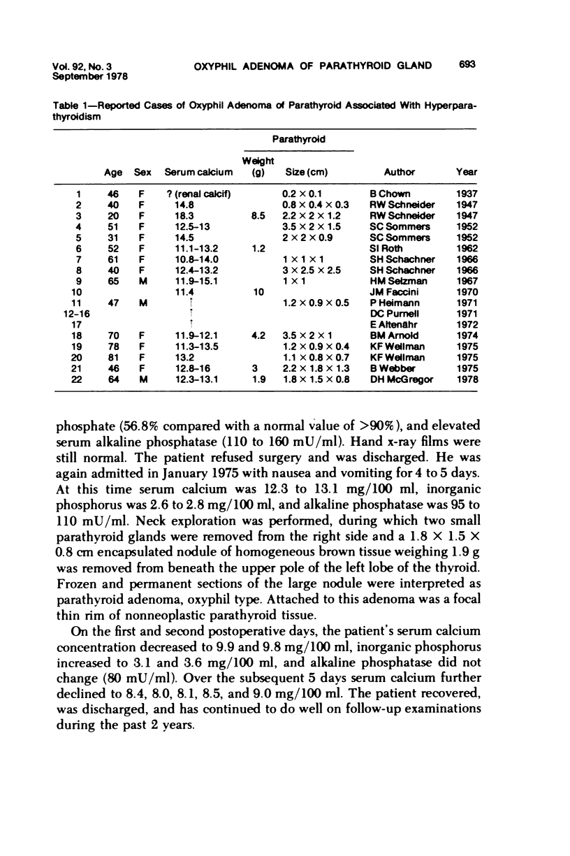
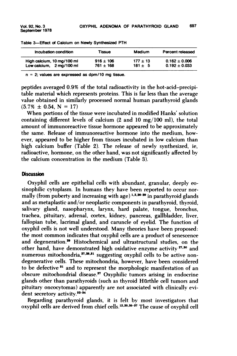
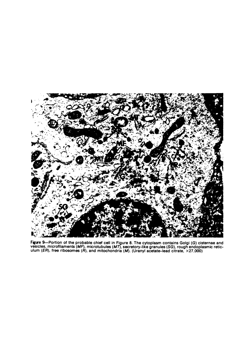
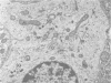
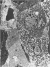
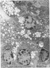
Oxyphil cells and oxyphil cell adenomas of parathyroid glands are, in most instances, regarded to be nonfunctioning. Although 21 cases of hyperparathyroidism associated with parathyroid oxyphil cell adenoma have been reported, secretion of hormone by these tumors has not been conclusively demonstrated. A parathyroid adenoma, diagnosed by light microscopy as oxyphil type, together with the results from ultrastructural and biochemical studies of the patient's adenomatous tissue, are reported here. The patient, a 64-year-old male, was found to have elevated serum calcium, low serum phosphorus, and elevated serum immunoreactive parathormone: findings consistent with hyperparathyroidism. After excision of two small normal-appearing glands and one greatly enlarged (1.9 g) parathyroid gland, those laboratory values returned to normal. Light microscopy of the enlarged parathyroid indicated that it consisted almost entirely of an oxyphil adenoma. Electron microscopy revealed that the adenoma was composed mainly of mitochondria-rich oxyphil cells but also of interspersed transitional oxyphil cells and rare scattered chief cells. Golgi zones, rough endoplasmic reticulum, and prosecretory and secretory-like granules were observed in some oxyphil cells, in most transitional oxyphil cells, and in the infrequent chief cells. Thus, many of these cells appear to contribute to the production and secretion of parathormone. Biochemical studies performed directly on the adenomatous tissue demonstrated that it was able to synthesize proparathormone and parathormone, although the proportion of hormonal peptide synthesis relative to that of the total protein synthesis in this tissue was much smaller (0.9%) than that found in normal parathyroid tissue (5.7%). There was a small increase in immunoreactive parathormone when the adenoma tissue was incubated in a low-calcium medium. These findings indicate that this oxyphil adenoma of the parathyroid gland synthesized and secreted parathormone, apparently to some extent autonomously, but suggest that its capacity to do so was largely dependent on its component of cells other than fully developed oxyphil cells, such as transitional oxyphil cells.
Full text
PDF


Images in this article


Selected References
These references are in PubMed. This may not be the complete list of references from this article.
- Altenähr E. Ultrastructural pathology of parathyroid glands. Curr Top Pathol. 1972;56:2–54. [PubMed] [Google Scholar]
- Arnold B. M., Kovacs K., Horvath E., Murray T. M., Higgins H. P. Functioning oxyphil cell adenoma of the parathyroid gland: evidence for parathyroid secretory activity of oxyphil cells. J Clin Endocrinol Metab. 1974 Mar;38(3):458–462. doi: 10.1210/jcem-38-3-458. [DOI] [PubMed] [Google Scholar]
- BALOGH K., Jr, ROTH S. I. HISTOCHEMICAL AND ELECTRON MICROSCOPIC STUDIES OF EOSINOPHILIC GRANULAR CELLS (ONCOCYTES) IN TUMORS OF THE PAROTID GLAND. Lab Invest. 1965 Mar;14:310–320. [PubMed] [Google Scholar]
- Boquist L., Lundgren E. Effects of variations in calcium concentration on parathyroid morphology in vitro. Lab Invest. 1975 Dec;33(6):638–647. [PubMed] [Google Scholar]
- Boquist L. Occurrence of oxyphil cells in suppressed parathyroid glands. Cell Tissue Res. 1975 Nov 19;163(4):465–470. doi: 10.1007/BF00218492. [DOI] [PubMed] [Google Scholar]
- Boquist L. Ultrastructural study of calcium-containing precipitation in human parathyroid glands. Virchows Arch A Pathol Anat Histol. 1975 Oct 20;368(2):99–108. doi: 10.1007/BF00432411. [DOI] [PubMed] [Google Scholar]
- Castleman B., Mallory T. B. The Pathology of the Parathyroid Gland in Hyperparathyroidism: A Study of 25 Cases. Am J Pathol. 1935 Jan;11(1):1–72.17. [PMC free article] [PubMed] [Google Scholar]
- Chown B. Minute Oxyphil Adenoma of the Parathyroid Associated with Calcium Deposits in the Kidney. Can Med Assoc J. 1937 Jul;37(1):16–18. [PMC free article] [PubMed] [Google Scholar]
- Christie A. C. The parathyroid oxyphil cells. J Clin Pathol. 1967 Jul;20(4):591–602. doi: 10.1136/jcp.20.4.591. [DOI] [PMC free article] [PubMed] [Google Scholar]
- Chu L. L., MacGregor R. R., Liu P. I., Hamilton J. W., Cohn D. V. Biosynthesis of proparathyroid hormone and parathyroid hormone by human parathyroid glands. J Clin Invest. 1973 Dec;52(12):3089–3094. doi: 10.1172/JCI107508. [DOI] [PMC free article] [PubMed] [Google Scholar]
- Chu L. L., Macgregor R. R., Hamilton J. W., Cohn D. V. Conversion of proparathyroid hormone to parathyroid hormone: the use of amines as specific inhibitors. Endocrinology. 1974 Nov;95(5):1431–1438. doi: 10.1210/endo-95-5-1431. [DOI] [PubMed] [Google Scholar]
- Faccini J. M. The ultrastructure of parathyroid glands removed from patients with primary hyperparathyroidism: a report of 40 cases, including four carcinomata. J Pathol. 1970 Dec;102(4):189–199. doi: 10.1002/path.1711020402. [DOI] [PubMed] [Google Scholar]
- Feldman P. S., Horvath E., Kovacs K. Ultrastructure of three Hürthle cell tumors of the thyroid. Cancer. 1972 Nov;30(5):1279–1285. doi: 10.1002/1097-0142(197211)30:5<1279::aid-cncr2820300521>3.0.co;2-q. [DOI] [PubMed] [Google Scholar]
- HAMPERL H. [Onkocytes and onkocytoma]. Virchows Arch Pathol Anat Physiol Klin Med. 1962;335:452–483. [PubMed] [Google Scholar]
- Hamilton J. W., Spierto F. W., MacGregor R. R., Cohn D. V. Studies on the biosynthesis in vitro of parathyroid hormone. II. The effect of calcium and magnesium on synthesis of parathyroid hormone isolated from bovine parathyroid tissue and incubation medium. J Biol Chem. 1971 May 25;246(10):3224–3233. [PubMed] [Google Scholar]
- Kovacs K., Horvath E. Pituitary "chromophobe" adenoma composed of oncocytes. A light and electron microscopic study. Arch Pathol. 1973 Apr;95(4):235–239. [PubMed] [Google Scholar]
- Landolt A. M., Oswald U. W. Histology and ultrastructure of an oncocytic adenoma of the human pituitary. Cancer. 1973 May;31(5):1099–1105. doi: 10.1002/1097-0142(197305)31:5<1099::aid-cncr2820310510>3.0.co;2-g. [DOI] [PubMed] [Google Scholar]
- MOLLENHAUER H. H. PLASTIC EMBEDDING MIXTURES FOR USE IN ELECTRON MICROSCOPY. Stain Technol. 1964 Mar;39:111–114. [PubMed] [Google Scholar]
- Mayer G. P., Habener J. F., Potts J. T., Jr Parathyroid hormone secretion in vivo. Demonstration of a calcium-independent nonsuppressible component of secretion. J Clin Invest. 1976 Mar;57(3):678–683. doi: 10.1172/JCI108324. [DOI] [PMC free article] [PubMed] [Google Scholar]
- McGregor D. H., Chu L. L., MacGregor R. R., Cohn D. V. Disruption of the Golgi zone and inhibition of the conversion of proparathyroid hormone to parathyroid hormone in human parathyroid tissue by tris(hydroxymethyl)aminomethane. Am J Pathol. 1977 Jun;87(3):553–568. [PMC free article] [PubMed] [Google Scholar]
- Purnell D. C., Smith L. H., Scholz D. A., Elveback L. R., Arnaud C. D. Primary hyperparathyroidism: a prospective clinical study. Am J Med. 1971 May;50(5):670–678. doi: 10.1016/0002-9343(71)90122-7. [DOI] [PubMed] [Google Scholar]
- REYNOLDS E. S. The use of lead citrate at high pH as an electron-opaque stain in electron microscopy. J Cell Biol. 1963 Apr;17:208–212. doi: 10.1083/jcb.17.1.208. [DOI] [PMC free article] [PubMed] [Google Scholar]
- ROTH S. I., MUNGER B. L. The cytology of the adenomatous, atrophic, and hyperplastic parathyroid glands of man. A light- and electron-microscopic study. Virchows Arch Pathol Anat Physiol Klin Med. 1962;335:389–410. doi: 10.1007/BF00957031. [DOI] [PubMed] [Google Scholar]
- ROTH S. I., OLEN E., HANSEN L. S. The eosinophilic cells of the parathyroid (oxyphil cells), salivary (oncocytes), and thyroid (Huerthle cells) glands. Light and electron microscopic observations. Lab Invest. 1962 Nov;11:933–941. [PubMed] [Google Scholar]
- ROTH S. I. Pathology of the parathyroids in hyperparathyroidism. Discussion of recent advances in the anatomy and pathology of the parathyroid glands. Arch Pathol. 1962 Jun;73:495–510. [PubMed] [Google Scholar]
- Roth S. I., Marshall R. B. Pathology and ultrastructure of the human parathyroid glands in chronic renal failure. Arch Intern Med. 1969 Oct;124(4):397–407. [PubMed] [Google Scholar]
- Rother P. Uber Vorkommen und Funktion der oxyphilen Welshschen Zellen in Glandulae parathyreoideae. Z Mikrosk Anat Forsch. 1968;79(4):533–556. [PubMed] [Google Scholar]
- SOMMERS S. C., YOUNG T. L. Oxyphil parathyroid adenomas. Am J Pathol. 1952 Jul-Aug;28(4):673–689. [PMC free article] [PubMed] [Google Scholar]
- Selzman H. M., Fechner R. E. Oxyphil adenoma and primary hyperparathyroidism. Clinical and ultrastructral observations. JAMA. 1967 Feb 6;199(6):359–361. [PubMed] [Google Scholar]
- Thiele J., Ries P., Georgii A. Spezielle und funktionelle Pathomorphologie der Epithelkörperchen in einem unausgewählten Obduktionsgut (589 Sektionen) Virchows Arch A Pathol Anat Histol. 1975 Aug 12;367(3):195–208. doi: 10.1007/BF00430707. [DOI] [PubMed] [Google Scholar]
- WATSON M. L. Staining of tissue sections for electron microscopy with heavy metals. J Biophys Biochem Cytol. 1958 Jul 25;4(4):475–478. doi: 10.1083/jcb.4.4.475. [DOI] [PMC free article] [PubMed] [Google Scholar]
- Webber B. Oxyphil adenoma of the parathyroid gland. S Afr J Surg. 1975 Jun;13(2):115–117. [PubMed] [Google Scholar]
- Wellmann K. F. Primärer Hyperparathyreoidismus und oxyphiles Nebenschilddrüsenadenom. Dtsch Med Wochenschr. 1975 May 16;100(20):1127–1132. doi: 10.1055/s-0028-1106346. [DOI] [PubMed] [Google Scholar]
- Weymouth R. J., Seibel H. R. An electron microscopic study of the parathyroid glands in man: evidence of secretory material. Acta Endocrinol (Copenh) 1969 Jun;61(2):334–342. doi: 10.1530/acta.0.0610334. [DOI] [PubMed] [Google Scholar]