

Abstract
In this study, a comparison between structure, chemical composition and mechanical properties of collagen fibers at three regions within a human periodontium, has enabled us to define a novel tooth attachment mechanism. The three regions include 1) the enthesis region: insertion site of periodontal ligament fibers (collagen fibers) into cementum at the root surface, 2) bulk cementum and 3) the cementum dentin junction (CDJ). Structurally, continuity in collagen fibers was observed from the enthesis, through bulk cementum and CDJ. At the CDJ the collagen fibers split into individual collagen fibrils and intermingled with the extracellular matrix of mantle dentin. Under wet conditions, the collagen fibers at the three regions exhibited significant swelling suggesting a composition rich in polyanionic molecules such as glycosaminoglycans. Additionally, site-specific indentation illustrated a comparable elastic modulus between collagen fibers at the enthesis (1 – 3 GPa) and the CDJ (2 – 4 GPa). However, the elastic modulus of collagen fibers within bulk cementum was higher (4 – 7 GPa) suggesting presence of extrafibrillar mineral.
It is known that the tooth forms a fibrous joint with the alveolar bone which is termed a gomphosis. Although narrower in width than the periodontal ligament space, the hygroscopic CDJ can also be termed as a gomphosis; a fibrous joint between cementum and root dentin capable of accommodating functional loads similar to that between cementum and alveolar bone. From an engineering perspective, it is proposed that a tooth contains two fibrous joints that accommodate the masticatory cyclic loads. These joints are defined by the attachment of dissimilar materials via graded stiffness interfaces, such as; 1) alveolar bone attached to cementum with the PDL and 2) cementum to root dentin with the CDJ. Thus, through variations in concentrations of basic constituents, distinct regions with characteristic structures and graded properties allow for attachment and the load bearing characteristics of a tooth.
1. INTRODUCTION
Biomechanical function of a load bearing system is defined by the physical properties of the tissues and their interfaces (interphases). However, inflammation due to disease progression leads to degraded physical properties resulting in loss of attachment and function. This study was performed to provide a foundation for tissue engineering formulations for tooth attachment by defining physical characteristics of tissues and interfaces within the human periodontium.
The periodontium of a human tooth includes three mineralized tissues: alveolar bone, cementum and root dentin, of which cementum and alveolar bone interface with a soft fibrous tissue; the periodontal ligament (PDL), forming a fibrous joint or gomphosis [1, 2]. Periodontal fibers and Sharpey’s fibers in acellular cementum seemingly attach to root dentin [3–5]. Additionally, they were reported to be calcified in acellular cementum [3, 4]. In addition, the acellular cementum fibers intermingle with the dentin matrix [6]. More recently, similar observations on acellular cementum were reported [7]. Although not clear in studies by Dewey, 1926 [5], results presented to date were only on acellular cementum.
Cementum is attached to root dentin via a hydrophilic fibrous cementum-dentin junction (CDJ) [8]. The CDJ is also known as the innermost cementum layer, intermediate cementum or collagen hiatus and consists of collagen fibrils and remnants of epithelial cells of Hertwig’s Epithelial root sheath (HERS) [9]. Additionally, it was suggested not to be a distinct matrix layer but a composite of non-collagenous proteins and collagen fibrils [10]. Despite the existing theories and these observations, no physical property associations between the fibrous CDJ and the PDL insertions into cementum, also known as Sharpey’s fibers (SFs) have been made.
In this study the attachment of bulk cementum to root mantle dentin was investigated with respect to the periodontal ligament (PDL) and previously reported hydrophilic CDJ [8]. This study sought to demonstrate that the collagen fibers within the CDJ are continuous with the Sharpey’s fibers in human cellular cementum, using various high-resolution imaging techniques and histology. The structure at micro- and nano-scales was investigated using light and atomic force microscopy techniques and the site-specific mechanical properties were studied using AFM-based nanoindentation.
2. MATERIALS AND METHODS
Specimen Preparation for Light Microscopy
Mandibular molars from varying ages (19 – 81 yrs) were sterilized using 0.26 Mrad of gamma-radiation [11]. Several 2–3 mm thick transverse sections perpendicular to the longitudinal axis of the tooth root were made using a diamond wafering blade and a low speed saw (ISOMET, Buehler, Lake Bluff, IL) under wet conditions. The specimens were ultrasonicated in deionized water for 10 seconds (L&R Ultrasonic Cleaning System, Kearny, NJ) after preparation to remove any abrasives from coarse sectioning. The transverse sections were fixed in 2.5% glutaraldehyde for 8 weeks followed by end-stage decalcification [12] using Cal-EX II decalcifying solution (Fisher Scientific, Fair Lawn, NJ). The specimens were embedded in paraffin and sectioned on a rotary microtome (Reichert-Jung Biocut, Vienna, Austria) using disposable steel blades (TBF™ Inc., Shur/Sharp™, Fisher Scientific, Fair Lawn, NJ). The paraffin sections were mounted on Superfrost Plus microscope slides (Fisher Scientific, Fair Lawn, NJ) deparaffinized with xylene followed by staining with either Safranin O’ or Masson’s trichrome. The stained tissues were characterized using a light microscope (BX 51, Olympus America Inc., San Diego, CA) and analyzed using Image Pro Plus v6.0 (Media Cybernetics, Inc., Silver Spring, MD). Cross polarized light was used to generate a strong contrast, thus enhancing the features of interest, including orientation of collagen fibers.
Specimen Preparation for Atomic Force Microscopy and AFM-based Nanoindentation
The specimens for AFM and AFM-based nanoindentation were prepared using an ultrasectioning method which was described previously [13]. Ultrasectioning was used to generate a relatively flat surface to maintain orthogonality between the nano-probes of the AFM and AFM-based nanoindenter and the specimen. In brief, unfixed transverse sections were glued to AFM steel stubs (Ted Pella, Inc., Redding, CA) using a cyanoacrylate adhesive (MDS Adhesive QX-4, MDS Products Inc., Anaheim, CA). The specimens were ultrasectioned using an ultramicrotome (Ultracut E, Reichert-Jung, Vienna, Austria) and trimmed with a glass knife (Electron Microscopy Laboratory – UCSF, San Francisco, CA). Final trimming of the specimens was performed using a diamond knife (Micro Star Technologies, Huntsville, TX) by removing 300 nm thick ultrasections at a speed of 0.75 mm/sec, thus creating a sectioned block surface for AFM and AFM-based nanoindentation under dry and wet conditions.
Atomic Force Microscopy and AFM-based nanoindentation of ultrasectioned cementum
The structure from the root surface of cementum to mantle dentin including the CDJ was characterized under dry and wet conditions using an AFM (Nanoscope III, Multimode, DI-Veeco Instruments Inc., Santa Barbara, CA). A Si3N4 tip attached to a ‘V-shaped’ type ‘D’ microlever with a nominal spring constant of 0.06 N/m (DNP, Veeco Probes, Camarillo, CA) at a scanning frequency of 1.5 Hz was used to scan the surfaces of the ultrasectioned block specimens. The nominal radius of curvature of the tip was less than 50 nm [13]. Areas as large as 100 × 100 μm2 were evaluated using a ‘J’ type piezo scanner. The structural changes of the Sharpey’s fibers (periodontal ligament inserts), along with the CDJ within each specimen block under dry and wet conditions were determined using the scanned areas and Nanoscope III version 5.2 software (Nanoscope III, Multimode, Veeco Instruments Inc., Santa Barbara, CA).
Nanomechanical tests on dry and wet specimens were performed using an AFM, to which a load-displacement transducer (Triboscope Micromechanical Test Instrument, Hysitron Incorporated, MN) was attached [8]. A sharp diamond Berkovich indenter with a radius of curvature less than 100 nm (Triboscope Micromechanical Test Instrument, Hysitron Incorporated, Minneapolis, MN) was fitted to the transducer. The scan speed, scan area and load displacement of the indenter on the specimen were controlled by a computer. After indentation, the AFM piezo was used to scan the indented area. Immersion of the specimen and indenter in deionized water allowed measuring the mechanical properties closer to in vivo conditions. The maximum load used was 500 μN, with load, hold, and unload for 3 seconds each when measuring the reduced elastic modulus ‘Er’. Fused silica was used as a calibration standard for the AFM-nanoindenter [14]. The Er of regions of interest was determined using the following equation:
where ‘S’ is the stiffness obtained from the load-displacement curve and ‘A’ is the contact area [15].
RESULTS
Structure Analysis using Light and Atomic Force Microscopies
These results are divided into sections demonstrating 1) that collagen fibers are continuous from root surface through bulk cementum, pass through the CDJ and enter the root dentin, 2) that the collagen fiber-rich region at the root surface and CDJ have similar compositions, and that 3) nanomechanical properties of the collagen rich region at the root surface and CDJ have similar values.
I. Collagen fibers are continuous from root surface through bulk cementum
Figures 1 – 3 illustrate representative light and atomic force micrographs of the collagen fibers within bulk cementum. Demineralized histology sections and ultrasectioned surface-block specimens illustrated the continuity and orientation of the collagen fibers from root surface through bulk cementum and CDJ (Figs. 1a, e). The continuity of the collagen fibers was more consistently observed in the demineralized histology sections (Fig. 1a) using polarized light microscopy. The 2 – 6 μm wide collagen fibers were oriented radially over the cross sections of the roots (Figs. 1c, 1d and 3). Additionally, these radial collagen fibers were perpendicular to the thinner and numerous 500 nm – 1 μm lamellae or circumferentially oriented collagen fibers (Figs. 1c–e).
Figure 1.

Structure of cementum; a-d) Polarized microscopy; a) demineralized section stained with Safranin O counterstained with fast green. Notice the continuous collagen fibers from the root surface to root mantle dentin (segmented curved arrows). a and b) Notice tufts of collagen fibers between lamellar cementum and mantle dentin (stars) form the hydrophilic CDJ. c and d) a demineralized section stained with Masson’s trichrome. Notice the two distinct orientations of collagen fibers; radial collagen fibers (PDL-inserts) or Sharpey’s fibers perpendicular (segmented arrows) to the circumferential or lamellar collagen fibers. e) 100 μm × 100 μm AFM micrograph from dry ultrasectioned surface-block specimen. Notice the 5 μm wide collagen fibers from the root surface through the CDJ into mantle dentin (segmented white arrow). The CDJ is a valley-like under dry conditions.
Figure 3.
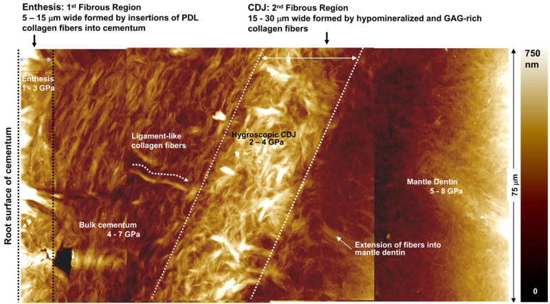
A composite of AFM micrographs of a wet ultramicrotomed surface-block. This image illustrates two distinct fibrous regions with corresponding ranges for modulus values; 1) enthesis and 2) the interface between cementum and root dentin (CDJ), illustrating ligament-like collagen fibers within bulk cementum extending into root mantle dentin.
An AFM of undemineralized cementum at higher resolution under wet conditions illustrated a 5 – 15 μm wide fibrous region (Fig. 3) at the root surface. Originating from the fibrous region were 2 – 6 μm wide partially swollen radial collagen fibers within bulk cementum which were similar in spatial distribution to those observed using a light microscope (Fig. 2b, 3). Closer examination of the collagen fibers illustrated fibrils (Fig. 4) with either an absence (Fig. 4a) or presence (Fig. 4b) of collagen periodicity.
Figure 2.

a) A demineralized section stained with Masson’s trichrome illustrating integration of terminal ends of the radial collagen fibers (PDL-inserts) with root mantle dentin. The exclusively radial collagen fiber rich region representative of CDJ is 20 μm wide. b) An AFM micrograph of undemineralized cementum illustrating similar termini of collagen fibers (stars). Note the radial and circumferential collagen fibers within bulk cementum. c1, 2 and 3). An AFM micrograph showing web-like PDL termini at the mantle dentin and intermingling of the 200 nm wide collagen fibrils with the extracellular matrix.
Figure 4.
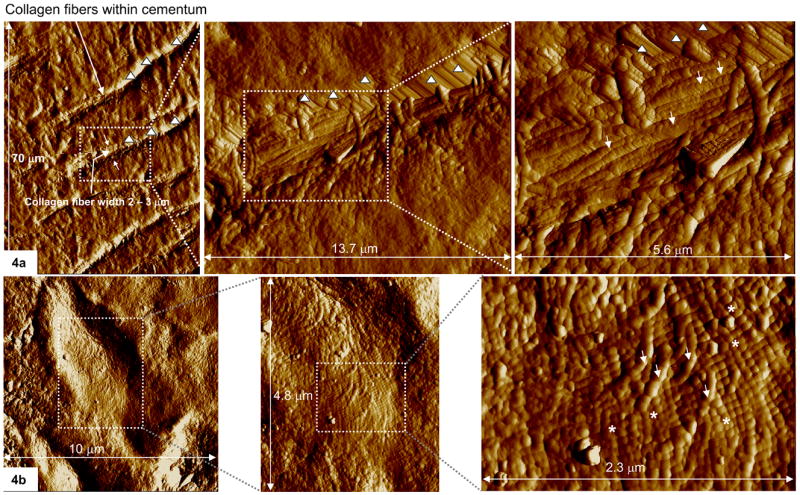
a) AFM deflection images in contact mode showing collagen fibrils (arrows) within undemineralized cementum. Notice collagen fibrils from adjacent cementum crossing over the lower fibrils at almost 90°. b) Notice the periodic pattern of the collagen fibrils (stars) with the collagen fiber. The absence of the periodic pattern of the collagen fibril (arrows) could be due to a coating of noncollagenous proteins and/or extrafibrillar mineral. Note: The fibrils shown in this figure are within a valley-like region. The regions represented by triangles are walls of the valley. It should be noted that the topography of the valley-walls could be an image artifact caused by specimen-surface and tip-geometry limitations [22].
II. Collagen fibers are continuous from cementum through the CDJ to root mantle dentin
The AFM micrograph shown in Figure 1e illustrates continuous collagen fibers from the root surface through the CDJ into mantle dentin. Notice the previously reported [8] valley-like CDJ under dry conditions (Fig. 1e). Traditional histology techniques, regardless of the type of stain illustrated a 15 to 35 μm wide collagen fiber-rich CDJ (Figs. 1b, 2a). Examination of a similar region under wet conditions at higher resolution using an AFM illustrated additional structural detail (Figs. 2b, 3). More importantly, collagen fibers were found to intermingle with root mantle dentin by forming web-like structures (Figs. 2b – 2c2) containing 200 nm single collagen fibrils (Fig. 2c3).
Chemical composition as related to hygroscopic nature of specific regions
Using an AFM, collagen fibers were distinctly observed under wet conditions (Fig. 3). Similar to the 5 – 15 μm wide hygroscopic fibrous regions at the root surface, a 15 – 35 μm wide hygroscopic fibrous region adjacent to mantle dentin was observed. Although hygroscopic collagen fibers were often observed within bulk cementum (Fig. 3), at times it was limited to partial swelling as indicated by the height variations (light and dark regions corresponding to Z scale) within the AFM micrographs (Fig. 3).
Site-specific elastic modulus using AFM-based nanoindentation
AFM-based nanoindentation provided elastic modulus values for collagen fiber-rich regions observed at the root surface, collagen fibers within bulk cementum and the hygroscopic CDJ. The elastic modulus varied from 1 – 3 GPa, while the indentation depths varied from 200 – 500 nm at the root surface and a higher range of 4 – 7 GPa, with an indentation depth of 200 – 350 nm within bulk cementum. The modulus range for the hygroscopic CDJ was 2 – 4 GPa, with an indentation depth of 220 – 500 nm. In this study, it is important to note that the maximum displacement of the indenter did not exceed 500 nm for fibers and fibrous regions equal to or greater than 6 μm wide.
DISCUSSION
The results of this study provided an association between the collagen-rich regions at the root surface, radially oriented collagen fibers within bulk cellular cementum and the CDJ by tracing their spatial distribution and comparing physical properties. The premise for this study was based on the following two concepts: 1) functionally these fibrous regions go through large displacements and strains thus accommodating and distributing cyclic masticatory forces during function. 2) The hygroscopic nature of the 5 – 15 μm wide fibrous region at the root surface, suggests that it may be hypomineralized, making it susceptible to degradation by the inflammatory products due to periodontitis.
The site of soft tissue insertions within mineralized tissues are termed entheses [16]. The physical properties (structure, chemical composition and mechanical properties) of entheses within the periodontium could facilitate distribution and transmission of functional loads, much like the osteoligamentous and the osteotendinous junctions in skeletal system. Traditionally, Sharpey’s fibers are defined as the collagen fibers of the PDL embedded in cementum [17] that form an enthesis. This implies that the spatial location of Sharpey’s fibers is limited to regions closer to the edges of the respective mineralized tissues; bone and cementum. However, results in this study, suggest that Sharpey’s fibers are not limited to root surface region of cementum, but appear to extend through bulk cementum into the CDJ and root mantle dentin, an inference first suggested by Dewey et al., 1926 [5]. In addition, similar observations were noted by Furseth, Selvig, and more recently by Raspanti et al., 2000 [7].
The attachment of cementum to periodontal ligament and mantle dentin is through the fibrous regions defined as the 1) enthesis and the 2) CDJ (Fig. 3). The basic load bearing structural components that define the extracellular matrices of the fibrous regions can be divided into soft and hard. The soft constituents are fibrous protein (collagen), and polyanionic molecules (glycosaminoglycans (GAGs)), that absorb water molecules. It is thought that the collagen-GAG interaction with water molecules contributes to the extracellular matrix swelling pressure or hydrostatic pressure which, in part can resist mechanical loads [18]. The hard constituent, mineral, (adjacent or within the collagen fibers) could provide an additional reinforcement, thus increasing the mechanical resistance of tissues. The local elastic modulus, which represents the response of the extracellular matrix to load within an indentation volume reflects the properties of these constituents and their interactions. The high GAG concentration and hypomineralization of the collagen fibers at the enthesis and CDJ yields lower modulus ranges of 1 – 3 GPa and 2 – 4 GPa (Fig. 3).
The observed ranges in modulus values can be explained as a possible combination of two factors: 1) local substrate effects when indenting the hypomineralized collagen-GAG network; 2) and mineral content variation in the extracellular matrix from root surface across bulk cementum. It should be noted that the general criterion for acceptable accuracy of stiffness values is that indentation depth should not be greater than 10% of the substrate thickness [19]. In this study, stiffness values were obtained for collagen-rich regions which were 6 μm or greater thickness using a 500 μN maximum load at indentation depths less than 500 nm in order to minimize possible substrate effects. This was particularly important when determining the modulus of elasticity of collagen fibers within bulk cementum. Although care was thus taken to minimize substrate effects when indenting collagen fibers within bulk cementum, it is possible that the stiffness could be influenced by the hypermineralized subsurface regions within bulk cementum. Secondly, mineral content variation within inhomogenous bulk cementum [20] could explain the observed range of 4 – 7 GPa. Additionally, the absence or presence (Fig. 4) of the noncollagenous proteins and extrafibrillar mineral within the collagen fibers can define imaging of collagen fibril periodicity and could contribute to the observed partial swelling of the collagen fibers (Fig. 3).
Although narrower in width than the PDL, the water-rich CDJ zone can also be termed as a gomphosis; a fibrous joint between the two mineralized tissues capable of accommodating functional loads similar to that between cementum and alveolar bone. From an engineering perspective, a tooth is a complex structure with two fibrous joints that accommodate the masticatory cyclic loads. These joints are defined by the attachment of dissimilar materials via graded stiffness interfaces, such as; 1) alveolar bone attached to cementum with the PDL and 2) cementum to root dentin with the CDJ (Fig. 5). Thus, through variations in concentrations of basic constituents, distinct regions with characteristic structures and graded properties allow for attachment and the load bearing characteristics of a tooth (Fig. 5).
Figure 5.

Schematic (not to scale) of the tissues and the interfaces responsible for tooth attachment illustrating the a) observed structure and b) variation in elastic modulus across alveolar bone, PDL, cementum and root dentin. The values for the PDL are from Cattaneo et al., 2005 [21], while the values for other materials were based on those observed in our laboratory. The wide range in elastic modulus is due to the viscoelastic nature of the fibrous PDL. Distinct regions with characteristic structures and graded properties allow for attachment and the load bearing characteristics of a tooth.
Figure 6 shows a schematic of a macroscale root illustrating the concept of collagen fiber continuity within cementum and the formation of the hygroscopic CDJ. The root schematic is surrounded by images illustrating the characteristics of the distinct microscale regions observed. Ligament-like hypomineralized GAG-rich collagen fibers emerge from the PDL, pass through the cementum (Fig. 6a, b) and CDJ and attach to root mantle dentin (Fig. 6c, d). An engineered scaffold that mimics this structure will include well-oriented collagen-like fibers interspaced with polyanionic matrix that duplicates the role of chondroitin-sulfated GAGs.
Figure 6.

A detail schematic (not to scale) illustrating the macroscale root of a tooth and the microscale regions involved in tooth attachment. The distinct microscale regions are shown from left to right a) light micrograph of circumferential and radial collagen fibers within bulk cementum, b) AFM micrograph illustrating hydrated PDL-inserts (collagen fiber) within bulk cementum, c) light micrograph illustrating the CDJ formed by collagen fibrils, d) an AFM micrograph illustrating hydrated CDJ and collagen fibers.
From a developmental biology perspective, PDL fibrils stitch with collagen fibrils secreted by cementoblasts during root formation, in particular for acellular cementum [6]. However, a similar explanation is likely to explain the observed spatial continuity of collagen fibers in cellular cementum.
CONCLUSIONS
It can be concluded that the physical properties of the two hygroscopic fibrous regions; at the enthesis and the CDJ are similar. Additionally, the ligament-like collagen fibers within bulk cementum exhibit similar modulus. These results are consistent with hygroscopic collagen fibers interspaced with GAGs at the enthesis running continuously through bulk cementum and CDJ, to attach within the mineralized root mantle dentin.
Acknowledgments
The authors thank Prof. Kuniko Saeki, DDS, Ph.D., DDS and Prof. Peter Loomer, DDS, PhD at UCSF, Prof. Thomas Diekwisch DMD, Ph.D. (sc.), Ph.D. (phil.), University of Illinois at Chicago and Prof. Songtao Shi, DDS, Ph.D., at University of Southern California, for technical discussions. The authors also thank Prof. Peter Sargent, Ph.D., Department of Cell and Tissue Biology, UCSF for the use of the ultramicrotome and Linda Prentice for her expert assistance with histology. Support was provided by Preventive and Restorative Dental Sciences, UCSF; School of Dentistry Creativity Fund Pilot Award #0601; NIH/NIDCR Grants T32DE07306, P01DE09859.
Support was provided by 1 K99 DE18212-01, Preventive and Restorative Dental Sciences, UCSF; School of Dentistry Creativity Fund Pilot Award #0601; NIH/NIDCR Grants T32DE07306, P01DE09859.
Footnotes
Publisher's Disclaimer: This is a PDF file of an unedited manuscript that has been accepted for publication. As a service to our customers we are providing this early version of the manuscript. The manuscript will undergo copyediting, typesetting, and review of the resulting proof before it is published in its final citable form. Please note that during the production process errors may be discovered which could affect the content, and all legal disclaimers that apply to the journal pertain.
LITERATURE
- 1.McIntosh JE, Anderton X, Flores-De-Jacoby L, Carlson DS, Shuler CF, Diekwisch TG. Caiman periodontium as an intermediate between basal vertebrate ankylosis-type attachment and mammalian “true” periodontium. Microsc Res Tech. 2002;59(5):449–59. doi: 10.1002/jemt.10222. [DOI] [PubMed] [Google Scholar]
- 2.Ten Cate R. Oral histology: development, structure, and function. Mosby-Year Book Inc.; St. Loius, MI: 1998. p. 236. [Google Scholar]
- 3.Selvig KA. The fine structure of human cementum. Acta Ondontol Scand. 1965;23:423–441. doi: 10.3109/00016356509007523. [DOI] [PubMed] [Google Scholar]
- 4.Furseth R. The fine structure of acellular cementum in human young premolars. Scand J Dent Res. 1974;82:437–441. doi: 10.1111/j.1600-0722.1974.tb00398.x. [DOI] [PubMed] [Google Scholar]
- 5.Dewey KW. Normal and pathological cementum formation. Dent Cosmos. 1926;68:560–585. [Google Scholar]
- 6.Bosshardt D, Schroeder HE. Cementogenesis Reviewed: A comparison between human premolars and rodent molars. Anat Rec. 1996;245:267–292. doi: 10.1002/(SICI)1097-0185(199606)245:2<267::AID-AR12>3.0.CO;2-N. [DOI] [PubMed] [Google Scholar]
- 7.Raspanti M, Cesari C, De Pasquale V, Ottani V, Strocchi R, Zucchelli G, Ruggeri A. A histological and electrón-microscopic study of the architecture and ultrastructure of human periodontal tissues. Arch Oral Biol. 2000;45:185–192. doi: 10.1016/s0003-9969(99)00145-4. [DOI] [PubMed] [Google Scholar]
- 8.Ho SP, Balooch M, Goodis HE, Marshall GW, Marshall SJ. Ultrastructure and nanomechanical properties of cementum dentin junction. J Biomed Mater Res A. 2004;68(2):343–351. doi: 10.1002/jbm.a.20061. [DOI] [PubMed] [Google Scholar]
- 9.El Mostehy MR, Stallard RE. Intermediate cementum. J Periodontal Res. 1968;3:24–29. [PubMed] [Google Scholar]
- 10.Nanci A. Ten Cate’s Oral Histology: Development, Structure and Function. 6. Mosby, Inc.: New York; 2003. pp. 256–257. [Google Scholar]
- 11.White JM, Goodis HE, Marshall SJ, Marshall GW. Sterilization of teeth by gamma-radiation. J Dent Res. 1994;73(9):1560–1567. doi: 10.1177/00220345940730091201. [DOI] [PubMed] [Google Scholar]
- 12.Sheehan D, Hrapchak BB. Theory and Practice of Histotechnology. 2. The C.V. Mosby Company; 1980. [Google Scholar]
- 13.Ho SP, Goodis HE, Balooch M, Nonomura G, Marshall SJ, Marshall GW. The effect of sample preparation technique on determination of structure and nanomechanical properties of human cementum hard tissue. Biomaterials. 2004;25(19):4847–57. doi: 10.1016/j.biomaterials.2003.11.047. [DOI] [PubMed] [Google Scholar]
- 14.Ho SP, Riester L, Drews MJ, Boland T, LaBerge M. Nanoindentation properties of compression-moulded ultra-high molecular weight polyethylene. Proc Inst Mech Eng [H] 2003;217(H5):357–366. doi: 10.1243/095441103770802522. [DOI] [PubMed] [Google Scholar]
- 15.Pharr GM, Oliver WC, Brotzen FR. 1992, On the generality relationship among contact stiffness, contact area, and elastic modulus during indentation. J Mater Res. 1992;7:613–617. [Google Scholar]
- 16.Benjamin M, Toumi H, Ralphs JR, Bydder G, Best TM, Milz S. Where tendons and ligaments meet bone: attachment sites. J Anat. 2006;208(4):471–90. doi: 10.1111/j.1469-7580.2006.00540.x. [DOI] [PMC free article] [PubMed] [Google Scholar]
- 17.Ten Cate R. Oral histology: development, structure, and function. Mosby-Year Book Inc.; St. Loius, MI: 1998. p. 267. [Google Scholar]
- 18.Ho SP, Sulyanto RM, Marshall SJ, Marshall GW. The cementum-dentin junction also contains glycosaminoglycans and collagen fibrils. J Struct Biol. 2005;151:69–78. doi: 10.1016/j.jsb.2005.05.003. [DOI] [PubMed] [Google Scholar]
- 19.Bueckle H. The Science of Hardness Testing and its Research Applications. In: Westbrook JW, Conrad H, editors. American Society for Metals, Materials Park. Nano-hardness investigations of thin films by an atomic force microscope. Microelec Eng. 1973. 1994. pp. 113–121. [Google Scholar]
- 20.Strocchi R, Raspanti M, Ruggeri A, Franchi M, Pasquale Vivian De, Strings L. Intertwined Sharpey fibers in human acellular cementum. Ital J Anat Embryol. 1999;104(4):175–183. [PubMed] [Google Scholar]
- 21.Cattaneo PM, Dalstra M, Melsen B. The finite element method: a tool to study orthodontic tooth movement. J Dent Res. 2005;84(5):428–33. doi: 10.1177/154405910508400506. [DOI] [PubMed] [Google Scholar]
- 22.Nanoscope® Command Reference Manual, Digital Instruments Inc., 1998, pg. 1–22 – 1–25.