

Abstract
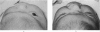
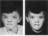
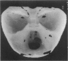
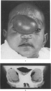
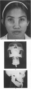
It is postulated that craniosynostosis is due to a growth abnormality in all or part of the cranial capsule. Release of the stenosed part in the first months of life will re-establish the balance between the rapidly growing brain and eye, and the cranial capsule. Three periods for operative treatment are described: early, intermediate and late. Only in the early period can operative treatment restore normal growth dynamics; in the late period the aim is correction of an established deformity. The relationship between cranial clefts and frontonasal encephaloceles is explored. If the space-occupying encephalocele is removed early, the distorted facial bones adopt a more normal position, whereas cranial clefts do not respond to early operation by remoulding. The treatment of the acquired deformities of acute cranio-facial trauma have taken on new perspectives with the application of the multi-disciplinary approach and surgical techniques developed in the treatment of congenital deformities resulting in considerable reduction in the period of hospitalisation.
Full text
PDF


Images in this article


Selected References
These references are in PubMed. This may not be the complete list of references from this article.
- Hoffman H. J., Mohr G. Lateral canthal advancement of the supraorbital margin. A new corrective technique in the treatment of coronal synostosis. J Neurosurg. 1976 Oct;45(4):376–381. doi: 10.3171/jns.1976.45.4.0376. [DOI] [PubMed] [Google Scholar]
- Marchac D. Radical forehead remodeling for craniostenosis. Plast Reconstr Surg. 1978 Jun;61(6):823–835. [PubMed] [Google Scholar]
- Mazzola R. F. Congenital malformations in the frontonasal area: their pathogenesis and classification. Clin Plast Surg. 1976 Oct;3(4):573–609. [PubMed] [Google Scholar]
- Persson K. M., Roy W. A., Persing J. A., Rodeheaver G. T., Winn H. R. Craniofacial growth following experimental craniosynostosis and craniectomy in rabbits. J Neurosurg. 1979 Feb;50(2):187–197. doi: 10.3171/jns.1979.50.2.0187. [DOI] [PubMed] [Google Scholar]
- Rahman N. U. Nasal encephalocoele. Treatment by trans-cranial operation. J Neurol Sci. 1979 Jun;42(1):73–85. doi: 10.1016/0022-510x(79)90153-9. [DOI] [PubMed] [Google Scholar]
- Suwanwela C., Suwanwela N. A morphological classification of sincipital encephalomeningoceles. J Neurosurg. 1972 Feb;36(2):201–211. doi: 10.3171/jns.1972.36.2.0201. [DOI] [PubMed] [Google Scholar]
- Tessier P. Anatomical classification facial, cranio-facial and latero-facial clefts. J Maxillofac Surg. 1976 Jun;4(2):69–92. doi: 10.1016/s0301-0503(76)80013-6. [DOI] [PubMed] [Google Scholar]
- Tessier P., Guiot G., Rougerie J., Delbet J. P., Pastoriza J. Ostéotomies cranio-naso-orbito-faciales. Hypertélorisme. Ann Chir Plast. 1967 Jun;12(2):103–118. [PubMed] [Google Scholar]