

Abstract
This short communication describes two normal variants of the accessory hemiazygous vein in a 15-year-old female. The article demonstrates that knowledge of the aberrant venous anatomy and the collateral pathway is important for the practising radiologist.
A 15-year-old female with malignant histocytoma underwent a CT scan of the thorax for evaluation of metastatic disease. The CT scan showed no evidence of metastatic disease. The CT images demonstrated a connection of the hemiazygos vein to the accessory hemiazygos vein (Figure 1). The accessory hemiazygos vein drained into the brachiocephalic vein (Figure 2). These are two normal variants of the accessory hemiazygos vein (Figure 3).
Figure 1.

Connection between the hemiazygos and the accessory hemiazygos, a rare but normal variant. (a,b) Sagittal CT images of the thorax demonstrate the accessory hemiazygos vein in continuity with the hemiazygos vein forming a single vessel (arrows). The level of communication T8 is shown by the arrow in (b). (c) Axial CT image at the level of T11 (lower arrow in Figure 1a) demonstrates the hemiazygos vein (white arrow).
Figure 2.

(a–e) The accessory hemiazygos drains into the left brachiocephalic vein (arrow). This is a normal variant.
Figure 3.
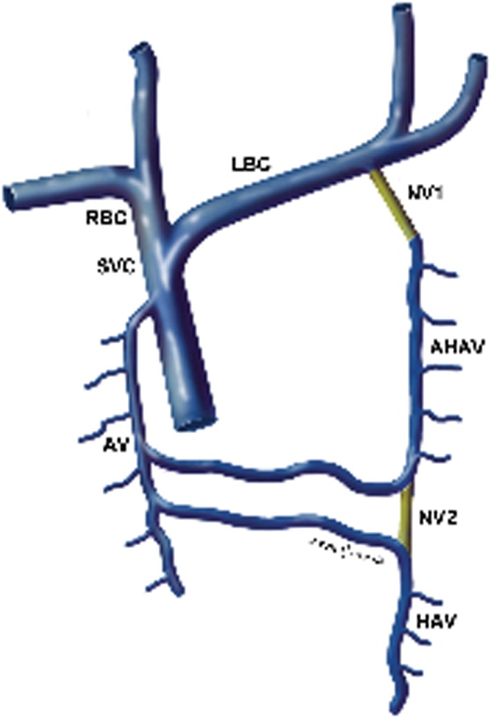
The diagram illustrates the normal anatomy of the azygos system. The blue represents normal venous anatomy. The yellow lines represent the normal variants. SVC, superior vena cava; RBC, right brachiocephalic vein; LBC, left brachiocephalic vein; AV, azygos vein; HAV, hemiazygos vein; AHAV, accessory hemiazygos vein; NV1, normal variant, the accessory hemiazygos drains into the left brachiocephalic vein; NV2, normal variant, the hemiazygos vein is in continuity with the accessory hemiazygos vein.
The azygos system forms from what remains of the posterior cardinal veins. The azygos vein has multiple tributaries including the hemiazygos, accessory hemiazygos and multiple lumbar and intercostal veins. The azygos vein drains into the superior vena cava. At approximately T8–T9 the hemiazygos veins join the azygos vein usually posterior to the aorta. The accessory hemiazygos joins the azygos at T7–T8 crossing posterior to the aorta [1].
The hemiazygos and the accessory hemiazygos are variable in their drainage and anatomy. Usually, the accessory hemiazygos and hemiazygos form common channels posterior to the aorta. In rare instances, 3.6% of the time, the hemiazygos and accessory hemiazygos veins form common channels ventral to the aorta called the interazygos veins [2]. A direct connection of the hemiazygos and accessory hemiazygos is thought to be very rare.
The accessory hemiazygos vein, also called the superior hemiazygos vein, drains the superior left hemithorax. In a majority of cases there is a small connection to the left superior intercostal vein, and rarely, 1–2% of the time, the accessory hemiazygos vein drains into the brachiocephalic vein [3].
The azygos system forms a connection between the superior vena cava and inferior vena cava and serves as an important collateral pathway when there is obstruction of the inferior vena cava or superior vena cava. Knowledge of the aberrant venous anatomy is also important in surgery and catheter placement. The hemiazygos system has also been mistaken for abnormal lymph nodes and knowledge of the azygos system and its variant anatomy is important.
References
- 1.Lawler LP, Corl FM, Fishman EK. Multi-detector row and volume-rendered CT of the normal and accessory flow pathways of the thoracic systemic and pulmonary veins. Radiographic 2002;22:S45–60 [DOI] [PubMed] [Google Scholar]
- 2.Das S, Paul S. Preaortic interazygos vein: A case report. Braz J Morphol Sci 2004;21:141–3 [Google Scholar]
- 3.Galwa RM, Prakash M, Khandelwal N 16-MDCT depiction of the accessory hemiazygos vein draining into the left brachiocephalic vein. Indian J Radiol Imaging 2007;17:50–1 [Google Scholar]