Abstract
Inguinal herniation of ureter is an uncommon finding that can potentially lead to obstructive uropathy. We report a case of inguinal herniation of ureter discovered incidentally during workup for acute renal failure and ultrasound finding of hydronephrosis.
Keywords: Computed tomography, genitourinary
CASE REPORT
The patient was a 76 year old male who presented with 5–6 week history of intermittent left upper quadrant, left flank pain, dysuria, nausea, and fever. Past medical history was significant for motor vehicle accident approximately 20 years ago resulting in multiple pelvic surgeries and left renal trauma that led to a non-functional left kidney after that. On admission, laboratory investigations showed an elevated creatinine level of 1.5 mg/dL, increased from his baseline creatinine of 1.1 mg/dL.
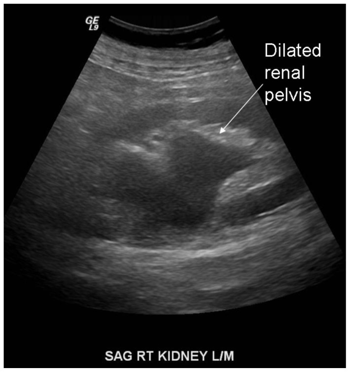
Ultrasound evaluation on admission showed an atrophic left kidney and a large left perinephric abscess as well as right renal pelviectasis (Figure 1). Percutaneous drainage of the left perinephric abscess was performed. Followup renal ultrasound performed 6 days later showed partial resolution of left perirenal fluid collection and redemonstration of right renal pelviectasis. A CT urogram was performed to further evaluate his right kidney and ureter.
Figure 1.
Dilated renal pelvis. Sagittal sonographic image was obtained with a 4 curved transducer at 4.0 MHz of the right kidney from a 76 year old male with acute renal failure, demonstrating dilated right renal pelvis and proximal ureter.
CT scan demonstrated atrophic left kidney with air-fluid collection inferior to the lower pole of the left kidney. A percutaneous drain had been previously placed within this fluid collection (Figure 6). Evaluation of the right kidney showed dilated renal pelvis without calyceal dilatation. There was no effacement of the renal pelvic fat. The proximal ureter appeared dilated but tapered down to normal caliber without evidence of obstruction. These findings were consistent with an extra-renal pelvis. Delayed phase evaluation of the distal ureter with 3-D reconstruction showed prolapse of the ureter into the right inguinal canal, where it made a U-turn back up towards and connecting to the bladder (Figures 2, 4, and 5). Bilateral inguinal hernia sacs were noted (figure 3), and the contrast-filled herniated ureter was seen medial to the hernia sac. This is consistent with a paraperitoneal, acquired type of inguinal ureteral hernia.
Figure 6.
The patient is a 76 year old male presenting with acute renal failure. Axial CT image shows placement of a percutaneous drain into a left renal abscess. Contrast excretion into the right renal pelvis was seen from previously administered contrast from a different study. This study was performed using GE LightSpeed 16-slice CT scanner using kV 140 and mA 379. No intravenous contrast was administered for this study.
Figure 2.
Extra renal pelvis and inguinal herniation of the right ureter. The patient is a 76 year old male presenting with acute renal failure. Here is an oblique reconstructed CT image showing a dilated renal pelvis with intact renal pelvic fat and gradual tapering of the proximal ureter consistent with an extrarenal pelvis. The ureter is seen going into the inguinal canal. There is no evidence of ureteral obstruction. This study was performed using GE LightSpeed 16-slice CT scanner using kV 140 and mA 379. 40 cc in intravenous Visipaque was injected at time 0. At 5 minutes an additional 120 cc of Visipaque was injected. At 7 minutes a combined nephrographic/execretory phase helical CT was performed of the abdomen and pelvis using 0.625 mm collimation with 2.5 mm axial reconstructions and 3 mm bilateral oblique reconstructions.
Figure 4.
Inguinal hernia of the ureter. The patient is a 76 year old male presenting with acute renal failure. 3-dimensional reconstruction image of CT urogram showing the right ureter going beneath the pelvic rim before making a hairpin turn up towards the bladder. This study was performed using GE LightSpeed 16-slice CT scanner using kV 140 and mA 379. 40 cc in intravenous Visipaque was injected at time 0. At 5 minutes an additional 120 cc of Visipaque was injected. At 7 minutes a combined nephrographic/execretory phase helical CT was performed of the abdomen and pelvis using 0.625 mm collimation with 2.5 mm axial reconstructions and 3 mm bilateral oblique reconstructions.
Figure 5.
Inguinal hernia of the ureter. The patient is a 76 year old male presenting with acute renal failure. 3-dimensional reconstruction image of CT urogram showing the right ureter going beneath the pelvic rim before making a hairpin turn up towards the bladder. This study was performed using GE LightSpeed 16-slice CT scanner using kV 140 and mA 379. 40 cc in intravenous Visipaque was injected at time 0. At 5 minutes an additional 120 cc of Visipaque was injected. At 7 minutes a combined nephrographic/execretory phase helical CT was performed of the abdomen and pelvis using 0.625 mm collimation with 2.5 mm axial reconstructions and 3 mm bilateral oblique reconstructions.
Figure 3.
Inguinal hernia of the ureter. The patient is a 76 year old male presenting with acute renal failure. Axial image of CT urogram with the patient in a prone position shows a contrast-filled right ureter within the right inguinal canal. Bilateral inguinal hernia sacs are present, and the herniated ureter is seen medial to the right inguinal hernia sac. Bilateral hip prostheses are present. This study was performed using GE LightSpeed 16-slice CT scanner using kV 140 and mA 379. 40 cc in intravenous Visipaque was injected at time 0. At 5 minutes an additional 120 cc of Visipaque was injected. At 7 minutes a combined nephrographic/execretory phase helical CT was performed of the abdomen and pelvis using 0.625 mm collimation with 2.5 mm axial reconstructions and 3 mm bilateral oblique reconstructions.
DISCUSSION
Herniation of ureter into the inguinal canal has been reported as a complication of renal transplant as well as spontaneous herniation of native ureter [1, 7]. Herniation of native ureter into the inguinal canal is a rare finding, with approximately 140 cases reported in literature as of 2009 [1]. It is usually an indirect inguinal hernia [5] and is divided into paraperitoneal type and extraperitoneal type, with the paraperitoneal type being more common (approximately 80% of all cases) [2].
In the paraperitoneal type, a loop of ureter is extended alongside a peritoneal sac. The herniated ureter is adherent to the posterior peritoneum, both of which are present in the hernia. It is a sliding type of hernia that is thought to be due to traction of underlying structures or adhesions that attach the ureter to the posterior peritoneum. Paraperitoneal type of ureteral hernia is believed to be acquired. Herniated bowel may also be present. It has been noted that the ureteral loop is located medial to the peritoneal sac, and when the ureter is extended into the scrotum it is more likely to be obstructed [3].
In contrast to the paraperitoneal type of ureteral hernia, the extraperitoneal type is thought to be due to a congenital embryonic defect that results in fusion between the ureter and the genitoinguinal ligaments. It is theorized to be the result of failure of separation of the ureteric bud from the Wolffian duct, both of which are then drawn down to the scrotum to form the epididymis and vas deferens [1, 3]. As such, in the extraperitoneal type of inguinal ureteral hernia the ureter herniates without peritoneum attached to it.
There has been one case report of an extraperitoneal ureteral hernia that was thought to be possibly due to adhesions from previous hernia repair [6], but in general the extraperitoneal type of inguinal ureteral hernia is thought to be congenital. It has been noted that in the extraperitoneal type of inguinal hernia there is often a large amount of fat tissue within the hernia [6]. While the distinction between paraperitoneal and extraperitoneal types of inguinal ureteral hernia is classically made during surgical exploration, CT urogram provides a non-invasive method to determine the hernia type.
Although rare, inguinal ureteral hernia can cause obstructive uropathy, and herinorrhaphy is indicated in these cases to relieve the obstruction. In our patient, the herniated ureter is not causing obstruction, with the proximal ureteral dilatation due to extra-renal pelvis. His symptoms and acute renal failure are attributable to his left renal atrophy and acute infection. His creatinine level gradually improved during his hospital stay until it was back to baseline. Thus a conservative approach with observation was chosen regarding his herniated right ureter. The patient was discharged in good condition. Subsequent followup showed that the patient is doing well with stable renal function.
TEACHING POINT
Inguinal hernia of the ureter is a rare finding that may be acquired or congenital and may lead to obstructive uropathy, and the inguinal ureter is prone to injury during herniorrhaphy. CT urogram with 3-D reconstruction provide good anatomic details that can diagnose the presence of a herniated ureter.
Table 1.
Summary table for inguinal hernia of the ureter
| Etiology | Extraperitoneal type – congenital; failure of separation of ureteric bud from Wolffian duct. Ureter descends with testes. Paraperitoneal type – slides along with inguinal hernia sac. Questionable relationship with prior surgery and trauma. |
| Incidence | Rare; approximately 140 cases reported to date. |
| Gender ratio | Extraperitoneal type only occurs in males. Paraperitoneal type likely reflects the gender ratio of inguinal hernias, which is 25:1. Femoral herniation of the ureter has been reported in female patients. |
| Age predilection | Extraperitoneal type is present since birth. However, they may not present until later in life. Paraperitoneal type tends to present in older patients. |
| Risk factors | Male gender. Prior surgery and trauma may be a questionable risk factor for paraperitoneal type. |
| Treatment | Signs of obstructive uropathy warrant surgical correction of the hernia. Watchful waiting is reasonable in cases with no evidence of obstruction. |
| Prognosis | Depends on the timing of surgery to relief obstructive uropathy; early diagnosis and treatment improves prognosis. |
| Findings on imaging | Ultrasound may show signs of obstruction (i.e. Pelvicaliectasis). CT urography will show ureter going into the inguinal canal. Presence of a hernia sac with herniated ureter medial to the sac is suggestive of paraperitoneal type of inguinal ureteral hernia. |
Table 2.
Differential diagnosis table for ureteral obstruction
| US | CT | Other imaging modalities (if applicable) | |
|---|---|---|---|
| Ureteral stone | Hydronephrosis, echogenic stone may be visualized | Hydronephrosis, dense stone, perinephric stranding | |
| Transitional cell carcinoma | Hydronephrosis. | Ureterectasis. Delayed phase may show intraluminal filling defect in the ureter. | |
| Prominent extrarenal pelvis | Dilated renal pelvis and proximal ureter | Prominent renal pelvis with no caliectasis, delayed excretion, or atrophy | |
| Ureteralpelvic junction obstruction | Pelvicaliectasis, may show abrupt narrowing at UPJ | Pyelocaliectasis with abrupt UPJ narrowing | Diuresis renography can help differentiate between obstruction and compliant hydronephrosis |
| Retrocaval ureter | Pelvicaliectasis and dilated proximal ureter | Will show the course of the ureter behind and medial to the mid inferior vena cava | Diuresis renography will show obstruction |
ABBREVIATIONS
- CT
computed tomography
- 3-D
three dimensional
REFERENCES
- 1.Odisho A, et al. Inguinal herniation of a transplant ureter. Kidney International. 2010;78:115. doi: 10.1038/ki.2010.65. [DOI] [PubMed] [Google Scholar]
- 2.Zarraonandia Andraca A, et al. Inguinal ureteral hernia: a clinical case. Arch Esp Urol. 2009;62(9):755–757. doi: 10.4321/s0004-06142009000900013. [DOI] [PubMed] [Google Scholar]
- 3.Schlussel RN, Retik AB. Ectopic ureter, ureterocele, and other anomalies of the ureter. In: Walsh PC, Wein AJ, Vaughan ED, et al., editors. Campbell’s urology. 8th ed. Oxford: WB Saunders; 2002. pp. 2047–8. [Google Scholar]
- 4.Roach SC, Moulding F, Hanbidge A. Inguinal herniation of the ureter. Am J Roentgenol. 2005 Jul;185(1):283. doi: 10.2214/ajr.185.1.01850283. [DOI] [PubMed] [Google Scholar]
- 5.Bertolaccini L, Giacomelli G, Bozzo RE, Gastaldi L, Moroni M. Inguinaoscrotal hernia of a double district ureter: case report and literature review. Hernia. 2005;9:291–293. doi: 10.1007/s10029-004-0296-4. [DOI] [PubMed] [Google Scholar]
- 6.Golgor E, Stroszczynski C, Froehner M. Extraperitoneal Inguinoscrotal herniation of the ureter: a rare case of recurrence after hernia repair. Urol Ing. 2009;83:113–115. doi: 10.1159/000224879. [DOI] [PubMed] [Google Scholar]
- 7.Pollack HM, Popky GL, Blumbert ML. Hernias of the Ureter - An anatomic Roentegenographic Study. Radiology. 1975 Nov;117:275–281. doi: 10.1148/117.2.275. [DOI] [PubMed] [Google Scholar]


