

Abstract
Infantile hemangiomas (IH) are the most common eyelid and orbital tumors of childhood. Although they are considered benign lesions that have a generally self-limited course, in the periocular region, they have the potential to cause amblyopia, strabismus, and severe disfigurement. The decision for treatment can be a source of anxiety for patients, parents, and physicians alike. There are numerous treatment modalities, including emerging therapies that may make treatment safer and more effective than ever before. This review discusses our current understanding of this disease, its management, and future therapies.
Keywords: Infantile hemangioma, Capillary hemangioma, Propranolol treatment
Introduction
Infantile hemangiomas (IH) are the most common tumors of the head and neck in childhood. These hamartomas vary greatly in phenotype and, consequently, their management and potential sequelae. Their significance is dependent on location, size, age of lesion and cosmetic implications. Although the exact etiology and pathophysiology of these lesions are not clear, insights have been gained during the preceding decades. Treatment strategies have been developed through research efforts and serendipitous breakthroughs, with particular progress and excitement in the past several years. Variability in phenotype and presentation requires an individualized approach to treatment. For proper management of these lesions, the clinical course, pathogenesis, diagnosis, and management strategies are detailed.
Nomenclature
Although, these lesions have been recognized for centuries, Lister was the first to formally characterize them.1 Colloquially referred to as “strawberry nevi”, these benign vascular hamartomas of early childhood have been given many names, including capillary hemangiomas, juvenile hemangiomas, hemangioblastomas, benign hemangioendotheliomas, and hypertrophic hemangiomas. More recently, investigators have referred to these lesions as infantile hemangiomas, and we have adopted this nomenclature.2 Historical ambiguity in naming and classification of “hemangiomas” ultimately required stricter categorization, yielding our current definition of infantile hemangiomas as unique vascular tumors that arise after birth during infancy, and undergo a characteristic proliferative phase followed by spontaneous involution. IH are distinct from congenital hemangiomas, which are fully-formed at birth, and should not be confused with the content of this review.
Epidemiology
The incidence of IH is referenced in the literature between 3% and 10%. A recent literature review confirmed that the incidence is likely 4–5%.3 A large, multi-center prospective study examining the demographics of IH found female gender, Caucasian ethnicity, prematurity, low birth weight, multiple gestations, and advanced maternal age as risk factors.4 Another potential risk factor includes chorionic villus sampling; however, the literature lacks robust confirmatory data and further studies are warranted. There is a racial predilection for Caucasians (10–12%) compared to blacks (1.4%) and Taiwanese and Japanese (0.2–1.7%).4
Clinical presentation
IH can occur anywhere on the skin, but are most common in the head and neck region.5 Rarely, periocular IH are noted at birth, and nearly all are identified by six months of age.6 Their clinical appearance can vary greatly in location, size, depth, and rate of growth. Lesions are categorized into three main subtypes based on depth of involvement. Superficial lesions appear as bright red papules or nodules that may be flat or have a bumpy appearance (Fig. 1A). They will blanch with pressure, which can help distinguish these lesions from port-wine stains (nevus flammeus), which are generally firm, rather than soft and easily compressible like IH. Deeper lesions cause variable changes in the skin color depending on the distance to the surface. Occasionally, they may have a blue or purple coloration, or they may have no discoloration at all (Fig. 1B). A third subtype contains components of both superficial and deep lesions (Fig. 1C). While lesions are often still described by their depth, morphological classification of lesions as localized (discrete) or segmental (involving a broad anatomic or developmental area) has been shown to be indicative of clinical course.7 The majority of lesions are localized (72%) with segmental and multifocal lesions comprising 18% and 3%, respectively.8
Figure 1.

Examples of different types of infantile hemangioma. (A) A superficial lesion affecting the left upper eyelid, forehead, and temple with a deep red, pebbly surface. (B) A deep lesion of the glabella (arrow) without any cutaneous discoloration. (C) A large mixed superficial and deep lesion of the scalp with features of both. (Courtesy of Timothy J. McCulley, MD).
Clinical course
IH are distinguished from other vascular malformations of early childhood by their characteristic clinical course. Although each lesion may have a distinct growth pattern, most follow a typical course divided into six proposed stages: (1) nascent, (2) early proliferative, (3) late proliferative, (4) plateau, (5) involution, and (6) abortive.7,9–11 The nascent stage is the newborn stage prior to emergence of lesions, which typically lasts 0–3 months. During the next 6–10 months, proliferation occurs in two stages: an early proliferative stage which brings rapid growth in the first few months of life during which lesions attain most of their size, followed by a late proliferative stage with less rapid growth. In general, 80% of IH attain their maximum size by five months. As compared to superficial or localized lesions, deep or segmental lesions appear to have slightly prolonged proliferative phases extending slightly beyond 6 months; however, most lesions still cease growth by nine months of age.7 The proliferative stage is followed by a plateau or stabilization phase of variable duration. Involution is typically heralded by a change in color from bright red to gray or purple, as well as a softening texture and flattening or diminution in size. Approximately half of IH involute by four years of age and three quarters by age seven.12
Although most IH have an uncomplicated clinical course, some can cause significant morbidity, necessitating treatment. Amblyopia is the most common ophthalmic complication of IH and affects 40–60% of IH patients.6,13 Lesions of the periocular area greater than 1 cm in greatest dimension may be predictive of amblyopia and a need to treat.14 In the eyelid, induced astigmatism causing anisometropic amblyopia is the most common complication, as well as occlusion of the visual axis resulting in deprivation amblyopia (Fig. 2). Orbital lesions are less common; however, secondary to their mass effect, they may result in strabismus, proptosis, exposure keratopathy or compressive optic neuropathy.6 The presence of any of these complications requires urgent intervention. Although not a functional impairment, the presence of early disfigurement or the potential for this complication also warrants treatment. In fact, 40–80% of IH can leave permanent residua even after tumor involution.15
Figure 2.
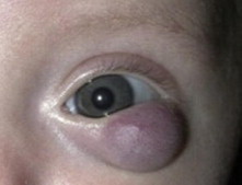
A left lower eyelid hemangioma causing induced astigmatism. (Courtesy of Timothy J. McCulley, MD).
IH in non-ocular locations are associated with other complications. Ulceration is the most frequent complication and occurs more commonly in ano-genital, lower lip, and neck regions. This may result in significant blood loss, pain, and predisposition to secondary infection. IH within the airway can cause airway obstruction and are therefore most worrisome; they are associated with concomitant IH in a beard distribution. Other systemic complications are associated with multifocal IH (more than five cutaneous lesions), visceral (e.g., liver) hemangiomas predisposing to high-output heart failure, and large segmental facial hemangiomas as part of the PHACE syndrome (Posterior fossa malformations, Hemangioma, Arterial abnormalities, Cardiac defects/aortic coarctation and Eye abnormalities).16 These lesions may be more resistant to typical treatments and require multidisciplinary consultation making referral to associated specialists essential.
Pathogenesis
Although the exact pathogenesis of hemangiomas is not completely understood, histopathologic and cell surface marker studies have improved our understanding of this entity. Genetic factors have been investigated, with twin studies suggesting that heredity does not play a large role. This is consistent with observations that IH usually occur sporadically. Others, however, have speculated a genetic contribution as evidenced by a doubling of risk if present in a family member,17 while the racial predilection in Caucasians compared to non-whites also suggests some genetic component.
Examination of molecular markers expressed by in vivo IH cells, cultured IH endothelial cells (HemEC), and hemangioma-derived stem cells (HemSC) have shed considerable light on possible mechanisms. Both angiogenesis (i.e., proliferation or migration from preexisting vessels) and vasculogenesis (i.e., de novo formation of vessels from progenitor cells) have been implicated in the pathogenesis of IH. In vivo IH cells express endothelial cell and immature vascular markers.18 Further study of expression patterns of cultured IH endothelial cells (HemEC) have discovered upregulated glucose transporter protein type-1 (GLUT-1), vascular endothelial growth factor (VEGF), basic fibroblast growth factor (bFGF), among others, with downregulation of other markers, such as CD146.2,17 GLUT-1 is a particularly useful cell surface marker in the diagnosis of these lesions as they are found exclusively in IH.19 This research on HemEC expression patterns suggests a clonal proliferation of partially differentiated, immature vascular cells, implicating angiogenesis in the formation of IH.
More recently, however, attention has turned toward angioproliferative signaling molecules such as VEGF, a key molecule in vessel formation and likely a fundamental part of IH formation. Increased serum VEGF-A levels have been noted in patients with proliferating IH compared to involuting IH as well as negative controls.20 Similarly, stem cells isolated from human IH demonstrate VEGF-A expression in the proliferating phase but not in the involuting phase of IH. This finding was further examined in the setting of the traditional therapeutic agent of choice, corticosteroids. In vivo mouse studies showed that dexamethasone treatment blocked secretion of VEGF-A from IH-derived stem cells.21 Related work provides evidence for a possible mechanism related to steroid inhibition of NF-κB, a molecule associated with both inflammation as well as angiogenesis.22 Additionally, HemSC have been shown to be capable of forming functional vessels with an IH-like phenotype without additional mesenchymal support that is required by other cell populations.23 Cumulatively, this implicates HemSC in vasculogenic formation of IH.
Research investigating the cellular origin of the hematopoietic IH stem cells points to a population of hematopoietic stem cells called endothelial progenitor cells (EPCs). This is supported by findings of increased EPCs in peripheral blood of IH patients, EPC-associated markers localized to endothelium of proliferating IH, and markers associated with primitive hematopoietic cells found in forming capillary endothelium.24 However, earlier studies implicate a possible placental origin based on research demonstrating cross-similarities in hemangioma endothelial cell and placental cellular markers.25,26
Ultimately, there is much still to be understood about the molecular pathogenesis of IH; however, the recent advances described above offer new targets for future therapies, most notably the VEGF cascade.
Histopathology
The clinical course of IH is mirrored by histopathological changes. Early proliferating IH are composed of cellular masses of compact immature capillaries lined by plump endothelial cells with high mitotic rates.27–29 By contrast, endothelial cells diminish in later stage lesions, which are composed of more formed vascular structures lined by flat endothelial cells, thereby conferring a more prominent vascular pattern.20 Finally, as lesions involute, mitosis gradually decreases, and vascular tissue is replaced by fibrofatty tissue, leading to fibrosis and eventual atrophy.27
Diagnosis
Most superficial lesions can be identified clinically. The clinical course, bright color and blanching with pressure usually make the diagnosis obvious. Moreover, IH deepen in color as they enlarge whereas other vascular malformations generally do not change in hue. When there are lesions that have an unusual appearance, growth pattern, or other phenotype, several diagnostic modalities have been utilized. Some have investigated urinary bFGF and serum VEGF as markers for IH.20,30 When lesions have deep or orbital components, imaging studies should be performed to support the diagnosis and examine the extent of involvement. Ultrasonography shows an increased vessel density and high-flow pattern in IH, which may be helpful in diagnosis, although imaging of its full extent and impact on surrounding structures may be limited with this modality.31 Magnetic resonance imaging (MRI) demonstrates well-circumscribed lesions that are isointense to extraocular muscles on T1 imaging and hyperintensity on T2. They enhance brightly with contrast (Fig. 3).32 Although generally more expensive and less available than computed tomography (CT), we recommend MRI to prevent radiation exposure to children in this vulnerable age group. If CT is necessary, the radiation dosage should be reduced to an age and size appropriate level, and the total number of studies should be considered. With CT imaging, IH appear well-circumscribed and enhance with contrast. Angiography using digital subtraction, magnetic resonance, or computed tomography may be useful in select cases.
Figure 3.

Magnetic resonance imaging. T1 post-contrast fat-suppressed axial (A) and sagittal (B) images demonstrate a well-circumscribed, brightly enhancing lesion of the left brow and upper eyelid.
It must be emphasized that biopsy of lesions of unclear etiology, unusual appearance, orbital location, or other atypical features should be performed to rule out other entities and arrive at a definitive diagnosis. GLUT-1 is expressed only in IH endothelium, and may confirm the diagnosis.
Differential diagnosis
There are several vascular tumors that can share a similar appearance to IH. Congenital hemangiomas are fully-formed at birth, which helps to distinguish them from IH. They are categorized into rapidly involuting and non-involuting forms. Kaposiform hemangioendotheliomas are rare vascular tumors which may be locally aggressive due to their rapid and massive growth pattern. They are generally formed at birth as well, although may appear shortly afterward. Treatment is generally aggressive to limit their destructive course. Tufted angiomas are skin or subcutaneous lesions that feature slow and progressive growth and a generally benign course. Both Kaposiform hemangioendothelioma and tufted angiomas can be associated with Kasabach-Merritt syndrome, a platelet sequestration phenomenon that can result in a severe, life-threatening coagulopathy.33
Vascular malformations are distinct from tumors in that they are formed from defects in vascular development, have non-proliferating endothelium, and generally persist without regression (though they may enlarge commensurately with growth of the patient). The most common head and neck vascular malformation is the capillary malformation, also known as nevus flammeus or port-wine stain. These lesions may be associated with Sturge-Weber syndrome. They have a clinical appearance similar to IH, but do not blanch with pressure.
In cases with diagnostic uncertainty, expert consultation and/or biopsy are recommended to distinguish IH from other lesions.
Management
One of the great difficulties in management of IH lies within the decision to initiate therapy. Since many IH may resolve spontaneously and interventions carry risk, the need to intervene is not always obvious. Parents and physicians alike approach the decision to intervene with anxiety and a degree of uncertainty. As per the “Guidelines of care for hemangiomas of infancy” published in the Journal of the American Academy of Dermatology, major goals of management are prevention or reversal of life-threatening or function-threatening complications, prevention of permanent disfigurement, diminution of psychosocial stress for the patient and family, avoidance of aggressive or detrimental interventions in lesions that may have excellent prognosis without treatment, and prevention or treatment of ulceration to lower scarring, infection, and pain.34 Appropriate counseling and education regarding the natural history, treatment options, and expected outcomes is mandatory for a well-informed and appropriate decision.
The medical implications of intervention depend on the size, location, age of the patient/lesion (indicative of the stage of the IH), and potential for sequelae of each lesion. All of these factors should be evaluated and the final decision for treatment should depend on the relative impact of each. Most IH run a benign course and involute spontaneously. Small lesions that are not causing abnormalities in vision or other vital function may be observed as the initial treatment of choice. However, existing or impending sequelae can necessitate intervention, despite the self-limited nature of the lesions. Amblyopia is a feared and common complication of periocular IH, making intervention mandatory in cases where anisometropia, occlusion, or strabismus are present.6,13 The effect of disfigurement should not be underestimated in the normal functional and psychological development of children. Some lesions, particularly those that are large and severe, cause damage to skin and surrounding structures, leaving behind a fibrofatty scar which remains even after resolution of the IH. Concern for continually enlarging, very large, or destructive lesions generally prompt intervention.
Steroids
Discovered serendipitously in 1963 while treating a thrombocytopenia in a child, systemic steroids (usually 2–5 mg/kg/day) have been shown to decrease IH and represent the historical mainstay of treatment.35 In larger studies, prednisone was given in large doses (20 mg daily for 3–8 weeks), which demonstrated an 84% response rate after two months of therapy. Others have needed up to 6 months of therapy. However, this relatively high response rate is accompanied by a 35% rate of side effects including behavioral changes, irritability, cushingoid appearance, and growth delay.36 In the event of growth delay, catch-up growth may occur in many cases, although the effect may be permanent or long-lasting (Fig. 4).
Figure 4.

Twin infants. The one on the right received oral corticosteroids and shows growth delay compared to his twin. (Courtesy of Timothy J. McCulley, MD).
Intralesional steroids
In an effort to reduce the systemic side effects of oral steroids, investigators began experimenting with locally applied steroid therapy. Kushner first reported the successful use of intralesional steroid injections in an initial four cases.37 Other case reports followed, as well as a more robust series of 25 patients in which Kushner confirmed his previous findings, demonstrating responses of varying degrees, without complications, in all patients.38,39 A mixture of betamethasone 6 mg/ml and triamcinolone 40 mg/ml in a 1:1 ratio is injected intralesional, with a volume of 1–2 ml, depending on the size of the lesion. Response may be drastic and rapid, with improvement in days (Fig. 5).
Figure 5.

Involution of hemangioma following intralesional steroid injection. (A) Prior to treatment. (B) One week following injection, there is a mild decrease in the size of the lesion. (C) Four weeks later, marked improvement in lesion size has occurred.
However, as subsequent ophthalmic literature has demonstrated, due to direct communication between lesion and systemic vasculature, periocular intralesional steroid injections carry with them a small but very real risk of ophthalmic artery embolization.40–42 Other local complications include cutaneous hypopigmentation, fat atrophy, calcification, and full-thickness eyelid necrosis.43–47 Although rare, adrenal dysfunction may occur with local steroids as well.43 These potential complications make careful patient selection and informed consent critical. Intralesional steroids are now generally second line therapy for refractory lesions.
Topical steroids
Topical steroids have also been investigated and have shown moderate effects. After initial publication in the mid 1990s,48,49 popularity of this modality failed to achieve high levels. In 2005, in a series of 34 patients using “ultrapotent” steroids (clobetasol propionate 0.05% once or twice daily, halobetasol propionate 0.05%, or betamethasone dipropionate), 1/3 had a good response, 1/3 a partial response, and the rest failed to respond.50 While this treatment modality avoids many more serious side effects of systemic and injected steroids, it still carries with it potential side effects of skin hypopigmentation, hypertrichosis, glaucoma, and cataract formation when used over time, and is only effective for superficial lesions.
Interferon-alpha
Capitalizing on its anti-angiogenic properties, interferon-alpha was also investigated as a possible steroid-sparing agent. Indeed, interferon-alpha induced regression of half or more in 90% of cases in one series.51 However, serious side effects occurred with its use including neutropenia and spastic diplegia. Later consensus was that despite its efficacy, interferon-alpha’s 10–30% risk of neurotoxicity was unacceptably high and the treatment was abandoned.52
Vincristine
Vincristine is an alkaloid cancer chemotherapy, which works by inducing apoptosis through mitotic spindle microtubule interference, and has been used as a steroid-sparing agent for IH. It carries many known side effects, including peripheral neuropathy, constipation, jaw pain, and hematologic toxicity. Although effective, these side effects have made it a third-line treatment for refractory cases.53
Laser treatments
One of the non-medicinal modalities of treatment employed in IH is laser. Several types of laser have been tried, including argon laser, Nd-YAG laser, carbon dioxide laser, fractional photothermolysis, and pulsed dye laser (PDL).54–57 Of these lasers, PDL is the most widely investigated and utilized. This works via destruction of the hemangioma using light of the same wavelength as vessels. With a laser penetration depth of approximately 1.2 mm, this modality has been shown to have efficacy in the treatment of superficial lesions. In general, treatments are performed with multiple passes, with up to 8 or more sessions every 1–2 months. In their series of 617 cases, Hohenleutner et al. found that PDL arrested progression of 97% of IH, completely resolved 14% and partially regressed another 15%.34
However, PDL also carries a risk of hyperpigmentation and atrophic scarring,35,58 although the development of dynamic surface cooling devices has resulted in better epidermal protection. Subsequent randomized control trials of PDL versus observation showed no significant difference in outcome at one year.59 Longer term follow up of these patients after 5 years again showed no difference in clearance, but did find a significant increase of scarring in the PDL cohort.36 Although PDL may primarily halt progression (rather than resolve the IH) and may have additional risk of long-term disfigurement, it may still prove effective for select lesions, particularly those with ulceration.60
Surgery
Surgery continues to be a viable alternative to all of the above modalities.61 Although it yields nearly instantaneous resolution of the lesion, it carries with it the risk of bleeding, despite the well-circumscribed nature of IH. Walker et al. demonstrated impressive clinical results following surgical removal of 12 visually significant hemangiomas, half of which had failed prior therapy; however, two of those cases also necessitated intraoperative blood transfusions due to hemorrhage.62 Patients must be selected very carefully for excision accounting for the size, location, and surgical risk to surrounding structures (Fig. 6). Surgery is generally reserved for lesions that are refractory to less invasive treatments. Various techniques and technologies have aided in limiting and controlling intraoperative bleeding,63 rendering surgical excision a more favorable option in appropriate clinical scenarios.
Figure 6.
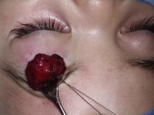
Intraoperative photograph. Despite repeated injection, the brow hemangioma continued to increase. It was excised with a subbrow incision without complication.
Beta-blockers
A recent paradigm shift in the treatment of IH occurred following Leaute-Labreze et al. serendipitous discovery of the efficacy of propranolol in 2008. In their landmark case series, they treated an infant with hypertrophic obstructive cardiomyopathy with oral propranolol, after which the infant’s coincident nasal tip IH was noted to regress. They subsequently confirmed this observation by treating an additional ten cases of IH (seven of which had some periorbital involvement) with oral propranolol.64 This systemic treatment showed promise for large, deep, and difficult to access lesions.
Subsequent to this, dozens of case reports and series demonstrating positive results have followed, including other periocular cases. Taban and Goldberg reported the successful treatment of a refractory left orbital and periorbital hemangioma, while Fay et al. reported the successful use of propranolol as primary therapy for a deep intraconal IH.65,66 Haider et al. reported a series of 17 patients with periocular IH showing 100% arrest of progression, more than 50% regression in 10 patients, and moderate regression in 6 patients.67 Missoi et al. demonstrated even more favorable results with reduction in size (median 61%) in all of 17 patients with periocular IH treated with oral propranolol.68 Dhaybi et al. echoed these findings in their case series of 18 patients with periocular IH and noted the tendency for greater regression to occur with earlier treatment.69 This series, as well as a smaller series by Fabian et al. further outlined the clinical significance of these results by documenting improved astigmatism (by greater than half the initial diopters) following treatment with oral propranolol.45,70 Although, not specific to periocular lesions, there is one randomized, double-blind, controlled trial to date comparing propranolol to placebo. The study found that growth stopped in all propranolol patients by week 4 (versus week 16 for placebo) and that there was a significant decrease in volume of the propranolol cohort at all weeks.71
Alongside great excitement over this new and effective modality in the treatment of IH came cautionary words as well. As warned by Siegfried et al., propranolol is not devoid of several very serious side effects including hypotension, bradycardia, a blunted hypoglycemic response, bronchospasm, congestive heart failure, hypothermia, and sleep disturbance, and therefore warrants close monitoring in infants.72 In the aforementioned case series, one patient was discontinued on therapy for symptomatic hypotension45 and one patient experienced a benign episode of bradycardia.44 Otherwise, propranolol was tolerated with only minor side effects in the majority of patients including gastrointestinal upset, reflux, and fatigue.43 Literature also points out that while many series report 100% efficacy in at least arresting lesions, cases of failure in which IH progress despite propranolol therapy are well-known, necessitating the use of alternate modalities of treatment.73
With the flurry of favorable reports with limited side effects, oral propranolol has become a first-line treatment for many institutions. It must be emphasized that although this modality may prove to be the best available treatment, further confirmatory studies must be conducted, and patients and families should be appropriately counseled. There are a number of ongoing trials currently, and more conclusive data will be forthcoming.
There are a number of protocols that have been used for the evaluation of patients, administration of medication, and surveillance for drug-related complications. At the Massachusetts Eye and Ear Infirmary and Massachusetts General Hospital in Boston, Massachusetts, USA, the combined Hemangiomas and Vascular Malformations Clinic utilizes the following protocol. Patient history is gathered, with special attention to previous reactive airway disease, asthma, hypotension, bradycardia, and diabetes mellitus. Parents are informed of and consented for off-label use of propranolol. Patients are admitted with pediatric consultation for 48 h, where vital signs and blood glucose are monitored. The dosing begins at 0.5 mg/kg/day either orally or intravenously divided into three daily doses. This is increased to a maximum dose of 2 mg/kg/day. Patients are followed up at week one and then monthly with medication adjustments made for changes in weight. The medication is continued for 1 year and then tapered over 10 days.
Topical beta-blockers
As with the use of steroids, in an effort to minimize the systemic side effects of oral propranolol, topical beta-blockers have been investigated. Guo reported the first successful case of topical timolol 0.5% applied twice daily for the treatment of IH causing significant astigmatism.74 After 7 weeks of treatment, astigmatic error was no longer anisometropic, and there was substantial improvement in the size, thickness, and color of the IH. Thereafter, the same group published an additional 7 cases, all of which responded within 4–8 weeks with a reduction in size ranging from 55–95%.75 None of these cases were associated with any side effects. A larger, multi-center, retrospective series examined 73 patients with smaller and superficial IH anywhere on the body treated with either 0.1% or 0.5% gel-forming timolol maleate twice daily.76 The only side effect was one patient who developed sleep disturbance requiring discontinuation of the medication. Overall, initial data suggests that top ical beta-blockers can be safe and effective for superficial lesions with longer duration of treatment yielding better effects.
Intralesional beta-blockers
There has been a single report of intralesional beta-blocker injection for IH.77 In this prospective, non-randomized study, ten consecutive patients received intralesional triamcinolone 40 mg/ml while the following 12 patients received intralesional propranolol 1 mg/ml (up to 1 ml). In this uncontrolled study, there were similar results with approximately 40% achieving an “excellent” response, 40–45% a “fair or good” response, and 17–20% that did not respond at all. No side effects were reported. This series merits further investigation to explore the safety and efficacy of intralesional/intravascular beta-blocker therapy.
Possible mechanisms of the effect of beta-blockers include an early (1–3 days) vasoconstrictive effect due to nitrous oxide (NO) release, an inhibition of proangiogenic factors (such as VEGF and basic fibroblast growth factor) that halt growth in intermediate phases, and induced apoptosis of proliferating endothelial cells resulting in regression in later stages.78 Research to further elucidate this mechanism continues.
Conclusion
Recent advances in the treatment of IH have inspired a renewed interest in the natural course, pathogenesis and successful treatment of this common tumor of childhood. Although most lesions have a benign and self-limited course, a significant number of lesions necessitate treatment to avoid permanent vision loss, disfigurement, or life-threatening complications from airway or visceral involvement. In this setting, expert consultation is recommended. The recent discovery of propranolol’s effect on IH has heralded a new era in IH management that is still in its early phase. All available interventions in the physician’s armamentarium should be considered to employ the strategy most likely to alleviate suffering from these lesions. The promise of further understanding of this tumor brings hope for even more effective and targeted treatment.
Acknowledgment
The authors thank Timothy J. McCulley, MD for providing figures for this manuscript.
Footnotes
Peer review under responsibility of King Saud University.
![]()
Contributor Information
Alison B. Callahan, Email: alison_callahan@meei.harvard.edu.
Michael K. Yoon, Email: michael_yoon@meei.harvard.edu.
References
- 1.Lister W.A. The natural history of strawberry naevi. Lancet. 1938;1:1429–1434. [Google Scholar]
- 2.Jinnin M., Ishihara T., Boye E., Olsen B.R. Recent progress in studies of infantile hemangioma. J Dermatol. 2010;37(11):939–955. doi: 10.1111/j.1346-8138.2010.00927.x. [DOI] [PubMed] [Google Scholar]
- 3.Kilcline C., Frieden I.J. Infantile hemangiomas: how common are they? A systematic review of the medical literature. Pediatr Dermatol. 2008;25(2):168–173. doi: 10.1111/j.1525-1470.2008.00626.x. [DOI] [PubMed] [Google Scholar]
- 4.Haggstrom A.N., Drolet B.A., Baselga E., Chamlin S.L., Garzon M.C. Prospective study of infantile hemangiomas: demographic, prenatal, and perinatal characteristics. J Pediatr. 2007;150(3):291–294. doi: 10.1016/j.jpeds.2006.12.003. Hemangioma investigator group. [DOI] [PubMed] [Google Scholar]
- 5.Léauté-Labrèze C., Taïeb A. Efficacy of beta-blockers in infantile capillary haemangiomas: the physiopathological significance and therapeutic consequences. Ann Dermatol Venereol. 2008;135(12):860–862. doi: 10.1016/j.annder.2008.10.006. [DOI] [PubMed] [Google Scholar]
- 6.Haik B.G., Jakobiec F.A., Ellsworth R.M., Jones I.S. Capillary hemangioma of the lids and orbit: an analysis of the clinical features and therapeutic results in 101 cases. Ophthalmology. 1979;86(5):760–792. doi: 10.1016/s0161-6420(79)35452-5. [DOI] [PubMed] [Google Scholar]
- 7.Chang L.C., Haggstrom A.N., Drolet B.A., Baselga E., Chamlin S.L. Hemangioma investigator group. Growth characteristics of infantile hemangiomas: implications for management. Pediatrics. 2008;122(2):360–367. doi: 10.1542/peds.2007-2767. [DOI] [PubMed] [Google Scholar]
- 8.Chiller K.G., Passaro D., Frieden I.J. Hemangiomas of infancy: clinical characteristics, morphologic subtypes, and their relationship to race, ethnicity, and sex. Arch Dermatol. 2002;138:1567–1576. doi: 10.1001/archderm.138.12.1567. [DOI] [PubMed] [Google Scholar]
- 9.Jacobs A.H. Strawberry hemangiomas: the natural history of the untreated lesion. Calif Med. 1957;86(1):8–10. [PMC free article] [PubMed] [Google Scholar]
- 10.Esterly N.B. Cutaneous hemangiomas, vascular stains and malformations, and associated syndromes. Curr Probl Dermatol. 1995;7(3):65–108. doi: 10.1016/s0045-9380(96)80023-5. [DOI] [PubMed] [Google Scholar]
- 11.Haggstrom A.N., Drolet B.A., Baselga E., Chamlin S.L., Garzon M.C., Horii K.A. Prospective study of infantile hemangiomas: clinical characteristics predicting complications and treatment. Pediatrics. 2006;118(3):882–887. doi: 10.1542/peds.2006-0413. [DOI] [PubMed] [Google Scholar]
- 12.Margileth A.M., Museles M. Cutaneous hemangiomas in children. Diagnosis and conservative management. JAMA. 1965;194(5):523–526. [PubMed] [Google Scholar]
- 13.Robb R.M. Refractive errors associated with hemangiomas of the eyelids and orbit in infancy. Am J Ophthalmol. 1977;83(1):52–58. doi: 10.1016/0002-9394(77)90191-x. [DOI] [PubMed] [Google Scholar]
- 14.Schwartz S.R., Blei F., Ceisler E. Risk factors for amblyopia in children with capillary hemangiomas of the eyelid and orbit. J AAPOS. 2006;10:262–268. doi: 10.1016/j.jaapos.2006.01.210. [DOI] [PubMed] [Google Scholar]
- 15.Boscolo E., Bischoff J. Vasculogenesis in infantile hemangioma. Angiogenesis. 2009;12:197–207. doi: 10.1007/s10456-009-9148-2. [DOI] [PMC free article] [PubMed] [Google Scholar]
- 16.Holland K.E., Drolet B.A. Infantile hemangioma. Pediatr Clin North Am. 2010;57(5):1069–1083. doi: 10.1016/j.pcl.2010.07.008. [DOI] [PubMed] [Google Scholar]
- 17.Drolet B.A., Swanson E.A. Hemangioma investigator group. Infantile hemangiomas: an emerging health issue linked to an increased rate of low birth weight infants. J Pediatr. 2008;153(5):712–715. doi: 10.1016/j.jpeds.2008.05.043. [DOI] [PubMed] [Google Scholar]
- 18.Takahashi K., Mulliken J.B., Kozakewich H.P., Rogers R.A., Folkman J., Ezekowitz R.A. Cellular markers that distinguish the phases of hemangioma during infancy and childhood. J Clin Invest. 1994;93:2357–2364. doi: 10.1172/JCI117241. [DOI] [PMC free article] [PubMed] [Google Scholar]
- 19.Leon-Villapalos J., Wolfe K., Kangesu L. GLUT-1: an extra diagnostic tool to differentiate between haemangiomas and vascular malformations. Br J Plast Surg. 2005;58:348–352. doi: 10.1016/j.bjps.2004.05.029. [DOI] [PubMed] [Google Scholar]
- 20.Zhang L., Lin X., Wang W., Zhuang X., Dong J., Qi Z. Circulating level of vascular endothelial growth factor in differentiating hemangioma from vascular malformation patients. Plast Reconstr Surg. 2005;116:200–204. doi: 10.1097/01.prs.0000170804.80834.5f. [DOI] [PubMed] [Google Scholar]
- 21.Greenberger S., Boscolo E., Adini I., Mulliken J.B., Bischoff J. Corticosteroid suppression of VEGF-A in infantile hemangioma-derived stem cells. N Engl J Med. 2010;362(11):1005–1013. doi: 10.1056/NEJMoa0903036. [DOI] [PMC free article] [PubMed] [Google Scholar]
- 22.Greenberger S., Adini I., Boscolo E., Mulliken J.B., Bischoff J. Targeting NF-κB in infantile hemangioma-derived stem cells reduces VEGF-A expression. Angiogenesis. 2010;13(4):327–335. doi: 10.1007/s10456-010-9189-6. [DOI] [PMC free article] [PubMed] [Google Scholar]
- 23.Khan Z.A., Boscolo E., Picard A., Psutka S., Melero-Martin J.M., Bartch T.C. Multipotential stem cells recapitulate human infantile hemangioma in immunodeficient mice. J Clin Invest. 2008;118:2592–2599. doi: 10.1172/JCI33493. [DOI] [PMC free article] [PubMed] [Google Scholar]
- 24.Itinteang T., Tan S.T., Brasch H., Day D.J. Haemogenic endothelium in infantile haemangioma. J Clin Pathol. 2010;63(11):982–986. doi: 10.1136/jcp.2010.081257. [DOI] [PubMed] [Google Scholar]
- 25.Barnés C.M., Huang S., Kaipainen A., Sanoudou D., Chen E.J., Eichler G.S. Evidence by molecular profiling for a placental origin of infantile hemangioma. Proc Natl Acad Sci U S A. 2005;102(52):19097–19102. doi: 10.1073/pnas.0509579102. [DOI] [PMC free article] [PubMed] [Google Scholar]
- 26.North P.E., Waner M., Mizeracki A., Mrak R.E., Nicholas R., Kincannon J. A unique microvascular phenotype shared by juvenile hemangiomas and human placenta. Arch Dermatol. 2001;137(5):559–570. [PubMed] [Google Scholar]
- 27.Mulliken J.B., Glowacki J. Hemangiomas and vascular malformations in infants and children: a classification based on endothelial characteristics. Plast Reconstr Surg. 1982;69(3):412–422. doi: 10.1097/00006534-198203000-00002. [DOI] [PubMed] [Google Scholar]
- 28.Pasyk K.A., Grabb W.C., Cherry G.W. Cellular haemangioma. Light and electron microscopic studies of two cases. Virchows Arch A Pathol Anat Histol. 1982;396(1):103–126. doi: 10.1007/BF00428503. [DOI] [PubMed] [Google Scholar]
- 29.Smoller B.R., Apfelberg D.B. Infantile (juvenile) capillary hemangioma: a tumor of heterogeneous cellular elements. J Cutan Pathol. 1993;20(4):330–336. doi: 10.1111/j.1600-0560.1993.tb01271.x. [DOI] [PubMed] [Google Scholar]
- 30.Przewratil P., Sitkiewicz A., Wyka K., Andrzejewska E. Serum levels of vascular endothelial growth factor and basic fibroblastic growth factor in children with hemangiomas and vascular malformations—preliminary report. Pediatr Dermatol. 2009;26:399–404. doi: 10.1111/j.1525-1470.2009.00910.x. [DOI] [PubMed] [Google Scholar]
- 31.Dubois J., Garel L., David M., Powell J. Vascular soft tissue tumors in infancy: distinguishing features on Doppler sonography. AJR Am J Roentgenol. 2002;178:1541–1545. doi: 10.2214/ajr.178.6.1781541. [DOI] [PubMed] [Google Scholar]
- 32.Moukaddam H., Pollak J., Haims A.H. MRI characteristics and classification of peripheral vascular malformations and tumors. Skeletal Radio. 2009;38:535–547. doi: 10.1007/s00256-008-0609-2. [DOI] [PubMed] [Google Scholar]
- 33.Kasabach H.H., Merritt K.K. Capillary hemangioma with extensive purpura: report of a case. Am J Dis Child. 1940;59:1063. [Google Scholar]
- 34.Frieden I.J., Eichenfield L.F., Esterly N.B., Geronemus R., Mallory S.B. Guidelines of care for hemangiomas of infancy: American Academy of Dermatology guidelines/outcomes committee. J Am Acad Dermatol. 1997;37:631–637. doi: 10.1016/s0190-9622(97)70183-x. [DOI] [PubMed] [Google Scholar]
- 35.Katz HP. Thrombocytopenia associated with hemangiomata: critical analysis of steroid therapy. In XI International congress of pediatrics, Tokyo; 1965, p. 336–7.
- 36.Bennett M.L., Fleischer A.B., Jr, Chamlin S.L., Frieden I.J. Oral corticosteroid use is effective for cutaneous hemangiomas: an evidence-based evaluation. Arch Dermatol. 2001;137(9):1208–1213. doi: 10.1001/archderm.137.9.1208. [DOI] [PubMed] [Google Scholar]
- 37.Kushner B.J. Local steroid therapy in adnexal hemangioma. Ann Ophthalmol. 1979;11:1005–1009. [PubMed] [Google Scholar]
- 38.Zak T.A., Morin J.D. Early local steroid therapy of infantile eyelid hemangiomas (local steroid therapy of lid hemangiomas) J Pediatr Ophthalmol Strabismus. 1981;18(1):25–27. doi: 10.3928/0191-3913-19810101-07. [DOI] [PubMed] [Google Scholar]
- 39.Kushner B.J. The treatment of periorbital infantile hemangioma with intralesional corticosteroid. Plast Reconstr Surg. 1985;76(4):517–526. doi: 10.1097/00006534-198510000-00005. [DOI] [PubMed] [Google Scholar]
- 40.Shorr N., Seiff S.R. Central retinal artery occlusion associated with periocular corticosteroid injection for juvenile hemangioma. Ophthalmic Surg. 1986;17(4):229–231. [PubMed] [Google Scholar]
- 41.Ruttum M.S., Abrams G.W., Harris G.J., Ellis M.K. Bilateral retinal embolization associated with intralesional corticosteroid injection for capillary hemangioma of infancy. J Pediatr Ophthalmol Strabismus. 1993;30:4–7. doi: 10.3928/0191-3913-19930101-03. [DOI] [PubMed] [Google Scholar]
- 42.Kushner B., Lemke B. Bilateral retinal embolization associated with intralesional corticosteroid injection for capillary hemangioma of infancy. J Pediatr Ophthalmol Strabismus. 1993;30:397–399. doi: 10.3928/0191-3913-19931101-14. [DOI] [PubMed] [Google Scholar]
- 43.Weiss A.H., Kelly J.P. Reappraisal of astigmatism induced by periocular capillary hemangioma and treatment with intralesional corticosteroids injection. Ophthalmology. 2008;115(2):390–397. doi: 10.1016/j.ophtha.2007.03.077. [DOI] [PubMed] [Google Scholar]
- 44.Cogen M., Elsas F. Eyelid depigmentation following corticosteroid injection for infantile ocular adnexal hemangioma. J Pediatr Ophthalmol Strabismus. 1989;26:35–38. doi: 10.3928/0191-3913-19890101-09. [DOI] [PubMed] [Google Scholar]
- 45.Sutula F.C., Glover A.T. Eyelid necrosis following intralesional corticosteroid injection for capillary hemangioma. Ophthalmic Surg. 1987;18:103–105. [PubMed] [Google Scholar]
- 46.Vasquez-Botet R., Reyes B.A., Vazquez-Botet M. Sclerodermiform linear atrophy after the use of intralesional steroids for periorbital hemangiomas: a review of complications. J Pediatr Ophthalmol Strabismus. 1989;26:124–127. doi: 10.3928/0191-3913-19890501-07. [DOI] [PubMed] [Google Scholar]
- 47.Droste P., Ellis F., Sondhi N., Helveston E. Linear subcutaneous fat atrophy after corticosteroid injection of periocular hemangiomas. Am J Ophthalmol. 1988;105:65–69. doi: 10.1016/0002-9394(88)90122-5. [DOI] [PubMed] [Google Scholar]
- 48.Elsas F.J., Lewis A.R. Topical treatment of periocular capillary haemangioma. J Pediatr Ophthalmol Strabismus. 1994;31:153–156. doi: 10.3928/0191-3913-19940501-06. [DOI] [PubMed] [Google Scholar]
- 49.Cruz O.A., Zarnegan S.R., Myers S.E. Treatment of periocular capillary haemangioma with topical clobetasol proprionate. Ophthalmology. 1995;102:2012–2015. doi: 10.1016/s0161-6420(95)30762-2. [DOI] [PubMed] [Google Scholar]
- 50.Garzon M.C., Lucky A.W., Hawrot A., Frieden I.J. Ultrapotent topical corticosteroid treatment of hemangiomas of infancy. J Am Acad Dermatol. 2005;52(2):281–286. doi: 10.1016/j.jaad.2004.09.004. [DOI] [PubMed] [Google Scholar]
- 51.Ezekowitz R.A., Mulliken J.B., Folkman J. Interferon alfa-2a therapy for life-threatening hemangiomas of infancy. N Engl J Med. 1992;326(22):1456–1463. doi: 10.1056/NEJM199205283262203. [DOI] [PubMed] [Google Scholar]
- 52.Frieden I.J., Haggstrom A.N., Drolet B.A., Mancini A.J., Friedlander S.F., Boon L. Infantile hemangiomas: current knowledge, future directions. Proceedings of a research workshop on infantile hemangiomas, April 7–9, 2005, Bethesda, Maryland, USA. Pediatr Dermatol. 2005;22(5):383–406. doi: 10.1111/j.1525-1470.2005.00102.x. [DOI] [PubMed] [Google Scholar]
- 53.Enjolras O., Brevière G.M., Roger G., Tovi M., Pellegrino B., Varotti E. Vincristine treatment for function- and life-threatening infantile hemangioma. Arch Pediatr. 2004;11(2):99–107. doi: 10.1016/j.arcped.2003.10.014. [DOI] [PubMed] [Google Scholar]
- 54.Gladstone G.J., Beckman H. Argon laser treatment of an eyelid margin capillary hemangioma. Ophthalmic Surg. 1983;14(11):944–946. [PubMed] [Google Scholar]
- 55.Al Buainian H., Verhaeghe E., Dierckxsens L., Naeyaert J.M. Early treatment of hemangiomas with lasers. A review. Dermatology. 2003;206(4):370–373. doi: 10.1159/000069960. [DOI] [PubMed] [Google Scholar]
- 56.Laubach H.J., Anderson R.R., Luger T., Manstein D. Fractional photothermolysis for involuted infantile hemangioma. Arch Dermatol. 2009;145(7):748–750. doi: 10.1001/archdermatol.2009.114. [DOI] [PubMed] [Google Scholar]
- 57.Hohenleutner S., Badur-Ganter E., Landthaler M., Hohenleutner U. Long-term results in the treatment of childhood hemangioma with the flashlamp-pumped pulsed dye laser: an evaluation of 617 cases. Lasers Surg Med. 2001;28(3):273–277. doi: 10.1002/lsm.1050. [DOI] [PubMed] [Google Scholar]
- 58.Batta K., Goodyear H., Moss C., Williams H., Hiller L., Waters R. Randomized controlled study of early pulsed dye laser treatment of uncomplicated infantile haemangiomas: results of a 5-year analysis. Br J Dermatol. 2008;159(Suppl. 1):113. doi: 10.1016/S0140-6736(02)09741-6. [DOI] [PubMed] [Google Scholar]
- 59.Batta K., Goodyear H.M., Moss C., Williams H.C., Hiller L., Waters R. Randomised controlled study of early pulsed dye laser treatment of uncomplicated childhood haemangiomas: results of a 1-year analysis. Lancet. 2002;360(9332):521–527. doi: 10.1016/S0140-6736(02)09741-6. [DOI] [PubMed] [Google Scholar]
- 60.Cantatore J.L., Kriegel D.A. Laser surgery: an approach to the pediatric patient. J Am Acad Dermatol. 2004;50:165–184. doi: 10.1016/j.jaad.2003.08.004. [DOI] [PubMed] [Google Scholar]
- 61.Kullbersh J., Hochman M. Serial excision of facial hemangiomas. Arch Facial Plast Surg. 2011;13(3):199–202. doi: 10.1001/archfacial.2011.23. [DOI] [PubMed] [Google Scholar]
- 62.Walker R.S., Custer P.L., Nerad J.A. Surgical excision of periorbital capillary hemangiomas. Ophthalmology. 1994;101(8):1333–1340. doi: 10.1016/s0161-6420(94)31165-1. [DOI] [PubMed] [Google Scholar]
- 63.Fay A., Nguyen J., Waner M. Conceptual approach to the management of infantile hemangiomas. J Pediatr. 2010;157(6):881–888. doi: 10.1016/j.jpeds.2010.08.013. [DOI] [PubMed] [Google Scholar]
- 64.Léauté-Labrèze C., Dumas de la Roque E., Hubiche T., Boralevi F., Thambo J.B., Taïeb A. Propranolol for severe hemangiomas of infancy. N Engl J Med. 2008;358(24):2649–2651. doi: 10.1056/NEJMc0708819. [DOI] [PubMed] [Google Scholar]
- 65.Taban M., Goldberg R.A. Propranolol for orbital hemangioma. Ophthalmology. 2010;117(1):195. doi: 10.1016/j.ophtha.2009.08.040. [DOI] [PubMed] [Google Scholar]
- 66.Fay A., Nguyen J., Jakobiec F.A., Meyer-Junghaenel L., Waner M. Propranolol for isolated orbital infantile hemangioma. Arch Ophthalmol. 2010;128(2):256–258. doi: 10.1001/archophthalmol.2009.375. [DOI] [PubMed] [Google Scholar]
- 67.Haider K.M., Plager D.A., Neely D.E., Eikenberry J., Haggstrom A. Outpatient treatment of periocular infantile hemangiomas with oral propranolol. J AAPOS. 2010;14(3):251–256. doi: 10.1016/j.jaapos.2010.05.002. [DOI] [PubMed] [Google Scholar]
- 68.Missoi T.G., Lueder G.T., Gilbertson K., Bayliss S.J. Oral propranolol for treatment of periocular infantile hemangiomas. Arch Ophthalmol. 2011;129(7):899–903. doi: 10.1001/archophthalmol.2011.40. [DOI] [PubMed] [Google Scholar]
- 69.Al Dhaybi R., Superstein R., Milet A., Powell J., Dubois J., McCuaig C. Treatment of periocular infantile hemangiomas with propranolol: case series of 18 children. Ophthalmology. 2011;118(6):1184–1188. doi: 10.1016/j.ophtha.2010.10.031. [DOI] [PubMed] [Google Scholar]
- 70.Fabian I.D., Ben-Zion I., Samuel C., Spierer A. Reduction in astigmatism using propranolol as first-line therapy for periocular capillary hemangioma. Am J Ophthalmol. 2011;151(1):53–58. doi: 10.1016/j.ajo.2010.07.022. [DOI] [PubMed] [Google Scholar]
- 71.Hogeling M., Adams S., Wargon O. A randomized controlled trial of propranolol for infantile hemangiomas. Pediatrics. 2011;128(2):e259–e266. doi: 10.1542/peds.2010-0029. [DOI] [PubMed] [Google Scholar]
- 72.Siegfried E.C., Keenan W.J., Al-Jureidini S. More on propranolol for hemangiomas of infancy. N Engl J Med. 2008;359(26):2846. doi: 10.1056/NEJMc086443. [DOI] [PubMed] [Google Scholar]
- 73.Meena M. Re: “propranolol for the treatment of orbital infantile hemangiomas”. Ophthal Plast Reconstr Surg. 2011;27(5):392. doi: 10.1097/IOP.0b013e31822671ae. [DOI] [PubMed] [Google Scholar]
- 74.Guo S., Ni N. Topical treatment for capillary hemangioma of the eyelid using beta-blocker solution. Arch Ophthalmol. 2010;128(2):255–256. doi: 10.1001/archophthalmol.2009.370. [DOI] [PubMed] [Google Scholar]
- 75.Ni N., Langer P., Wagner R., Guo S. Topical timolol for periocular hemangioma: report of further study. Arch Ophthalmol. 2011;129(3):377–379. doi: 10.1001/archophthalmol.2011.24. [DOI] [PubMed] [Google Scholar]
- 76.Chakkittakandiyil A., Phillips R., Frieden I.J. Timolol maleate 0.5% or 0.1% gel-forming solution for infantile hemangiomas: a retrospective, multicenter cohort study. Pediatr Dermatol. 2012;29:28–31. doi: 10.1111/j.1525-1470.2011.01664.x. [DOI] [PubMed] [Google Scholar]
- 77.Awadein A., Fakhry M.A. Evaluation of intralesional propranolol for periocular capillary hemangioma. Clin Ophthalmol. 2011;5:1135–1140. doi: 10.2147/OPTH.S22909. [DOI] [PMC free article] [PubMed] [Google Scholar]
- 78.Storch C.H., Hoeger P.H. Propranolol for infantile haemangiomas: insights into the molecular mechanisms of action. Br J Dermatol. 2010;163(2):269–274. doi: 10.1111/j.1365-2133.2010.09848.x. [DOI] [PubMed] [Google Scholar]