

Abstract
Developmental dysplasia of hip (DDH) is one entity one occasionally comes across while in a busy orthopaedic or paediatric outpatient department. The knowledge of risk factors and awareness of the condition is must for every orthopaedic surgeon and paediatrician as well lest the diagnosis will be missed. An early diagnosis can alter the prognosis of the disease and prevent late disabilities.
Keywords: Developmental dysplasia of hip, Congenital dislocation of hip, Diagnosis, Risk factors
1. Introduction
Developmental (congenital) dysplasia of hip (DDH) generally includes subluxation (partial dislocation) of the femoral head, acetabular dysplasia, and complete dislocation of femoral head from the true acetabulum. The original term ‘Congenital dislocation of the hip (CDH)’ dates back to the time of Hippocrates. Since then, significant progress has been made in the evaluation and treatment of DDH.1,2 The older term congenital dislocation of hip has been gradually replaced by developmental dysplasia of hip (DDH), which was introduced to include in the disorder, infants normal at birth but in whom the hip dysplasia or dislocation subsequently developed or vice versa.3 The term developmental displacement of the hip, thus, indicates a dynamic disorder potentially capable of getting better or worse as the child develops depending on the multidisciplinary care provided.4
2. Risk factors
Developmental dysplasia of the hip affects 1–3% of newborns and is responsible for 29% of primary hip replacements in people up to the age of 60 years.5 In India, the incidence has been reported to be 1.0–9.2 per 1000 in various studies with the incidence being more in northern region.6–8
95–98% of DDH cases are possibly reversible. 2% of DDH cases may have teratologic dislocation which is generally not reversible. 60% will normalize with no treatment after 1 month. 88% will normalize with no treatment after 2 months. This comes to about 1–2 patients per 1000 which have a true DDH and which will go on to produce the pathological changes of DDH.
The left hip is dislocated more often than the right and 20% of cases are bilateral. It is more common in cultures that use swaddling of babies, a manoeuvre that forces the hips into extension and adduction.9 There is a 9:1 female predominance. Apparently the baby's own female hormones aggravate and contribute to the abnormal looseness of the hip ligaments. Of children with DDH, approximately 60% are firstborn and about 20% are born breech. In breech presentation, the hip position tends to force the hip out of the socket, predisposing to dislocation after birth. Highest risk is with extended breech position with the hips flexed and the knees extended and feet reaching the shoulders. There is also an association of congenital muscular torticollis (“wry neck”), metatarsus adductus (toes bent inward) or talipes equinovarus (club foot) with DDH. There is an increased incidence of DDH among the offspring of mother's with DDH from 3 to 5 times above normal. Identical twins have a 34% chance of both having DDH, and among fraternal twins a 3–4% incidence is present.
3. Diagnosis
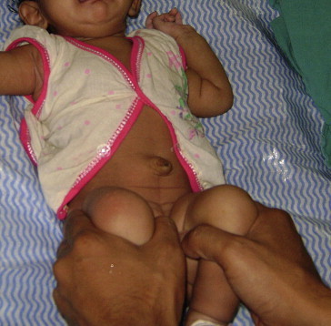
Screening for DDH is usually a part of the physical examination of newborn and later follow ups. For at risk patients i.e. first born, female, breech presentation, and those with a positive family history, the physical examination of the hip at birth is mandatory. Asymmetrical skin folds are found in 25% of normal babies and therefore not an important clinical finding in isolation. Under 3 months old, the Ortolani and Barlow tests are the most common clinical tests for newborn babies. In the Ortolani test, the examiner applies forward pressure to each femoral head in turn, in an attempt to move a posteriorly dislocated femoral head forwards into the acetabulum. The child is rested supine and pelvis is stabilised with opposite hand. The examining hand grasps the affected hip with the thumb on the medial aspect of the thigh and the middle and index finger along the greater trochanter. The hip and knee is then brought to 90° flexion and then abducted with middle and index finger pushing the trochanter forward. Palpable movement suggests that the hip is dislocated or subluxed, but reducible (Fig. 1). In the Barlow test, backward pressure is applied to the head of each femur in turn, and a subluxtable hip is suspected on the basis of palpable partial or complete displacement.10 In performing a Barlow test, the child is rested supine with pelvis stabilized with one hand. The hip is flexed 90° and adducted from abducted position. Simultaneously a backward pressure in the line of femur is applied to displace the femoral head from the acetabular socket. The palpable clunk of dislocatable head moving out is appreciated in a positive Barlow test (Fig. 2). Both the Barlow and Ortolani tests detect an unstable hip but do not detect a dislocated, irreducible hip, (which is best detected by identifying limited abduction of the flexed hip) or a stable hip with abnormal anatomy, e.g. acetabular dysplasia. Benign hip clicks, resulting from soft tissues snapping over bony prominences during hip movement, should be carefully distinguished from the clunks produced during the Ortolani manoeuvre as the dislocated femoral head is reduced and from the subluxation felt during the Barlow test. The Barlow and Ortolani tests are useful in neonates but become difficult by 2–3 months of age.
Fig. 1.

Ortolani manoeuvre.
Fig. 2.
Barlow manoeuvre.
In children aged 3–6 months, the hip laxity reduces and the hip may remain in dislocated position outside the acetabular socket. Thus, the utility of Ortolani and Barlow tests wanes. The Galeazzi sign may be positive in the older child. The Galeazzi sign is performed with the child lying down supine with the hips and knees flexed to 90°, the feet flat on the table and the heels touching the buttocks. The height of each knee is compared. Unilateral femoral shortening may signify hip dislocation or rarer abnormalities of the femur (Fig. 3). False negative results may occur with bilateral hip dysplasia or when the pelvis is not level. A positive sign is that one leg appears shorter than the other. This is usually due to dislocation of the hip; however, any discrepancy of limb length will produce a positive sign. Other physical signs for late dislocation include asymmetry of the gluteal thigh or labral skin folds, a widened perineum on the affected side, buttock flattening, decreased abduction on the affected side, and standing or walking with external rotation of the affected leg. Hip abduction of less than 60° is highly suspicious of a dislocated hip. The elder child may walk on toes on the affected side or present with a painless limp. Bilateral dislocation of the hip can be quite difficult to diagnose, especially after the neonatal period and an index of suspicion is necessary to make a diagnosis. There is often an associated waddling gait with hyperlordosis.
Fig. 3.

Galeazzi manoeuvre. Note that skin creases may look similar on both sides. The right hip is dislocated.
In doubtful cases and equivocal cases, dynamic ultrasound is the imaging tool of choice.11 Both coronal and transverse plane images are taken and stress views are done. The abnormal anatomy visualized on a hip ultrasound could include subluxation or dislocation of the femoral head, abnormal alpha angle, abnormal thickness of labrum, presence of pulvinar, or asymmetry of the ossification centers (Fig. 4).12,13 Ultrasound helps diagnosis in children under 5 months but pelvic X-rays are more useful in older infants and children (Figs. 5, 6). Arthrography, CT and MRI scanning may also be useful. Ultrasound evaluation has rendered arthrographic investigation of the hip largely redundant for diagnostic purposes.14 But because of problems of observer dependency and a poor relationship to the ultimate development of the hip, universal ultrasound screening of hip at birth for DDH is not advocated.15 The main role of ultrasound evaluation of the hip thus, remains in high risk and doubtful cases.16
Fig. 4.

Ultrasound interpretation of DDH.
The alpha and beta angle measurements are used to indicate the degree of acetabular development. A line is drawn parallel to the ossified lateral wall of the ilium (baseline). A second line is drawn along the roof of the cartilaginous acetabulum (from the lateral bony edge of the acetabulum to the labrum) to give the beta angle; this denotes the slope of the cartilaginous acetabulum. A third line is drawn from the inferior edge of the bony acetabulum, at the triradiate cartilage, to the distal part of the ilium, tangential to the slope of the bony acetabulum (roof line). The angle between the 1st and 3rd line is the alpha angle. The alpha angle denotes the slope of the bony acetabulum. Normally, the alpha angle is greater than 60° and an angle less than 55° is considered abnormal.
Fig. 5.

Radiographic findings in DDH.
The triradiate cartilage lies between the ilium, the ischium, and the pubis. Hilgenreiner's line passes through the triradiate cartilages. Perkin's line, drawn at the lateral margin of the acetabulum, is perpendicular to the Hilgenreiner's line. Shenton's line is a curved line that begins at the lesser trochanter, goes up the femoral neck, and connects to a line along the inner margin of the pubis. Acetabular index is an angle formed by the juncture of Hilgenreiner's line and a line drawn along the acetabular surface. Centre edge (CE) angle (used in older children) is formed at the juncture of Perkin's line and a line connecting the lateral margin of acetabulum to the centre of the femoral head. In normal hip, the medial beak of the femoral physis lies in the lower inner quadrant formed by the juncture of Perkin's and Hilgenreiner's line, the Shenton's line is not broken, acetabular index is less than 30°, the CE angle is more than 19° (6–13 years).
Fig. 6.

Same patient as Figs. 1–3 above. The plain radiographs show the dislocated right hip.
4. Management
It is important to diagnose developmental dysplasia of the hip early to improve treatment results and to decrease the risk of complications.17 However, early detection and treatment does not entirely avoid the need for subsequent surgery, and surgery is needed by up to 5% of infants treated with abduction splinting.18 The indications for the various procedures and the most effective management interventions remain controversial. Several small studies have shown that stable hips with mild dysplasia can be observed safely for six weeks before a decision to treat is made. Other studies have referred abduction splinting as the first-line treatment in children younger than 6 months in nonsurgical management. A commercially available dynamic flexion-abduction orthosis (Pavlik® harness), left in place at all times, is used to maintain hip reduction. The harness can be adjusted as the child grows and the hip stabilises. A harness is more likely to succeed when applied in a reducible hip. One study showed that when harness treatment was started by 90 days of age, only 5.7% of babies required further treatment. In another study on 546 hips, the harness failed to reduce 3.3% of DDH. These required surgical treatment. Of those dysplastic hips which were successfully reduced in the harness, 2.4% showed persistent significant late dysplasia and 0.2% persistent severe late dysplasia. All of these could be identified at 5 years of age, and many by 18 months. Avascular necrosis of femoral head was seen in 1% of hips treated in the harness.19 Surgery is only indicated for those who do not respond to early splint or harness treatment, and those who are diagnosed late and are not suitable for splint or harness treatment. The most common operation is closed reduction with adductor or psoas tenotomy, followed by 3–4 months in a plaster cast or abduction brace. The older the child, the more likely an extensive procedure will be required with open reduction and soft tissue stabilisation of the joint, followed by a cast. Over the age of 18–24 months, an additional pelvic and/or femoral osteotomy is often required.
Developmental dysplasia of the hip is an orthopaedic condition usually first encountered by the paediatrician. Hence, paediatrician is an essential part of the team approach required for the management of DDH. A timely diagnosis and prompt management can prevent the long term morbidity associated with the DDH. Identifying the risk factors, performance of clinical tests like Ortolani and Barlow, and at times USG is all that is required to initiate the management till the child is referred to the orthopaedic surgeon.
Conflicts of interest
No benefits in any form have been received or will be received from a commercial party related directly or indirectly to the subject of this article.
References
- 1.Sankar W.N., Weiss J., Skaggs D.L. Orthopaedic conditions in the newborn. J Am Acad Orthop Surg. 2009;17(2):112–122. doi: 10.5435/00124635-200902000-00007. [DOI] [PubMed] [Google Scholar]
- 2.AIUM practice guideline for the performance of an ultrasound examination for detection and assessment of developmental dysplasia of the hip. J Ultrasound Med. 2009;28(1):114–119. doi: 10.7863/jum.2009.28.1.114. [DOI] [PubMed] [Google Scholar]
- 3.Davies S.J., Walker G. Problems in the early recognition of hip dysplasia. J Bone Joint Surg Br. 1984;66:479–484. doi: 10.1302/0301-620X.66B4.6746677. [DOI] [PubMed] [Google Scholar]
- 4.Klisic P.J. Congenital dislocation of the hip– a misleading term: a brief report. J Bone Joint Surg Br. 1989;71:136. doi: 10.1302/0301-620X.71B1.2914985. [DOI] [PubMed] [Google Scholar]
- 5.Sewell M.D., Rosendahl K., Eastwood D.M. Developmental dysplasia of the hip. BMJ. 2009 Nov 24;339:b4454. doi: 10.1136/bmj.b4454. [DOI] [PubMed] [Google Scholar]
- 6.Singh M., Sharma N.K. Spectrum of congenital malformations in the newborn. Ind J Pediatr. 1980;47:239–244. doi: 10.1007/BF02758201. [DOI] [PubMed] [Google Scholar]
- 7.Gupta A.K., Kumar S., Arora P.L., Kumar R., Methani A.K., Sood L.K. Hip instability in newborns in an urban community. Nat Med J India. 1992;5:269–272. [PubMed] [Google Scholar]
- 8.Kaushal V., Kaushal S.P., Bhakoo O.N. Congenital dysplasia of the hip in Northern India. Int Surg. 1976;61:29. [PubMed] [Google Scholar]
- 9.Kutlu A., Memik R., Mutlu M., Kutlu R., Arslan A. Congenital dislocation of the hip and its relation to swaddling used in Turkey. J Pediatr Orthop. 1992;12(5):598–602. [PubMed] [Google Scholar]
- 10.Barlow T.G. Early diagnosis and treatment of congenital dislocation of the hip. J Bone Joint Surg Br. 1962;44:292–301. [Google Scholar]
- 11.Kocher M.S. Ultrasonographic screening for developmental dysplasia of the hip: an epidemiologic analysis (Part I) Am J Orthop. 2000;29(12):929–933. [PubMed] [Google Scholar]
- 12.Graf R. The ultrasound examination of the hip. In: Tonnis D., editor. Congenital Dysplasia and Dislocation of the Hip in Children and Adults. Springer-Verlag; Berlin: 1987. pp. 172–229. [Google Scholar]
- 13.Graf R., Tschauner C., Klapsch W. Progress in prevention of late developmental dislocation of the hip by sonographic newborn “screening”: results of a comparative follow-up study. J Pediatr Orthop. 1993;2:115–121. [Google Scholar]
- 14.McEvoy A., Paton R.W. Ultrasound compared with radiographic assessment in developmental dysplasia of the hip. J R Coll Surg Edinb. 1997;42:254–255. [PubMed] [Google Scholar]
- 15.Castelein R.M., Sauter A.J., de Vlieger M., van Linge B. Natural history of ultrasound hip abnormalities in clinically normal newborns. J Pediatr Orthop. 1992;12:423–427. doi: 10.1097/01241398-199207000-00001. [DOI] [PubMed] [Google Scholar]
- 16.Sochart D.H., Paton R.W. Role of ultrasound assessment and harness treatment in the management of developmental dysplasia of the hip. Ann R Coll Surg Engl. 1996;78:505–508. [PMC free article] [PubMed] [Google Scholar]
- 17.Storer S.K., Skaggs D.L. Developmental dysplasia of the hip. Am Fam Physician. 2006;74(8):1310–1316. [PubMed] [Google Scholar]
- 18.Dezateux C., Rosendahl K. Developmental dysplasia of the hip. Lancet. 2007;369(9572):1541–1552. doi: 10.1016/S0140-6736(07)60710-7. [DOI] [PubMed] [Google Scholar]
- 19.Cashman J.P., Round J., Taylor G., Clarke N.M.P. The natural history of developmental dysplasia of the hip after early supervised treatment in the Pavlik harness. J Bone Joint Surg Br. 2002;84:418–425. doi: 10.1302/0301-620x.84b3.12230. [DOI] [PubMed] [Google Scholar]