

Abstract
Blue Rubber Bleb Nevus Syndrome is a rare disorder that is characterized by multiple recurrent vascular malformations of skin and gastrointestinal tract. The affected patients may present with diverse manifestations including iron deficiency anemia.
We report this syndrome in a 22-year-old man that was referred to our hospital for iron deficiency anemia with unknown cause and vascular malformations in the skin and gastrointestinal tract. Because of stable hemoglobin level, we decided to treatment him by iron supplementation and close follow up. We report this case along with a review of literature.
Keywords: Blue rubber bleb nevus syndrome, Iron deficiencyAnemia, Vascular malformations
INTRODUCTION
Blue Rubber Bleb Nevus Syndrome (BRBNS) or Bean’s syndrome is a disorder characterized by distinctive vascular malformations that mostly involve cutaneous and gastrointestinal tract .1Occult bleeding and iron deficiency anemia (IDA) are the most common clinical presentation of BRBNS but melena ,hematochezia and involvement of internal organs has also been reported.1, 2It is a very rare syndrome and only about 200 cases have been reported till 2012.
Here, we report a case of BRBNS that present with IDA. To the best of our knowledge this is the second case reported from Iran.3
CASE REPORT
A 22 years old man was referred to the GI clinic of Afzalpour Hospital for evaluation of IDA. There was no history of frank bleeding including melena, hematemesis or rectal bleeding. History of abdominal pain, prolonged diarrhea and/or constipation, vomiting, flatulence and prolonged fever were negative. Drug history was negative including for non–steroidal anti- inflammatory agents. He has been receiving occasionally intravenous iron during the last three years. He suffered from a vascular mass on right supraclavicular region at birth with gradual enlargement. At the childhood, surgical resection of the mass was performed and histology showed irregular cavernous channels in both cutaneous and subcutaneous tissue compatible with venous malformation. At the age of 15, oral iron intake was started because of IDA. At about the same time, multiple bluish colored nodular lesions appeared on his lower extremities. Familial history was negative for vascular lesions resembling BRBNS and any other remarkable diseases. He was not drug abuser and drug history included only oral and intravenous iron supplementation since 7 years ago. In physical examination he appeared well nourished but pale. Multiple compressible bluish nodules on lower extremities were seen (Figure1). These nodules measured from 0.5 to 1.5 cm and tended to refill after compression. Scar of a previous incision was present on the right supraclavicular region (Figure2). Otherwise, the physical examination was unremarkable.
Fig.1.

Multiple compressible bluish nodules on the patient foot.
Fig.2.
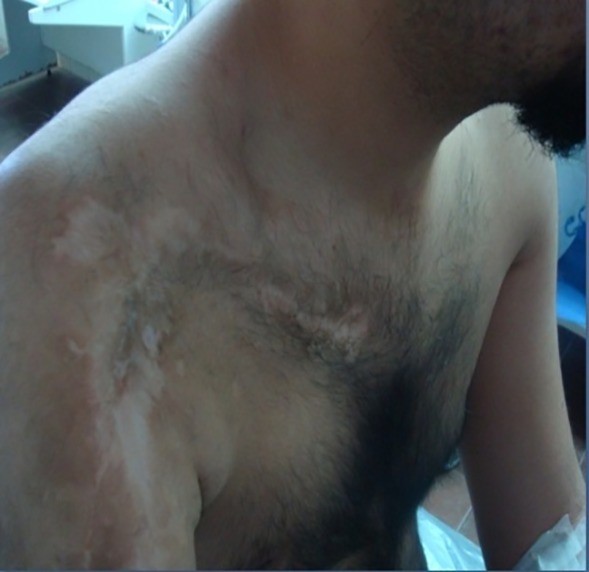
Scar of previous surgical incision on the right supraclavicular region.
Laboratory findings revealed a hemoglobin level of 10.5 g/dL. Other lab results are shown in table 1. The results of routine laboratory tests were within normal limits. Upper and lower gastrointestinal endoscopy along with duodenal biopsy had been performed 4 years ago and was normal.Serum immunoglobulin A(Ig A) and anti-tissue transglutaminase antibody (TTG) -Ig A levels were also normal.
Table 1. Results of patient laboratory tests on admission .
| Variable |
Reference Range,
Age-Adjusted |
On admission |
| White-cell count (per mm3) | 4000–11,000 | 6500 |
|
Hematocrit (%) Hemoglobin (g/dl) MCV (μm3) MCH (pg) MCHC (g/dL) |
38-46 14–16.5 (men) 89-98 27-30 32-35 |
31.7 10.5 75 20 25.7 |
| Platelet count (per mm3) | 150000-450000 | 230000 |
| Ferittin( µg/dl) | 30-250 | 10 |
| Creatinine (mg/dl) | 0.5 -1 | 0.8 |
| Albumin(g/dl) | 3.5-4.5 | 4 |
| Lactate dehydrogenase(U/liter) | 150-230 | 210 |
| Erythrocyte sedimentation rate (mm/hr) | Less than 20 | 12 |
| Anti-tissue transglutaminase antibody(Ig A) IU/ml | Under 10 | Neg |
| Prothrombin time (sec) | 12-13 | 14 |
| International normalized ratio for prothrombin time | 12-13 | 1.2 |
| Alanine aminotransferase(U/liter) | 18-34 | 22 |
| Aspartate aminotransferase(U/liter) | 12-30 | 18 |
| Alkaline phosphatase(U/liter | 68-240 | 88 |
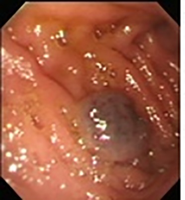
In our center the patient underwent upper and lower GI endoscopy for second time. In upper endoscopy, a bluish vascular lesion with 10 mm diameter and wrinkled surface was seen in proximal part of duodenum (Figure3-a).Colonoscopy also revealed a similar lesion in the descending part of colon (Figure3-b). The patient refused assessment of the small intestine tract.
Fig. (3-a, 3 -b) .
3-a (left) shows a bluish vascular lesion with wrinkled surface in proximal part of duodenum. 3-b (Right) a similar lesion in the descending part of colon.

DISCUSSION
Important diseases that involve the skin and gastrointestinal tract with vascular malformations include BRBNS, Mafucci’s syndrome and Osler-Weber- Rendu syndrome. However, Rendu-Osler-Weber syndrome is an inherited autosomal dominant condition and these patients often experience recurrent episodes of epistaxis with telangiectasia in histologic examination 4 and patients with Mafucci’s syndrome also have bone malformations and chondrodysplasias.5
A diagnosis of BRBNS was made on the basis of the typical skin lesions which were rubbery discrete compressible blebs increasing in number over the years, multiple bluish nevi in duodenum and colon on GI endoscopy, and histological picture of venous malformation on skin biopsy.1-3,6-7, Therefore, we were able to diagnose our patient with BRBNS.
BRBNS (or Bean’s syndrome) is a rare disorder that is characterized by multiple recurrent vascular malformations that primarily involve the skin and the gastrointestinal tract .1The disease and its association with hemagniomas in the skin and gastrointestinal tract was first reported in 1860 by Gascoyen.8A century later in 1958, William Bean named it as BRBNS.9
Skin lesions in BRBNS are often first noticed at birth or in the neonatal period, but can present later .7The size ranges from a few millimeters to many centimeters. Lesions mainly appear in the trunk and upper extremities. Three types of skin lesions have been described .3The most common type is red to blue, nipple-like skin lesion that is easily compressible and refills slowly on release of pressure. This is an important diagnostic sign .8The skin lesions in this case were also small, bluish, soft and compressible, and they occurred in the upper and lower extremities; these are the typical features of BRBNS.
The gastrointestinal tract is the most common organ involved in this disorder, small bowel and distal colon are predominantly affected .9 Bleeding from vascular malformations in gasterointestinal tract often cause IDA that is the most common clinical manifestation of BRBNS but rarely presented as melena or hematochezia.1, 2 In endoscopy, the mucosal nodules are either flat, polypoid or have central bluish nipples.1, 7 In our case, the gastrointestinal lesions were limited to two protuberant lesions with wrinkled surface in duodenum and distal part of colon.
Rarely, involvement of other internal organs occurs and can result in hemothorax, hemopericardium, pulmonary hypertension and central nervous system symptoms.10-12
Often, BRBNS develops sporadically, but sometimes is inherited as an autosomal dominant pattern and its association with chromosome 9p has already been established .13Our case seems to have developed sporadically because no family member had history of BRBNS.
In most cases,only asymptomatic skin lesions are present and patients have a normal life span without any malignant transformation. Extent of visceral organ involvement determines the prognosis of disease.
GI bleeding and rarely, central nervous system involvement can befatal and skeletal lesions are able to cause functional and physical inability.12 Intussusceptions, volvulus, and bowel infarction should be considered in patient with BRBNS and abdominal pain.14
The treatment of BRBNS is mainly conservative. When the patient is asymptomatic and only has mild anemia, observation and iron supplements are adequate.1- 3
Several therapeutic modalities have been attempted to date for treating BRBNS. Antiangiogenetic agents such as steroids, interferon α-2a and octreotide have been applied to reduce the frequency and severity of bleeding episodes.15,16Unfortunately, in most cases, discontinuing administration of these medical treatments has led to recurrence of the disease .15,17
Although endoscopic laser photocoagulation, sclerosis, band ligation and polypectomy have been successfully utilized, they may be dangerous in transmural lesions.18,19
Laparatomy with intraoperative endoscopy may be the best approach for patients with refractory anemia and multiple lesions in GI tract especially small bowel.15- 20In the largest study on treatment of BRBNS published so far, ten patients with BRBNS and IDA with prior history of at least one blood transfusion, underwent whole GI endoscopy. All GI lesions were removed and patients were followed for five years. Subsequent gastrointestinal bleeding recurred in only one patient who had received less extensive procedures.20
Our patient was consulted about the natural history of the disease and the chronicity of the condition which is disabling but not fatal in majority of cases. He was discharged on iron supplements. He did well during 2 years of follow up and his hemoglobin level was maintained without any need to transfusions. Long term follow up is required to evaluate the effect of conservative treatment with iron supplementation in this patient and determine surgical intervention will be necessary or not.
CONFLICT OF INTEREST
The authors declare no conflict of interest related to this work.
Please cite this paper as:
Zahedi MJ,DarvishMoghadam S, SeyedMirzaei SM, DehghaniM, Shafiei pour S,RastiA. Blue Rubber Bleb Nevus Syndrome as a rare Cause of Iron Deficiency Anemia: a Case Report and Review of Literature. Middle East J Dig Dis 2013;5:235-9.
References
- 1.Oksuzoglu BC, Oksuzoglu G, Cakir U, Bayir T, Esen M. Blue rubber bleb nevus syndrome. Am J Gastroenterol. 1996;91:780–2. [PubMed] [Google Scholar]
- 2.Rodrigues D, Bourroul ML, Ferrer AP, Monteiro Neto H, Gonçalves ME, Cardoso SR. Blue rubber bleb nevus syndrome. Rev Hosp ClinFac Med Sao Paulo. 2000;55:29–34. doi: 10.1590/s0041-87812000000100006. [DOI] [PubMed] [Google Scholar]
- 3.Akhiani M, Fateh S, Ghanadan A, Lajevardi V. Extensive Blue Rubber Bleb Nevus Syndrome with Multiple Gastrointestinal Venous Malformations: A Case Report. Iran J Dermatology. 2009;12:99–102. [Google Scholar]
- 4.Shovlin CL, Guttmacher AE, Buscarini E, Faughnan ME, Hyland RH, Westermann CJ. et al. Diagnostic criteria for hereditary hemorrhagic telangiectasia (Rendu-Osler-Weber syndrome) Am J Med Genet. 2000;91:66–7. doi: 10.1002/(sici)1096-8628(20000306)91:1<66::aid-ajmg12>3.0.co;2-p. [DOI] [PubMed] [Google Scholar]
- 5.Shepherd V, Godbolt A, Casey T. Maffucci’s syndrome with extensive gastrointestinal involvement. Australas J Dermatol. 2005;46:33–37. doi: 10.1111/j.1440-0960.2005.00133.x. [DOI] [PubMed] [Google Scholar]
- 6.McKinlay JR, Kaiser J, Barrett TL, Graham B. Blue rubber bleb nevus syndrome. Cutis. 1998;62:97–8. [PubMed] [Google Scholar]
- 7.Beck PL, Aspinall AI, Kilvert VM, Dort J. Blue rubber bleb nevus syndrome. GastrointestEndosc. 2002;56:598–600. doi: 10.1067/mge.2002.127750. [DOI] [PubMed] [Google Scholar]
- 8.Gascoyen M. Case of naevus involving the parotid gland, and causing death from suffocationNaevi of the viscera. Trans PatholSoc (Lond) 1860;11:267. [Google Scholar]
- 9. Bean WB. Blue Vascular Spiders and Related Lesions of the Skin.Springfield, Illinois: Charles C Thomas. 1958;:17-85.
- 10.Giordano C, Battagliese A, di Gioia CR, Campagna D, Benedetti F, Travaglini C, Et al. Blue rubber bleb nevus syndrome and pulmonary hypertension: an unusual association. Cardiovasc Pathol. 2004;13:317–22. doi: 10.1016/j.carpath.2004.07.004. [DOI] [PubMed] [Google Scholar]
- 11.Langleben D, Wolkove N, Srolovitz H, Billick RC, Sheiner NM. Hemothorax and hemopericardium in a patient with Bean's blue rubber bleb nevus syndrome. Chest. 1989;95:1352–3. doi: 10.1378/chest.95.6.1352. [DOI] [PubMed] [Google Scholar]
- 12.Tzoufi MS, Sixlimiri P, Nakou I, Argyropoulou MI, Stefanidis CJ, Siamopoulou-Mavridou A. Blue rubber bleb nevus syndrome with simultaneous neurological and skeletal involvement. Eur J Pediatr. 2008;167:897–901. doi: 10.1007/s00431-007-0615-8. [DOI] [PubMed] [Google Scholar]
- 13.Gallione CJ, Pasyk KA, Boon LM, Lennon F, Johnson DW, Helmbold EA. et al. A gene for familial venous malformations maps to chromosome 9p in a second large kindred. J Med Genet. 1995;32:197–9. doi: 10.1136/jmg.32.3.197. [DOI] [PMC free article] [PubMed] [Google Scholar]
- 14.Gallo SH. McClave SABlue rubber bleb nevus syndrome: gastrointestinal involvement and its endoscopic presentation. Gastrointest Endosc. 1992;38:72–6. doi: 10.1016/s0016-5107(92)70339-3. [DOI] [PubMed] [Google Scholar]
- 15.Hansen LF, Wewer V, Pedersen SA, Matzen P, Paerregaard A. Severe blue rubber bleb nevus syndrome in a neonate. Eur J Pediatr Surg. 2009;19:47–9. doi: 10.1055/s-2008-1038367. [DOI] [PubMed] [Google Scholar]
- 16.Gonzalez D, Elizondo BJ, Haslag S, Buchanan G, Burdick JS, Guzzetta PC. et al. Chronic subcutaneous octreotide decreases gastrointestinal blood loss in bluerubber-bleb nevus syndrome. J Pediatr Gastroenterol Nutr. 2001;33:183–8. doi: 10.1097/00005176-200108000-00017. [DOI] [PubMed] [Google Scholar]
- 17.Boente MD, Cordisco MR, Frontini MD, Asial RA. Blue rubber bleb nevus (Bean syndrome): evolution of four cases and clinical response to pharmacologic agents. PediatrDermatol. 1999;16:222–7. doi: 10.1046/j.1525-1470.1999.00065.x. [DOI] [PubMed] [Google Scholar]
- 18.Dwivedi M, Misra SP. Blue rubber bleb nevus syndrome causing upper GI hemorrhage: a novel management approach and review. Gastrointest Endosc. 2002;55:943–6. doi: 10.1067/mge.2002.124212. [DOI] [PubMed] [Google Scholar]
- 19.Ng EK, Cheung FK, Chiu P. Blue rubber bleb nevus syndrome: treatment of multiple gastrointestinal hemangiomas with argon plasma coagulator. Dig Endosc. 2009;21:40–2. doi: 10.1111/j.1443-1661.2008.00817.x. [DOI] [PubMed] [Google Scholar]
- 20.Fishman SJ, Smithers CJ, Folkman J, Lund DP, Burrows PE, Mulliken JB. et al. Blue rubber bleb nevus syndrome: surgical eradication of gastrointestinal bleeding. Ann Surg. 2005;241:523–8. doi: 10.1097/01.sla.0000154689.85629.93. [DOI] [PMC free article] [PubMed] [Google Scholar]