

Abstract
The aim of our study was to identify all previously reported cases of phenytoin- or fosphenytoin-associated purple glove syndrome (PGS) and summarize the most current understanding of the pathophysiology, clinical presentation, diagnosis, and treatment of the disease. We searched the English language references from MEDLINE, EMBASE, CINAHL, TOXNET, and gray literature that featured one or more case descriptions of phenytoin- or fosphenytoin-associated PGS after administration and provided information on the clinical setting of the event and associated outcome(s). Descriptive statistics were employed to summarize relevant facts about the cases. We identified 82 unique cases of parenteral phenytoin-associated PGS and 5 cases of fosphenytoin-associated PGS that were published from 1984 to 2015. Additionally, we found two cases of PGS associated with oral formulation of phenytoin published from 1999 to 2015. The spectrum of tissue injury ranged from mild local cutaneous reactions around the infusion site to frank limb ischemia. Just over a half of cases reported symptoms after one dose of IV phenytoin. Pathologic findings included evidence for microvascular thrombosis and possible microvascular or subclinical extravasation as a contributing mechanism. Dopper ultrasound and conventional angiography were used in some patients to identify arterial or venous thrombosis. Various treatments were documented including the use of supportive care such as limb elevation and heat or cold application, utilization of systemic antibiotics, anticoagulants, or vasodilators, and local infiltration of hyaluronidase, heparin, or other compounds. In a small number of patients, invasive interventions such as regional anesthesia, thrombectomy, fasciotomy, and debridement were described. Time to resolution varied from days to weeks. Resolution of PGS without deficits was documented in the majority of cases. Skin changes followed by sensory and motor deficits were described in 16, 6, and 5 cases, respectively. Four patients underwent skin grafting and eight patients required limb amputation. Death as a result of PGS was documented in two patients. PGS associated with oral and injectable phenytoin or parenteral fosphenytoin has been documented in the literature and sometimes includes significant vascular thrombosis and potentially limb-threatening ischemia. Avoidance of small hand veins, adherence to recommended IV administration guidelines and monitoring of the infusion site for reactions should be considered to decrease the morbidity of IV phenytoin or fosphenytoin use. Patients with PGS and evidence of decreased distal perfusion should undergo prompt vascular imaging and potential intervention to avoid ischemic sequelae. Alternative anticonvulsant drugs should be considered in patients at risk for PGS when possible.
Keywords: Phenytoin, Adverse drug events, Purple glove syndrome, Fosphenytoin, Hand injuries, Forearm injuries, Vascular injuries, Emergency services
Patient Case
A 48-year-old man with history of obesity, hypertension with cerebral multifocal microhemorrhages, right parietal hemorrhage, and secondary seizure disorder presented with partial left-sided motor seizure progressing to generalized tonic-clonic activity in the emergency department. After treatment with 4 mg of intravenous (IV) lorazepam, a load of 2300-mg phenytoin (20 mg/kg) in the form of an undiluted infusion was initiated through a 20-gauge peripheral IV catheter in the dorsal left proximal thumb at a rate not exceeding 25 mg/min using Baxa Microfuse® Infuser.
Within 15 min (250 mg infused), the patient complained of pain in his left hand and forearm, and the infusion of phenytoin was suspended. His left fingers became dusky in color and painful, progressing over 3 h to sensory loss, cyanosis, mottling, and paralysis of the left hand (Figs. 1 and 2). The IV catheter site in the dorsal left proximal thumb area did not have signs of infiltration and had good blood return and flow. It was immediately removed due to ongoing severe pain in the left hand and forearm. An 18-gauge peripheral IV catheter was placed in his right forearm, and the remainder of the phenytoin load was successfully infused through this cannula. One hour post-load blood phenytoin concentration was 12.3 mcg/mL.
Fig. 1.
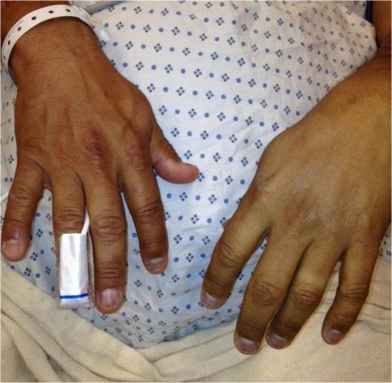
Photograph of patient’s hands 2 h after intravenous infusion of undiluted phenytoin 250 mg infused using Baxa Microfuse® Infuser over 15 min through a 20-gauge peripheral intravenous catheter in the dorsal left proximal thumb at a rate not exceeding 25 mg/min. Left hand with diminished perfusion, skin pallor, and dusky and cyanotic nailbeds
Fig. 2.
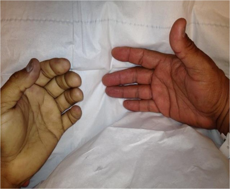
Photograph of patient’s hands 4 h after intravenous infusion of undiluted phenytoin 250 mg. Progression of ischemia to sensory loss and paralysis of left hand. Left hand and fingers with extensive pallor and cyanosis
Left hand films revealed mild diffuse swelling about the hand without osseous abnormalities or gas. Ulnar and palmar pulses were non-palpable and progressively unable to be obtained by Doppler sonography. A diagnosis of purple glove syndrome (PGS) was suspected. After consultation with hand surgery, vascular surgery and interventional radiology experts, it was decided to proceed with conservative management (serial exams, nicardipine, and heparin) in light of uncontrolled hypertension and history of hemorrhagic stroke. Ongoing severe pain in the left hand and forearm was treated with IV morphine and hydromorphone with minimal relief. Intravenous nicardipine and heparin were initiated within 4 and 6 h, respectively. CT angiography of the left hand, obtained 20 h later, revealed proximal ulnar artery and distal artery occlusion with no run-off to the hand, edema of the multiple compartments and soft tissues in the forearm and hand, reflecting ischemia, and several foci of gas in the palmar aspect of the hand, concerning for necrosis (Fig. 3).
Fig. 3.

CT angiography of the left upper extremity performed after loss of left distal pulses obtained 20 h after intravenous infusion of undiluted phenytoin 250 mg. Proximal ulnar artery and distal radial artery occlusion with no run-off to the hand. Edema of the multiple muscle compartments and soft tissues in the forearm and hand, reflecting ischemia. Several foci of gas in the palmar aspect of the hand, concerning for necrosis
The patient was then immediately taken to the operating room and ulnar and radial thrombectomies with forearm fasciotomy were performed. Vascular flow was initially restored but then failed within the next 24 h. Six days later, the patient underwent split-thickness skin grafting from his left thigh to left ventral forearm fasciotomy incision. A wound VAC was placed on the skin graft at 75 mm Hg and removed a week later. Twenty-four days later, the patient underwent a left hand and distal forearm amputation for ischemic sequelae.
Introduction
Phenytoin sodium (Dilantin®, Pfizer Inc., NY, NY) is an anticonvulsant drug used for the treatment and prevention of seizures since 1956 [1]. It is recommended by the Epilepsy Foundation of America (EFA) and European Federation of Neurological Societies (EFNS) practice guidelines as the second-line agent for treatment of status epilepticus after IV benzodiazepines [2]. Phenytoin has been associated with a number of serious adverse drug reactions (ADR) including hypotension and arrhythmias with rapid IV administration, dermatologic reactions ranging from rashes to Stevens-Johnson syndrome or toxic epidermal necrolysis, severe hepatotoxicity, and other hypersensitivity events [3]. Extravasation or soft tissue injuries following administration of IV phenytoin have been reported or documented since 1950s. In 1984, Comer et al. first described the association between IV phenytoin administration and rapid onset of discoloration, pain, and tissue necrosis affecting the distal limb through which the drug was intravenously administered. In her 1992 publication, Hanna named a delayed, soft tissue injury of the hand and forearm following IV administration of phenytoin “purple glove syndrome” (PGS) [4, 5]. Increasing reports of this condition have recently emerged, some involving permanent tissue damage or loss, leading to questions regarding the safety and risk-benefit profile of IV phenytoin [6]. PGS has also been associated with fosphenytoin (Cerebyx®, Pfizer Inc., NY, NY), the water-soluble pro-drug of phenytoin approved by the FDA in 1996, although at a lower estimated incidence [3, 6]. Use of fosphenytoin has been limited, in part due to cost, however now a less expensive generic formulation has led to further debate about the continued role of phenytoin in the treatment of seizures, particularly status epilepticus [7].
Indications for Use of Phenytoin in the Emergency Department
Phenytoin is commonly utilized in several clinical settings within the emergency department. Patients with a history of seizures who are on daily phenytoin therapy can present with uncomplicated seizures after noncompliance or subtherapeutic administration of the drug. In this situation, an oral or parenteral loading dose is often administered. Patients presenting with status epilepticus refractory to benzodiazepine administration require prompt IV phenytoin administration [2, 7, 8]. Finally, patients with traumatic intracranial injury or symptomatic mass lesions are frequently prescribed with phenytoin for prophylaxis against seizure activity in either oral or parenteral form [9–11]. While the dosing of the drug is similar for all of the above situations, the route and timing of administration vary and influence the predisposition to possible adverse reactions such as PGS.
Objective
PGS is uncommon, and without surveillance mechanism for adverse effects, current literature on PGS is limited primarily to individual reports or single-center case series. Our objective was to identify all previously reported cases of phenytoin- or fosphenytoin-associated PGS and summarize the most current understanding of the pathophysiology, clinical presentation, diagnosis, and treatment. We submit recommendations for future safe use of phenytoin or fosphenytoin and the prevention and treatment of subsequent cases of PGS.
Methods
We searched for English language references from the following online databases: MEDLINE (1950 to present), EMBASE, CINAHL (Cumulative Index to Nursing and Allied Health), and TOXNET. The following search terms and combination were used: (phenytoin$ OR Dilantin$ OR fosphenytoin$ OR Cerebyx$) AND (purple$ glove$ OR adverse event$ (MeSH) OR adverse drug reaction (MeSH) OR adverse reaction (MeSH) OR hand injury (MeSH) OR forearm injury (MeSH) OR finger injury (MeSH) OR arterial injury (MeSH)). We also conducted a gray literature search of Federal and State government web sites, professional organizations, the Internet with Google and Google Scholar for any publications describing or mentioning “purple glove syndrome” or “PGS”. The titles and abstracts of the articles were screened for relevance, and their bibliographies were examined for further relevant studies. We included any references that featured one or more case descriptions of phenytoin or fosphenytoin-associated PGS after intravenous or oral administration and provided information on the clinical setting of the event and associated outcome(s). All references were reviewed by three study authors (LAG, BCD, and JMP), and any disagreements were settled by consensus. Descriptive statistics were employed to summarize relevant facts about the cases.
Purple Glove Syndrome: Pathophysiology
Descriptions of PGS have included a wide range of symptoms, timing, and recovery, suggesting that the underlying mechanism(s) of tissue damage are complex [12]. In terms of site, PGS is most commonly associated with upper extremities; however, it has been also reported in lower extremities and referred to as purple sock syndrome [13–16]. Bhattacharjee et al. summarized the disease course into three temporal stages of injury: initial painful blue-purple discoloration and edema around the IV catheter site, subsequent worsening of pain, edema, and discoloration with development of epidermal sloughing, ulceration, or bullae formation, and neuromuscular symptoms such as paresthesias or weakness, and eventually resolution of edema and discoloration starting from the periphery of the injury and moving toward the site of catheter insertion [17]. Timing of these stages appears quite variable, with initial discoloration occurring from minutes to days after phenytoin administration and tissue recovery spanning days to months [18, 19]. There are also a subset of more severe cases in which the second stage of expanding edema and superficial tissue damage is followed by dermal and subcutaneous tissue necrosis and ischemia leading to permanent injury, surgical debridement, or amputation. Tissue samples obtained both early and late in the course of symptoms have shown the common presence of epidermal necrosis, subcutaneous edema, perivascular lymphoid infiltration, and local vascular thrombosis [17, 20, 21].
The mechanism by which PGS occurs remains speculative. Many reports have proposed that the combination of small vascular tearing or micro-extravasation at the site of IV catheter insertion allow for leakage of the severely alkaline (pH = 12) phenytoin solution into surrounding tissue, causing necrosis, disruption of endothelial barriers, and vasoconstriction [22–24]. An additional theory regarding the unique alkalinity of IV phenytoin solution argues that contact with blood leads to precipitation of certain compounds, leading to catheter obstruction and solution extravasation [24–26]. The observation that multiple cases of PGS have been reported without IV catheter extravasation suggests the presence of an additional underlying pathophysiology [4, 5, 13, 15–17, 19, 21, 23, 27–42]. The repeated finding of microvascular thrombosis on histopathology and the association between documented large vessel thromboses in the most severe cases of PGS suggests that an unidentified procoagulant mechanism may be responsible for the extent of tissue destruction [17, 20].
Review of Reported Cases
Our search identified 13 English-language citations, including 10 individual case reports, 1 publication of two individual cases, and 2 multiple-patient case series [5, 15, 17–19, 22, 28, 31, 34, 37, 40, 43, 44]. After review of the associated bibliographies, an additional 26 articles were found [4, 13, 16, 20, 21, 23, 27, 29, 30, 32, 33, 35, 36, 38, 39, 41, 42, 45–54]. In total, 72 patients experienced 82 cases of PGS following IV infusion of phenytoin described from 1984 to 2015. Six patients had reactions in both extremities, and one patient developed reactions in all four extremities following IV phenytoin administration in each extremity [16, 23, 32, 40, 41].
Table 1 displays a summary of demographic and descriptive characteristics of the 82 cases of PGS. The number of cases that reported each of the individual variables is listed. The mean age of adult patients was 61.5 ± 17.1 years and pediatric patients 3.4 ± 5.2 years. Seventy percent of all adult patients were female. The most common treatment indication was acute seizure without status epilepticus in 41 (64.1 %) patients [5, 13, 17, 18, 21, 22, 33–35, 37, 38, 40, 41, 50, 51]. Twelve (18.8 %) patients were receiving phenytoin for seizure prophylaxis [4, 15, 20, 22, 32, 41, 46]. Over one half (22/43, 64.1 %) had a history of epilepsy, though patient comorbidities were described in only 43 of 82 cases. One patient was noted to have a known history of peripheral arterial disease [37].
Table 1.
Demographic and descriptive characteristics
| Variables | Results | Number of PGS cases providing information (n = 82) |
|---|---|---|
| Adult, n (%) | 70 (85.4) | |
| Age (years) | 61.5 ± 17.1 | |
| Child, n (%) | 8 (9.8) | |
| Age (years) | 3.4 ± 5.2 | |
| Gender, n (%) | ||
| Adult | 80 (97.6) | |
| Female | 56 (70.0) | |
| Child | 8 (9.8) | |
| Male | 5 (62.5) | |
| Clinical scenario, n (%) | 64 (78.0) | |
| Seizure | 41 (64.1) | |
| Prophylaxis | 12 (18.8) | |
| Status epilepticus | 11 (17.2) | |
| At least one comorbidity present, n (%) | 43 (52.4) | |
| Epilepsy | 22 (51.2) | |
| Brain tumora | 7 (16.3) | |
| Hypertension | 5 (11.6) | |
| Ischemic stroke | 4 (9.3) | |
| Traumatic brain injury | 3 (7.0) | |
| Hemorrhagic stroke | 2 (4.7) | |
| Encephalitis | 1 (2.3) | |
| Cardiac arrest | 1 (2.3) | |
| Cerebral sinus thrombosis | 1 (2.3) | |
| Peripheral arterial disease | 1 (2.3) | |
| Diabetes mellitus | 1 (2.3) | |
| Microcephaly | 1 (2.3) | |
| Dosing—adult | ||
| Weight-based loading dose, n (%) | 6 (7.3) | |
| Dose (mg/kg) | 17.5 (14–21.5) | |
| Total loading dose, n (%) | 53 (64.6) | |
| Dose (mg) | 659 (400–900) | |
| Number of loading doses, n (%) | 53 (64.6) | |
| 1 dose | 47 (88.7) | |
| 2–3 doses | 5 (9.4) | |
| 10 dosesb | 1 (1.9) | |
| Number of maintenance doses, n (%) | 32 (39.0) | |
| 1–2 doses | 26 (81.3) | |
| 3–6 doses | 4 (12.5) | |
| 7–10 doses | 2 (6.2) | |
| Dosing—pediatric | ||
| Weight-based loading dose, n (%) | 6 (7.3) | |
| Dose (mg/kg) | 11 (10–14.25) | |
| Total loading dose, n (%) | 5 (6) | |
| Dose (mg) | 99 (32.8–200) | |
| Number of loading doses, n (%) | 8 (9.8) | |
| 1 dose | 7 (87.5) | |
| 2 doses | 1 (12.5) | |
| Administration, n (%) | 49 (59.8) | |
| Infusion rate (mg/min) | 32.1 ± 16.2 | |
| Dilution, n (%) | 58 (70.7) | |
| Undiluted phenytoin | 41 (70.7) | |
| Diluted phenytoin | 17 (29.3) | |
| Diluent type | 17 (20.7) | |
| Normal saline | 14 (82.4) | |
| Plasmalyte | 2 (11.8) | |
| Sterile water for injection | 1 (5.8) | |
| Concentration, n (%) | 8 (9.8) | |
| Concentration (mg/mL) | 6.3 (4 – 10.5) | |
| Infusion location, n (%) | 82 (100) | |
| Intravenous | 78 (95) | |
| Intra-arterial | 4 (5) | |
| Extravasation, n (%) | 58 (70.7) | |
| Present | 22 (37.9) | |
| Absent | 36 (62.1) | |
| IV catheter gauge, n (%) | 33 (40.2) | |
| ≤18 gauge | 13 (39.4) | |
| 19–20 gauge | 10 (30.3) | |
| >20 gauge | 10 (30.3) | |
| IV catheter location, n (%) | 56 (62.3) | |
| Hand | 35 (62.5) | |
| Forearm | 7 (12.5) | |
| Wrist | 6 (10.7) | |
| Antecubital fossa | 5 (8.9) | |
| Leg | 3 (5.4) | |
| Time to onset, n (%) | 71 (86.6) | |
| ≤15 min | 16 (22.5) | |
| >15 min − ≤2 h | 12 (17.0) | |
| >2 h − ≤12 h | 37 (52.1) | |
| >12 h − ≤24 h | 3 (4.2) | |
| >24 h | 3 (4.2) | |
| Time to onset (minutes) | 360 (70–390) |
Numerical data expressed as median (interquartile range) or mean ± standard deviation
IV intravenous
aPrimary brain malignancy or brain metastatic lesion
bLoading dose 1 g given undiluted as 100 mg IV push every 1 h
In adults, the median loading dose received prior to symptom onset was 659 mg (400–900 mg). The majority of adult patients received a single dose with the remainder receiving multiple doses for both loading and maintenance phenytoin regimens, 47/53 (88.7 %) and 26/32 (81.3 %), respectively. Similarly, 7/8 (87.5 %) children received a single phenytoin dose for loading purposes. Of those studies that described the rate of intravenous infusion, the mean value was 32.1 ± 16.2 mg/min. Four (6.7 %) cases acknowledged, confirmed, or strongly suspected accidental intra-arterial drug administration [29–31, 45].
The majority of cases (41/58, 70.7 %) reported use of undiluted phenytoin as initially recommended by the drug maker. Approximately one third (17/58, 29.3 %) of the PGS cases described dilution of phenytoin, with 14/17 (82.4 %) in normal saline for IV administration with diluent volumes ranging from 10 to 500 mL [13, 15, 16, 19, 29, 34, 35, 38, 40, 45, 46]. Two cases reported inadvertent dilution of phenytoin in plasmalyte and one in sterile water for injection, with both solutions not compatible with phenytoin [16, 32]. Dilution to a final median concentration of 6.3 mg/mL (4–10.5 mg/mL) was described in eight cases [15, 19, 34, 35, 38, 41, 45, 46].
The IV catheter gauge was reported in 33/82 (40.2 %) of PGS cases, with a third of subjects having a 20-gauge or smaller catheter in place. The majority of catheters were placed in the hand (35/56, 62.5 %) or forearm (7/56, 12.5 %). Three cases reported catheter placement and subsequent symptoms in the foot or distal lower extremity, two involving an asphyxiated term baby girl and a 6-month-old male and the third a 16-year-old male. Extravasation around the catheter site was described in 22/58 (37.9 %) cases, though the details of how this was classified were generally not reported [5, 16, 21, 29, 32, 33, 46–49, 54].
In most cases, signs or symptoms occurred within 12 h of drug administration, including 16 (22.5 %) within 15 min, of which 15 reportedly occurred immediately after drug infusion [4, 5, 16, 21, 22, 33, 46, 47]. About one fifth (12/71, 17 %) described development of PGS within 15 min to 2 h and one half (37/71, 52.1 %) within 2 to 12 h after IV administration of phenytoin. Three (4.2 %) cases reported a time to onset between 12 to 24 h and three (4.2 %) greater than 24 h, with one subject developing symptoms 5 days after the last recorded dose of medication [19, 22, 44, 52]. The median time to symptom onset was 360 min (70–390 min).
A small number of cases described use of various concomitant medications including dexamethasone, mannitol, remifentanil, lidocaine, plasmalyte, benzodiazepines, barbiturates, or neuromuscular blockers [4, 18, 32, 34, 37, 39, 46, 47, 50, 52]. Visible phenytoin precipitation due to medication incompatibilities was reported in one case resulting in full recovery [32]. One of two patients who received concomitant mannitol and phenytoin, both medications with strong vesicant properties, developed frank necrosis of the right hand necessitating hand and distal forearm fasciotomy [46]. Two cases of improper phenytoin dilution and resultant drug precipitation described in one pediatric patient led to immediate onset of intense bluish discoloration around the cannula site leading to “partial thickness dermal injury” characterized by blistering, mottling, and darkening of the skin [16]. Concurrent administration of phenytoin with benzodiazepines or barbiturates led to PGS with ulcerations at the site of injection and one patient requiring surgical debridement and repair with a skin graft [47, 52].
Table 2 displays information on the presentation, diagnosis, and treatment of PGS. The vast majority of cases described a combination of purple discoloration (76/81, 93.8 %) and edema (75/81, 92.6 %), with other common findings including erythema (42/81, 51.9 %), pain (28/81, 34.6 %), and coldness (17/81, 21 %). Notably, 24/81 (29.6 %) of patients were unable to verbalize pain. Physical findings included blistering (29/74, 39.2 %), necrosis (14/74, 18.9 %), and ulceration (13/74, 17.6 %). One third (17/52, 32.7 %) noted a decrease in amplitude or lack of palpable distal pulses in the affected extremity [18, 20, 27–32, 37–39, 41, 45, 47]. Advanced imaging was employed in one fifth (18/82, 22.0 %) of all patients. Of these, 17 (94.4 %) patients had Doppler ultrasound, 1 (5.6 %) had immediate conventional angiography, and 3 (16.7 %) cases had both Doppler ultrasound and conventional angiography. Twelve out of the 18 (66.7 %) patients that had imaging found a thrombus. Of these, seven and five patients developed arterial and venous thrombosis, respectively [4, 18, 27–32, 36, 45, 46].
Table 2.
Presentation, diagnosis, and treatment modalities
| Variables | Results | Number of PGS cases providing information (n = 82) |
|---|---|---|
| Signs and symptoms, n (%) | 81 (98.8) | |
| Purple discoloration | 76 (93.8) | |
| Edema | 75 (92.6) | |
| Erythema | 42 (51.9) | |
| Pain | 28 (34.6) | |
| Unable to verbalize pain | 24 (29.6) | |
| Coldness | 17 (21.0) | |
| Weakness | 5 (6.2) | |
| Sensory changes | 4 (4.9) | |
| Physical findings, n (%) | 74 (90.2) | |
| Blistering | 29 (39.2) | |
| Necrosis | 14 (18.9) | |
| Ulceration | 13 (17.6) | |
| Pulse deficit evaluated, n (%) | 52 (63.4) | |
| Pulse deficit present | 17 (32.7) | |
| Diagnostic modality used, n (%) | 18 (22.0) | |
| Doppler ultrasound only | 17 (94.4) | |
| Conventional angiography only | 1 (5.6) | |
| Doppler ultrasound and conventional angiography | 3 (16.7) | |
| Presence of a thrombus evaluated, n (%) | 18 (22.0) | |
| Arterial | 7 (38.9) | |
| Venous | 5 (27.8) | |
| None | 6 (33.3) | |
| At least one treatment modality used, n (%) | 44 (53.7) | |
| Limb elevation | 26 (59.1) | |
| Local heat application | 20 (45.5) | |
| Systemic antibiotics | 11 (25.0) | |
| Systemic anticoagulation | 6 (13.6) | |
| Local nerve block | 6 (13.6) | |
| Infiltrationa | 4 (9.1) | |
| Skin graft | 4 (9.1) | |
| Debridement | 3 (6.8) | |
| Fasciotomy | 3 (6.8) | |
| Topical nitroglycerin | 3 (6.8) | |
| Systemic vasodilators | 1 (2.3) | |
| Thrombectomy | 1 (2.3) | |
| At least one outcome reported, n (%) | 79 (96.3) | |
| No deficits | 54 (68.4) | |
| Skin changes | 16 (20.3) | |
| Amputationb | 8 (10.1) | |
| Sensory deficits | 6 (7.6) | |
| Motor deficits | 5 (6.3) | |
| Mortality, n (%) | 80 (97.6) | |
| Death related to PGS | 2 (2.5) | |
| Time to reported outcome, n (%) | 68 (82.9) | |
| Days to resolution/amputation/death | 14 (8–21) |
Numerical data expressed as median (interquartile range) or mean ± standard deviation
aInfiltration (four cases): hydrocortisone (two), heparin (one), hyaluronidase (one)
bAmputation (eight cases): thumb and digits (three), hand and distal forearm (two), limb (one), below elbow (one), disarticulation of wrist (one)
A wide variety of treatment modalities were used in the identified cases, ranging from simple elevation of the affected extremity to prophylactic systemic antibiotics, systemic anticoagulation, intravenous or topical vasodilators, and regional nerve blocks or various surgical techniques. On follow-up evaluation documented in 79 of 82 cases, fifty-four (68.4 %) subjects demonstrated full recovery of function and appearance of the affected extremity. Of those reports that specified duration, resolution of symptoms occurred after a median of 14 (8–21) days. Sixteen (20.3 %) cases noted permanent skin changes without functional motor or sensory deficits. There were eight (10.1 %) reports of amputations all involving the upper extremity [20, 23, 28, 30, 41, 44, 46, 53]. Two deaths were attributed to the described case of PGS. One involved an 83-year-old female with accidental intra-arterial phenytoin administration leading to limb ischemia that, despite embolectomy and intraoperative heparin and papaverine administration, progressed to involve the lateral chest wall. The patient rapidly developed multisystem organ failure and died 4 days after drug administration [45]. The second case described by Weinstein et al. in 1989 reported immediate development of hand ischemia after intravenous phenytoin administration associated with death of the patient, but no further details were provided by the authors [49].
Overview of Pediatric Cases
Eight cases were identified describing PGS in children with ages ranging from newborn to 14 years. Over one half (5/8, 62.5 %) patients were male. Median phenytoin dose administered was 11 mg/kg (10–14.25 mg/kg) [15, 16, 19, 27–29, 46]. Six children had IV access placed in the hand and two in the foot. In six cases reporting preparation, phenytoin was diluted to a final concentration ranging from 5 to 30 mg/mL. The rate of administration was 0.3–0.75 mg/kg/min in three cases and not reported in the remaining cases [15, 16]. Resolution occurred within 7 h in two cases and within 1 to 3 weeks in five. One case report described PGS resulting in loss of brachial, radial, ulnar, and superficial palmar pulses in the right extremity within 4 h after administration of 15 mg/kg IV phenytoin through a dorsal metacarpal vein in a 14-month-old boy. His right hand and forearm were erythematous and markedly swollen. After administration of subcutaneous hyaluronidase (0.2 mL aliquots of 15 units/mL solution) into four sites in the dorsum of the right hand, there was an immediate blanching of the overlying erythematous skin and dramatic decrease in swelling. The same dose was repeated leading to a return of radial and superficial palmar pulses and reduction of his pain. Assessment at the 5-month follow-up appointment revealed full recovery with return to the baseline use of the patient’s right hand [27].
Sharief et al. reported a newborn female baby with two different cases of PGS, one occurring with phenytoin 10 mg/kg erroneously diluted in sterile water for injection and administered through a 24-gauge cannula in the left hand and 2 days later with phenytoin properly diluted in normal saline and administered through a cannula in the left foot. Both cases presented similarly with intense blue discoloration, edema, and blister formation with full recovery within a week [16]. Prince et al. described PGS with distal forearm venous occlusion and subsequent left index finger and thumb gangrene. A 9-year-old girl with tuberous sclerosis, recent resection of a giant cell astrocytoma, epilepsy, autistic spectrum disorder, and development delay was admitted to a pediatric intensive care unit with status epilepticus and Escherichia coli septic shock. Intravenous phenytoin was administered as an 18 mg/kg load followed 90 min later by a half re-load. Seven hours later, the patient’s left hand was noted to be swollen, purple, and cold with very prolonged capillary refill time. There was no clinical evidence of phenytoin extravasation. Anticoagulant therapy could not be instituted because of concomitant disseminated intravascular dissemination (DIC). Over the course of the next 3 weeks, the left index finger and thumb demarcated with the distal portions non-viable. The author concluded that presence of DIC and absence of clinical extravasation findings supported thrombosis as an important etiological factor in the development of PGS in this patient [28].
Discussion
Although PGS was first described over 30 years ago, it remains poorly understood and incompletely characterized. Furthermore, predicting which patients with PGS will suffer adverse outcomes and implementing effective treatment strategies to improve recovery has been based on isolated reports or small series. We sought to synthesize information from published cases of PGS in order to provide a better understanding of this rare and potentially serious complication.
Incidence
In a first prospective case study in 1983, Earnest et al. described burning and pain with IV phenytoin infusions in 29/200 (14.5 %) patients. Two thirds (65.6 %) required adjustments in administration techniques (slower rate of administration or change in dilution concentration) due to severe pain. Local symptoms were positively correlated with both the concentration of phenytoin and the rate of infusion. No case of PGS was mentioned in their study. The authors could have missed PGS, as the study was designed to evaluate complications during administration of phenytoin only [55].
During a 26-month period in a single hospital in 1988, Spengler et al. investigated 11 patients who developed 17 cases of severe limb edema and discoloration within 24 h of their IV phenytoin infusion. Two patients had reactions in both upper extremities. One patient, readmitted to hospital 40 days after her initial reaction, developed reactions in all four extremities following IV phenytoin administration in each extremity. She required below-the-elbow amputation in one extremity. The incidence of PGS in this retrospective chart review was approximated from 3 to 7 % [23].
In 1998, O’Brien et al. sought to prospectively evaluate PGS (defined as the progressive development of edema, discoloration, and pain in the limb) in 179 consecutive patients who had IV phenytoin ordered during a 3-month period. A total of 152 patients received IV phenytoin, and nine (5.9 %) developed PGS. PGS patients received a greater median initial dose of phenytoin, total 24-h dose, and total number of doses (all p < 0.05). PGS in three patients resulted in skin ulceration. In eight of the nine patients (88.8 %), PGS resolved completely within 1 month and surgical intervention was not necessary. One of the nine patients (11.1 %) developed a large area of ulceration necessitating skin grafting with successful recovery. PGS was recognized by the treating physicians as being associated with IV phenytoin administration in five of the patients (55.6 %). This association was first recognized at the onset of the clinical features in two of these patients, 1 day after onset in two patients, and 2 days after onset in the remaining patient [22].
In 2001, Burneo et al. prospectively evaluated the incidence of PGS in 157 patients who received 179 consecutive exposures of intravenous phenytoin. PGS (defined as edema, discoloration, and pain around the injection site) was observed in three of the 179 exposures (1.7 %). Two patients were affected with one having two different events of PGS. Both patients experienced edema, discoloration, and mild pain without skin ulcerations. Lower incidence of PGS at this hospital was attributed to a standardized phenytoin administration protocol. Phenytoin was reconstituted in normal saline by pharmacy and administered via IV piggyback at a rate not to exceed 20 mg/min by using an electronic infusion control device and a 0.22-μm in-line filter [40].
The range of occurrence in the above studies (1–7 %) is significantly higher than in our clinical experience and that reported to the FDA in controlled trials of intravenous phenytoin [6]. Whether this is due to aggressive screening and identification, selection bias in published reports, or other reasons is unclear. Further prospective studies are needed to better understand the true incidence of PGS.
Presentation
PGS affects a wide spectrum of patients receiving intravenous phenytoin, from the very young to elderly, and occurs in any of the more frequent scenarios in which phenytoin is administered in the acute care setting. The hallmark of PGS is purplish discoloration and edema of the distal extremity, seen in greater than 90 % of reported cases. Pain is also likely to be present, but its presence may often be obscured by pathologic or iatrogenic-induced alteration in consciousness after a seizure. Alterations in appearance including blistering, erythema, or necrosis are more common than deficits in function, such as paralysis or sensory changes, potentially allowing for a more rapid diagnosis [1, 6, 12, 17]. Time to onset of symptoms was generally rapid, consistent with a potentially ischemic pathology, though cases were reported up to 5 days after the last dose of phenytoin [52].
Just over half of cases reported symptoms after one dose of IV phenytoin. Four cases of PGS were associated with documented or strongly suspected intra-arterial administration of phenytoin and tended to display more severe injury and worse outcomes, with one amputation and one death directly associated with PGS [29–31, 45]. While previous studies have emphasized an association between extravasation of drug and PGS, only 37.9 % of cases we reported had documented or suspected extravasation, and more than one half (62.1 %) specifically described a lack of extravasation on inspection of the affected limb. Nonetheless, visible and tactile inspection cannot completely exclude any contact between the highly alkaline IV phenytoin solution and surrounding tissue, as microscopic vascular injury after intravenous catheter placement leading to extravasation is still possible.
Administration of IV Phenytoin
Phenytoin injection is poorly soluble in water. Several vehicles must be employed to improve solubility to allow the drug to be administered parenterally. Phenytoin injection is commercially available as a ready-mixed solution that contains 50 mg of phenytoin per milliliter in a vehicle of 40 % propylene glycol and 10 % alcohol in water. This solution is then adjusted to a pH 12 with sodium hydroxide and is not compatible with dextrose or dextrose-containing fluids due to lack of solubility and resultant precipitation [1, 6, 12].
Analysis of the phenytoin label approval history by Pfizer available from the Drugs@ FDA Web Site revealed an interesting observation. From 1956 to 2001, Pfizer recommended use of a straight or undiluted phenytoin for IV administration due to lack of phenytoin solubility and stability data. In November of 2001, based on a number of phenytoin-associated extravasation injuries and availability of several in vitro studies of phenytoin solubility and stability in normal saline, a request was made by the sponsor to add instructions for the administration of parenteral phenytoin via bolus and infusion methods to the product label. After its review, the updated recommendations for the administration of IV phenytoin via infusion method and further dilution in normal saline were shortly approved by the Food and Drug Administration.
Ten years later, in November of 2011, local toxicity information including PGS information was added to the Pfizer’s Dilantin label: “Soft tissue irritation and inflammation has occurred at the site of injection with and without extravasation of intravenous phenytoin. Edema, discoloration and pain distal to the site of injection (described as “purple glove syndrome”) have also been reported following peripheral intravenous phenytoin injection. Soft tissue irritation may vary from slight tenderness to extensive necrosis, and resolution of symptoms may be spontaneous, skin necrosis and limb ischemia have occurred and required such interventions as fasciotomies, skin grafting, and, in rare cases, amputation” [1, 6, 12]. This information is also reflected in current generic phenytoin labels [1, 56–58].
At the same time in November 2011, the following recommendations for dilution and administration were added to the product label: “Because of the risk of local toxicity, intravenous Dilantin should be administered directly into a large peripheral or central vein through a large-gauge catheter. Prior to administration, the patency of the IV catheter should be tested with a flush of sterile saline. Each injection of parenteral Dilantin should then be followed by a flush of sterile saline through the same catheter to avoid local venous irritation due to the alkalinity of the solution…For infusion administration, parenteral Dilantin should be diluted in normal saline with the final concentration of Dilantin in the solution no less than 5 mg/mL. Administration should commence immediately after the mixture has been prepared and must be completed within 1 to 4 h (the infusion mixture should not be refrigerated). An in-line filter (0.22–0.55 μm) should be used” [1]. In 2010, Pfizer ceased its production of parenteral Dilantin in the USA. The instructions for IV infusion administration of phenytoin listed in Pfizer’s Dilantin label are however not reflected in the current generic phenytoin labels [56–58].
It is unclear from our data exactly how the administration techniques of intravenous phenytoin may have contributed to the development of PGS. Given that the majority of cases occurred before the 2011 and do not consistently describe the details of administration, we cannot determine the effectiveness of the above recommendations in preventing occurrence of PGS. We suggest following the instructions included in Pfizer’s Dilantin label and strongly encourage generic manufacturers of phenytoin to include this information in their drug labeling.
Risk Factors
Several authors have attempted to identify risk factors for PGS after intravenous phenytoin administration. Spengler et al. identified 11 patients with 17 cases of PGS at a single hospital over a 2-year period and performed a case-control study using unaffected patients that received IV phenytoin over the same period. They identified older age, female gender, and history of cardiovascular disease, number of doses, infusion rate, and smaller gauge IV catheters as factors associated with PGS in a univariate analysis [23]. O’Brien et al. performed a retrospective cohort study that identified 152 consecutive patients who had received intravenous phenytoin over a 3-month period at several hospitals, of whom 9 developed PGS. Older age, more frequent and higher absolute doses, and use in the setting of acute seizure or outside the operating room were found to be associated with PGS [22].
While our data is limited by the lack of a controlled comparison, we found that 70 % of all reported cases involved females. This suggests that female gender is a risk factor, though we cannot comment on whether this is independent of other potentially coincident factors. The median infusion rate was 32.1 mg/min similar to that from Spengler et al. of 38 mg/min, though 13 patients had phenytoin administered at a rate of 50 mg/min, the maximum recommended by the manufacturer of IV phenytoin [1, 5, 42, 46, 49, 53]. We found a majority of IV catheters were 19 gauge or smaller or placed in the hand, consistent with the hypothesis that PGS is associated with infusion into smaller, more fragile veins that are more prone to trauma and possibly microscopic or visible extravasation. Nonetheless, 39.4 % of cases occurred with an 18 gauge or larger catheter and 12.5 and 8.9 % with IV catheters in the forearm and antecubital fossa, respectively, suggesting that no size or location is exempt from occurrence. While it has been hypothesized that comorbid peripheral arterial disease may predispose patients to PGS, we were only able to document one case in which the subject had a known history [37]. This is limited, however, by the lack of consistent documentation of patient comorbidities among the reviewed studies as well as a likely under-appreciation of the prevalence of this disease among patients.
Diagnosis and Treatment
While the diagnosis is often considered when a purplish discoloration is noted following infusion of phenytoin, the natural history from the published literature and FDA summary does not guide clinicians adequately for the potentially serious vascular complications that may ensue, as evidenced by our case [6, 12]. It appears that either evidence or suspicion of vascular thrombosis is associated with a decreased likelihood of full recovery. Of the 12 cases of documented thrombosis, only 6 (50 %) made a complete recovery [4, 18, 27–32, 36, 45, 46]. Of the 6 additional cases where advanced imaging was employed but no clot was identified, 3 (50 %) recovered completely [13, 35, 38]. In comparison, of the remaining 64 % cases that did not report use of diagnostic imaging, 39 (63.8 %) cases reported a full recovery of symptoms. Although pain is a cardinal symptom of limb ischemia, this finding may be absent in patients with altered mentation due to seizures or traumatic brain injury. We would recommend immediate limb angiography for patients with any change in signs or symptoms of altered perfusion of the affected extremity, as this finding may progress rapidly in a proportion of patients. If vascular imaging is not performed, frequent neurovascular assessments must be initiated to identify ischemic progression to prevent subsequent tissue compromise. Once vascular compromise is suspected, consultation or transfer for evaluation by a hand or vascular surgery expert is highly recommended.
A variety of treatment modalities were employed among the reviewed cases, including simple non-pharmacological therapies such as elevation and more advanced systemic and local interventions. Systemic antibiotics were the most commonly administered medication, prescribed in 25 % of cases [21, 22, 29, 31, 33, 35, 40, 43]. We would recommend not treating with antibiotics, as there is no evidence that routine prophylactic antimicrobial therapy improves outcome in the setting of intravenous drug infiltration, unless the diagnosis of PGS is clouded by other factors suggestive of local or systemic infection. If vascular thrombosis is identified in association with PGS, treatment with systemic anticoagulation and possible catheter-directed or open thrombectomy or thrombolysis should be considered in the absence of contraindications and in consultation with a vascular surgeon. Of the six cases that reported use of systemic anticoagulation (four with documented arterial thrombosis), three eventually resulted in full recovery, two in limb amputation, and one in death [29–31, 44, 45, 50]. Only one case reported use of thrombectomy or thrombolysis, which was unsuccessful and eventually resulted in patient death [45].
Two additional novel therapies, local infiltration, and regional anesthesia via local nerve block, were employed in several cases. Four cases described infiltration of the affected area with a variety of compounds, including hyaluronidase, hydrocortisone, triamcinolone, or heparin [4, 15, 19, 27]. Three cases described use of topical nitroglycerin [32, 38]. Of the seven that provided patient follow-up, five resulted in full recovery and two with skin or sensory deficits. Hyaluronidase, an enzymatic compound that increases the permeability of connective tissue by hydrolyzing hyaluronic acid contained within the extracellular matrix and along the epidermal skin border, has been used to treat a variety of extravasation injuries from chemotherapy agents and other vesicants [59]. Given the paucity of data, its use or the infiltration of any other compound(s) in the treatment of PGS should be considered experimental. An additional six cases described using regional nerve blockade via either singular injection or continuous infusion of local anesthetic, with three demonstrating full recovery, two with skin or sensory deficits, and one amputation [18, 30, 31, 35, 38, 43]. This therapy should also be considered experimental and performed in consultation with an experienced anesthesiologist.
Prevention
Although not all cases progress to serious vascular compromise, the morbidity can be significant and prevention may be the most expedient recommendation. Use of oral phenytoin, whenever possible, would be preferred over the risk of parenteral infusion, including limiting IV fosphenytoin [6, 12]. When administered parenterally, a large bore IV in a proximal site avoiding the distal hand and forearm should be preferred. If large bore access is unable to be obtained, a smaller bore catheter can be employed with caution. Catheter insertion sites in close proximity to extremity arteries such as ventral distal wrist or the medial antecubital fossa should also be used with caution, as intra-arterial administration of phenytoin appears to be associated with the most devastating complications [29–31, 45]. Manufacturer’s recommendations for phenytoin dilution and administration should be followed to minimize or prevent PGS [1]. The elderly and very young have more fragile tissue and vasculature and may be poor candidates for routine IV phenytoin therapy. Concomitant phenytoin administration with arterial cannulation in the same limb should also be cautioned. Mahajan et al. reported a case of rapidly progressing distal limb ischemia resulting in compartment syndrome with arterial occlusion and thrombosis [31]. Finally, alternative drug regimens should be considered in situations in which efficacy has been shown to be non-inferior to phenytoin [2, 10, 60]. Any intravenous insertion sites should be monitored closely for pain, signs of extravasation, discoloration, or changes in perfusion and documented thoroughly [1, 3]. Of the 82 cases of PGS that we identified, only 4 (4.9 %) occurred without any potentially preventable risk factors (intra-arterial administration, extravasation, undiluted suspension, concomitant vesicants, 22 gauge or smaller catheter, IV placement in the hand, foot or wrist) [13, 22, 32].
Association with Fosphenytoin
Fosphenytoin (Cerebyx®), a water-soluble phosphate ester pro-drug of phenytoin with pH of 8.6–9, was developed by Pfizer to overcome infusion complications associated with phenytoin. Cerebyx® has been available for use in the USA since 1996. Several other manufacturers have begun to market generic fosphenytoin since 2007. Because of the fosphenytoin’s higher cost initially and sporadic availability later, parenteral phenytoin continues to be primarily utilized primarily in hospitals nationwide.
In 2008, Food and Drug Administration (FDA) issued a product safety alert about PGS associated with IV phenytoin based on the review of the Adverse Event Reporting System (AERS). Additionally, a search of the FDA AERS safety database identified four cases of possible PGS and four of several injection site reactions with fosphenytoin from an overall total of 575 reports for this drug. Pfizer identified eight potential case reports of PGS or local cutaneous reactions with fosphenytoin, of which five cases were considered to be possibly or probably consistent with a diagnosis of PGS [6, 12]. Limited information was reported about relevant medical history, concomitant medications, IV site and cannula size, administration procedures, treatment options, and outcomes of the events. Two patients received 600 mg, one patient 1000 mg, and the remainder unknown doses of fosphenytoin. Extravasation was noted in the two cases. Outcomes were reported in two patients, one was improving at 5 days and the other at 2 weeks. One patient required surgical debridement and hyperbaric treatment [12].
Full investigation was commenced leading to the joint meeting of FDA’s Central Nervous System Drugs Advisory Committee and the Drug Safety and Risk Management Advisory Committee. Two years later, members of these committees and a large group of advisors to FDA met and discussed safety concerns with IV administration of phenytoin and fosphenytoin including PGS and made a number of labeling recommendations to encourage the safe use of phenytoin or fosphenytoin. As a result, parenteral phenytoin and fosphenytoin product labels were updated in November 2011 with additional safety information including information about PGS.
Association with Non-parenteral Phenytoin
Our search identified two case reports of PGS associated with non-parenteral or oral formulation of phenytoin.
In 1999, Yoshikawa et al. described development of bilateral PGS in the upper and lower extremities, which is more accurately termed purple glove and sock syndrome, in a 10-year-old boy with spastic quadriplegia, severe mental retardation, and epilepsy. The patient was unintentionally treated with an overdose of oral phenytoin suspension (55 mg/kg/day) for 4 days instead of his usual maintenance dose (5.5 mg/kg/day) through a nasogastric tube. The purple discoloration and swelling of his hands and feet began within several hours after the first toxic dose and disappeared 11 days after the drug was discontinued. Blood phenytoin concentrations were measured 2 and 6 days after the drug discontinuation and were 78.12 and 19.5 mcg/mL, respectively. It was not reported whether these phenytoin values were corrected for the patient’s albumin concentration. The authors concluded that a high blood concentration of phenytoin and chronic illness could have played a role in causing bilateral PGS in both hands and feet in this patient [61].
In 2015, Jain et al. reported a case of bilateral PGS with onset after 20 days of oral phenytoin therapy with therapeutic concentrations in a 35-year-old man with status epilepticus. The patient was initially treated with 4 mg of IV lorazepam using a 20-gauge cannula on the lateral aspect of right hand. Then, 1000 mg of phenytoin diluted in 100 mL normal saline was administered over 50 min at a rate of 20 mg/min using infusion pump through the same IV cannula. Empiric therapy for chronic tubercular meningitis was initiated, and IV phenytoin was replaced by with oral formulation (300 mg/day) on the very next day. Twenty days after the oral phenytoin intake, he developed pain and purple discoloration of both hands raising a possibility of PGS. There was no skin excoriation, ulcer, or elevated temperature in affected limbs. Capillary refill under the nail bed was normal, and all the pulses in the limbs were equally well palpable with normal arterial and venous Doppler studies. Serum phenytoin levels were 12 mcg/mL. After consultation with a dermatology expert, phenytoin was replaced by sodium valproate (1000 mg/day) in divided doses. Conservative management consisting of monitoring, limb elevation and anti-inflammatory therapies were implemented. Patient’s signs and symptoms of PGS completely improved over the next 10 days. The authors postulated that accumulation of free phenytoin in small veins and capillaries of both hands, despite that the first dose of IV phenytoin was administered in the right hand, could have induced vasoconstriction and micro-thrombi formation, further leading to skin discoloration and edema. Additionally, presence of chronic illness resulting in impaired vascular integrity might have been a contributing risk factor in this patient [14].
The etiology of PGS with therapeutic and toxic doses of oral phenytoin still remains unclear. Although extremely rare, monitoring for PGS during oral phenytoin therapy is warranted as suggested by these two case reports.
Summary
Local cutaneous reactions are common during intravenous administration of phenytoin or fosphenytoin. PGS, a severe form of soft tissue injury, has been defined as progressive skin discoloration, edema, and pain. Additionally, skin discoloration can lead to blistering, necrosis, or compartment syndrome. In susceptible patients, PGS can result in significant microvascular thrombosis and subsequent limb-threatening ischemia; interventions such as fasciotomies, skin grafts, or amputation may be required.
When intravenous phenytoin or fosphenytoin treatment is warranted, a number of safety considerations should be implemented. First, small peripheral veins in the hand, wrist, or antecubital fossa should be avoided; large peripheral and central lines are preferred. Second, strict adherence to recommended IV administration guidelines such as use of an appropriate diluent, dilution to optimal concentration to minimize drug precipitation, and infusion of diluted phenytoin using an in-line 0.22–0.55 μm filter due to the potential for precipitation of the solution should be followed. Following IV administration, normal saline flush should be injected through the same needle or IV catheter to minimize irritation from drug pooling at the infusion site. Care should be taken to absolutely avoid intra-arterial phenytoin administration or intravenous administration in close proximity to concomitant intra-arterial cannulas. Third, monitoring of the infusion site for reactions especially in patients unable to verbalize pain should be considered to decrease the morbidity of IV phenytoin or fosphenytoin use. And lastly, administration of multiple vesicant medications through the same IV cannula should be avoided to minimize severity and extent of soft tissue injury around the infusion site.
When signs or symptoms of soft tissue injury or infusion pain out of proportion to clinical findings in patients able to verbalize discomfort are present, the offending agent needs to be immediately discontinued and frequent neurovascular assessments implemented to identify ischemic progression to prevent subsequent tissue compromise. We would recommend immediate limb angiography for patients with any change in signs or symptoms of altered perfusion of the affected extremity, as this finding may progress rapidly in a proportion of patients. Once vascular compromise is suspected, consultation or transfer for evaluation by a hand or vascular surgery expert is highly recommended.
Limitations
There are several limitations to our review of reported cases of PGS. It is a weakness that we have not been able to review all the relevant literature. The number of publications of case reports or case report series has not been readily available and although we have attempted to identify those publications relevant for our purpose, we might have missed some. It was difficult to find good search terms for our review in light of loose definition of PGS ranging from mild soft tissue irritation or inflammation to frank skin necrosis and limb ischemia. Still, after repeated electronic searches and manual searches in reference lists, we had a corpus of literature comprised of previously reported cases of phenytoin or fosphenytoin associated PGS and were able to present the most current understanding of the pathophysiology, clinical presentation, diagnosis, and treatment. Our recommendations for future safe use of phenytoin/fosphenytoin are derived from lessons learned during critical appraisal of the retrospective data.
Conclusion
Increased awareness, early recognition, a strong index of suspicion, and timely management of purple glove syndrome can minimize morbidity of a rare and serious adverse effect of commonly used parenteral and non-parenteral formulations of phenytoin or intravenous fosphenytoin.
Acknowledgments
Conflict of interest
The authors declare that they have no competing interest.
References
- 1.Parenteral Dilantin (Phenytoin Sodium Injection, USP) [prescribing information]. Revised February 2013. Parke Davis: New York. http://www.accessdata.fda.gov/drugsatfda_docs/label/2013/010151s037lbl.pdf. Accessed 6 June 2015.
- 2.Meierkord H, Boon P, Engelsen B, Gocke K, Shorvon S, Tinuper P, et al. EFNS guideline on the management of status epilepticus in adults. Eur J Neurol. 2010;17(3):348–55. doi: 10.1111/j.1468-1331.2009.02917.x. [DOI] [PubMed] [Google Scholar]
- 3.Cerebyx (Fosphenytoin Sodium Injection) [prescribing information]. Pfizer Labs: New York. Revised April 2014. http://labeling.pfizer.com/ShowLabeling.aspx?id=749. Accessed 6 June 2015.
- 4.Comer J. Extravasation from intravenous phenytoin. Am J Intraven Ther Clin Nutr. 1984;11:23–9. [Google Scholar]
- 5.Hanna D. Purple glove syndrome: a complication of intravenous phenytoin. J Neurosci Nurs. 1992;24(6):340–5. doi: 10.1097/01376517-199212000-00011. [DOI] [PubMed] [Google Scholar]
- 6.Intravenous Phenytoin and Fosphenytoin Safety Concerns Background Package. Available at: http://www.fda.gov/downloads/advisorycommittees/committeesmeetingmaterials/drugs/peripheralandcentralnervoussystemdrugsadvisorycommittee/ucm231776.pdf. Accessed 6 June 2015.
- 7.Brophy G, Bell R, Claassen J, Alldredge B, Bleck T, Glauser T, et al. Guidelines for the evaluation and management of status epilepticus. Neurocrit Care. 2012;17(1):3–23. doi: 10.1007/s12028-012-9695-z. [DOI] [PubMed] [Google Scholar]
- 8.Huff J, Melnick E, Tomaszewski C, Thiessen M, Jagoda A, Fesmire F. Clinical policy: critical issues in the evaluation and management of adult patients presenting to the emergency department with seizures. Ann Emerg Med. 2014;63(4):437–447.e15. doi: 10.1016/j.annemergmed.2014.01.018. [DOI] [PubMed] [Google Scholar]
- 9.Brain Trauma Foundation. American Association of Neurological Surgeons. Congress of Neurological Surgeons. Joint Section on Neurotrauma and Critical Care, AANS/CNS. Bratton SL, Chestnut RM, et al. Guidelines for the management of severe traumatic brain injury. XIII. Antiseizure prophylaxis. J Neurotrauma. 2007;24(Suppl 1):S83–6. doi: 10.1089/neu.2007.9983. [DOI] [PubMed] [Google Scholar]
- 10.Shorvon S, Baulac M, Cross H, Trinka E, Walker M, et al. The drug treatment of status epilepticus in Europe: consensus document from a workshop at the first London colloquium on status epilepticus. Epilepsia. 2008;49(7):1277–85. doi: 10.1111/j.1528-1167.2008.01706_3.x. [DOI] [PubMed] [Google Scholar]
- 11.Birbeck GL, French JA, Perucca E, Simpson DM, Fraimov H, George JM, et al. Evidence-based guidelins: antiepileptic drug selection for people with HIV/AIDS: report of the quality standards subcommittee of the American Academy of Neurology and the Ad Hoc Task Force of the Commission on Therapeutic Strategies of the International League Against Epilepsy. Neurology. 2012;78(2):139–45. doi: 10.1212/WNL.0b013e31823efcf8. [DOI] [PMC free article] [PubMed] [Google Scholar]
- 12.Summary of information about Purple Glove Syndrome in association with intravenous administration of phenytoin and fosphenytoin. Available at: http://www.fda.gov/downloads/AdvisoryCommittees/CommitteesMeetingMaterials/Drugs/PeripheralandCentralNervousSystemDrugsAdvisoryCommittee/UCM231777.pdf. Accessed 6 June 2015.
- 13.Chhabra P, Gupta N, Kaushik A. Compartment syndrome as a spectrum of purple glove syndrome following intravenous phenytoin administration in a young male: a case report and review of literature. Neurol India. 2013;61(4):419–20. doi: 10.4103/0028-3886.117611. [DOI] [PubMed] [Google Scholar]
- 14.Jain RS, Nagpal K, Kumar S, Prakash S, Handa R. Purple glove syndrome occurring after oral administration of phenytoin in therapeutic doses: mechanism still a dilemma. Am J Emerg Med. 2015;33(1):123.e5–123.e6. doi: 10.1016/j.ajem.2014.05.039. [DOI] [PubMed] [Google Scholar]
- 15.Helfaer M, Ware C. Purple glove syndrome. J Neurosurg Anesthesiol. 1994;6(1):48–9. doi: 10.1097/00008506-199401000-00008. [DOI] [PubMed] [Google Scholar]
- 16.Sharief N, Goonasekera C. Soft tissue injury associated with intravenous phenytoin in a neonate. Acta Paediatr. 1994;83(11):1218–9. doi: 10.1111/j.1651-2227.1994.tb18288.x. [DOI] [PubMed] [Google Scholar]
- 17.Bhattacharjee P, Glusac E. Early histopathologic changes in purple glove syndrome. J Cutan Pathol. 2004;31(7):513–5. doi: 10.1111/j.0303-6987.2004.00224.x. [DOI] [PubMed] [Google Scholar]
- 18.Santoshi J, Justin A, Jacob J, Pallapati S, Thomas B. Purple glove syndrome: a case report. Hand surgeons and physicians be aware. J Plast Reconstr Aesthet Surg: JPRAS. 2010;63(3):e340–2. doi: 10.1016/j.bjps.2009.06.021. [DOI] [PubMed] [Google Scholar]
- 19.Sonohata M, Asami A, Tsunoda K, Hotokebuchi T. Purple glove syndrome associated with intravenous phenytoin administration in a patient with severe mental and motor retardation. J Orthop Sci. 2006;11(4):409–11. doi: 10.1007/s00776-006-1024-y. [DOI] [PubMed] [Google Scholar]
- 20.Hayes A, Chesney T. Necrosis of the hand after extravasation of intravenously administered phenytoin. J Am Acad Dermatol. 1993;28(6):360–3. doi: 10.1016/0190-9622(93)70055-X. [DOI] [PubMed] [Google Scholar]
- 21.Hunt S. Cutaneous necrosis and multinucleate epidermal cells associated with intravenous phenytoin. Am J Dermatopathol. 1995;17(4):399–402. doi: 10.1097/00000372-199508000-00017. [DOI] [PubMed] [Google Scholar]
- 22.O’Brien T, Cascino G, So E, Hanna D. Incidence and clinical consequence of the purple glove syndrome in patients receiving intravenous phenytoin. Neurology. 1998;51(4):1034–9. doi: 10.1212/WNL.51.4.1034. [DOI] [PubMed] [Google Scholar]
- 23.Spengler R, Arrowsmith J, Kilarski D, Buchanan C, Von Behren L, Graham D. Severe soft-tissue injury following intravenous infusion of phenytoin. Patient and drug administration risk factors. Arch Intern Med. 1988;148(6):1329–33. doi: 10.1001/archinte.1988.00380060093019. [DOI] [PubMed] [Google Scholar]
- 24.Jamerson B, Dukes G, Brouwer K, Donn K, Messenheimer J, Powell J. Venous irritation related to intravenous administration of phenytoin versus fosphenytoin. Pharmacother: J Hum Pharmacol Drug Ther. 1994;14(1):47–52. doi: 10.1002/j.1875-9114.1994.tb02788.x. [DOI] [PubMed] [Google Scholar]
- 25.Doellman D, Hadaway L, Bowe-Geddes L, Franklin M, LeDonne J, Parke-O’Donnell L, et al. Infiltration and extravasation. Update on prevention and management. J Infus Nurs. 2009;32(4):203–11. doi: 10.1097/NAN.0b013e3181aac042. [DOI] [PubMed] [Google Scholar]
- 26.Hannon M, Lee S. Extravasation injuries. J Hand Surg. 2011;36A:2060–5. doi: 10.1016/j.jhsa.2011.10.001. [DOI] [PubMed] [Google Scholar]
- 27.Sokol D, Dahlmann A, Dunn D. Hyaluronidase treatment for intravenous phenytoin extravasation. J Child Neurol. 1998;13(5):246–7. doi: 10.1177/088307389801300512. [DOI] [PubMed] [Google Scholar]
- 28.Prince N, Hill C. Purple glove syndrome following intravenous phenytoin administration. Arch Dis Child. 2011;96(8):734. doi: 10.1136/archdischild-2011-300236. [DOI] [PubMed] [Google Scholar]
- 29.Prasad R, Mishra O, Gupta A. Gangrene of left hand following accidental intra-arterial injection of phenytoin sodium. Pediatric Oncall [serial online] 2012. doi: 10.7199/ped.oncall.2012.28.
- 30.Sintenie J, Tuinebreijer W, Kreis R, Breederveld R. Digital gangrene after accidental intra-arterial injection of phenytoin (Epanutin) Eur J Surg. 1992;158(5):315–6. [PubMed] [Google Scholar]
- 31.Mahajan R, Batra Y, Rajeev S. Intravenous phenytoin and percutaneous arterial cannulation: the purple-glove syndrome. Eur J Anaesthesiol. 2007;24(10):900–1. doi: 10.1017/S0265021507001007. [DOI] [PubMed] [Google Scholar]
- 32.Edwards J, Bosek V. Extravasation injury of the upper extremity by intravenous phenytoin. Anesth Analg. 2002;94(3):672–3. doi: 10.1097/00000539-200203000-00035. [DOI] [PubMed] [Google Scholar]
- 33.Hagan H, 3rd, Hastings H. Extravasation of phenytoin in the hand. J Hand Surg - Am. 1988;13(6):942–3. doi: 10.1016/0363-5023(88)90276-6. [DOI] [PubMed] [Google Scholar]
- 34.Snelson C, Dieckman B. Recognizing and managing purple glove syndrome. Crit Care Nurse. 2000;20(3):54–61. [PubMed] [Google Scholar]
- 35.Singh G, Cherian V, Thomas B. Low-concentration, continuous brachial plexus block in the management of purple glove syndrome: a case report. J Med Case Rep. 2010 doi: 10.1186/1752-1947-4-48. [DOI] [PMC free article] [PubMed] [Google Scholar]
- 36.Twardowschy C, De Paola L, Germiniani F, Werneck LC, Silvado C. Pearls & oy-sters: soft-tissue necrosis as a result of intravenous leakage of phenytoin. Neurology. 2009;73(19):e94–5. doi: 10.1212/WNL.0b013e3181c0d401. [DOI] [PubMed] [Google Scholar]
- 37.Chokshi R, Openshaw J, Mehta N, Mohler E., 3rd Purple glove syndrome following intravenous phenytoin administration. Vasc Med. 2007;12(1):29–31. doi: 10.1177/1358863X07076551. [DOI] [PubMed] [Google Scholar]
- 38.Senthilkumaran S, Balamurgan N, Suresh P, Phirumalaikolundusubramanian P. Purple glove syndrome: a looming threat. J Neurosci Rural Pract. 2010;1(2):121–2. doi: 10.4103/0976-3147.71732. [DOI] [PMC free article] [PubMed] [Google Scholar]
- 39.Lalla R, Malhotra H, Garg R, Sahu R. Purple glove syndrome: a dreadful complication of intravenous phenytoin administration. BMJ Case Rep. 2012 doi: 10.1136/bcr-2012-006653. [DOI] [PMC free article] [PubMed] [Google Scholar]
- 40.Burneo J, Anandan J, Barkley G. A prospective study of the incidence of the purple glove syndrome. Epilepsia. 2001;42(9):1156–9. doi: 10.1046/j.1528-1157.2001.12901.x. [DOI] [PubMed] [Google Scholar]
- 41.Grinder D, Guastella CP, Pellegrino M. Soft-tissue damage and intravenous phenytoin. Drug Intell Clin Pharm. 1988;22(9):725–6. doi: 10.1177/106002808802200925. [DOI] [PubMed] [Google Scholar]
- 42.Fishel M, Sauer S, Allen J. When you give phenytoin i.v. RN. 1990;53(9):58–9. [PubMed] [Google Scholar]
- 43.Cadenbach A, Rottger K, Muller M. “Purple glove syndrome.” Severe soft tissue reaction following phenytoin infusion. Dtsch Med Wochenschr. 1998;123(11):318–22. doi: 10.1055/s-2007-1023967. [DOI] [PubMed] [Google Scholar]
- 44.Scumpia A, Yahsou J, Cajina J, Cao C. Purple glove syndrome after intravenous phenytoin administration presenting in the emergency department. J Emerg Med. 2013;44(2):e281–3. doi: 10.1016/j.jemermed.2012.07.057. [DOI] [PubMed] [Google Scholar]
- 45.McLean C, Cheng K, Clifton M. Fatal case of accidental intra-arterial phenytoin injection. Eur J Vasc Endovasc Surg. 2002;23(4):378–9. doi: 10.1053/ejvs.2001.1586. [DOI] [PubMed] [Google Scholar]
- 46.Rao V, Feldman P, Dibbell D. Extravasation injury to the hand by intravenous phenytoin. Report of three cases. J Neurosurg. 1988;68(6):967–9. doi: 10.3171/jns.1988.68.6.0967. [DOI] [PubMed] [Google Scholar]
- 47.Kasdan M, June L. Extravasation of phenytoin and diazepam requiring surgical debridement and skin grafting. Orthopedics. 1993;16(12):1355–7. doi: 10.3928/0147-7447-19931201-13. [DOI] [PubMed] [Google Scholar]
- 48.Marders J. Sounding the alarm for I.V. infiltration. Nursing. 2005;35(4):18–20. doi: 10.1097/00152193-200504000-00012. [DOI] [PubMed] [Google Scholar]
- 49.Weinstein M. Severe soft-tissue injury following intravenous infusion of phenytoin. Arch Intern Med. 1989;149(8):1905. doi: 10.1001/archinte.1989.00390080147038. [DOI] [PubMed] [Google Scholar]
- 50.Keane M, Shirazi H, Marsh P, Khushal A. Purple glove syndrome following intravenous phenytoin infusion. Br J Surg. 2009;96(9):1065. [Google Scholar]
- 51.Kirsch S, Bayard M, Darraj K. Distal upper extremity edema and discoloration. Am Fam Physician. 2007;75(6):889–91. [PubMed] [Google Scholar]
- 52.Rajabally H, Nageshwaran S, Russell S. An atypical case of purple glove syndrome: an avoidable adverse event. BMJ Case Reports. 2012 doi: 10.1136/bcr.01.2012.5653. [DOI] [PMC free article] [PubMed] [Google Scholar]
- 53.Kilarski D, Buchanan C, Von Behren L. Soft-tissue damage associated with intravenous phenytoin. N Engl J Med. 1984;311(18):1186–7. [PubMed] [Google Scholar]
- 54.McDonnell P. Purple glove syndrome. PA PSRS Patient Saf Advis. 2006;3(4):12–3. Available at:http://patientsafetyauthority.org/ADVISORIES/AdvisoryLibrary/2006/Dec3%284%29/documents/12.pdf. Accessed 6 June 2015.
- 55.Earnest M, Marx J, Drury L. Complications of intravenous phenytoin for acute treatment of seizures. Recommendations for usage. JAMA. 1983;249(6):762–5. doi: 10.1001/jama.1983.03330300046032. [DOI] [PubMed] [Google Scholar]
- 56.Phenytoin sodium injection, USP [prescribing information]. Hospira Inc: Lake Forrest. Revised November 2010. http://www.hospira.com/Images/EN-2681_81-5775_1.pdf. Accessed 6 June 2015.
- 57.Phenytoin Sodium Injection, USP [prescribing information]. West-Ward Pharmaceutical Corps: Eatontown. Revised April 2015. http://dailymed.nlm.nih.gov/dailymed/drugInfo.cfm?setid=035a8d4e-2063-4240-83cb-d7eebcabe301. Accessed 6 June 2015.
- 58.Phenytoin sodium injection, USP [prescribing information]. X-Gen Pharmaceuticals. Big Flats: NY. Revised February 2011. http://x-gen.us/wp-content/uploads/2014/03/phenytoin_pi.pdf. Accessed 6 June 2015.
- 59.Beaulieu M. Hyaluronidase for extravasation management. Neonatal Netw - J Neonatal Nurs. 2012;31(6):413–8. doi: 10.1891/0730-0832.31.6.413. [DOI] [PubMed] [Google Scholar]
- 60.Inaba K, Menaker J, Branco B, Gooch J, Okoye O, Herrold J, et al. A prospective multicenter comparison of levetiracetam versus phenytoin for early posttraumatic seizure prophylaxis. J Trauma Acute Care Surg. 2013;74(3):766–71. doi: 10.1097/TA.0b013e3182826e84. [DOI] [PubMed] [Google Scholar]
- 61.Yoshikawa H, Abe T, Oda Y. Purple glove syndrome caused by oral administration of phenytoin. J Child Neurol. 2000;15(11):762. doi: 10.1177/088307380001501110. [DOI] [PubMed] [Google Scholar]