

Transcatheter aortic valve replacement (TAVR) has been a breakthrough therapeutic advance in the treatment of patients with symptomatic severe aortic stenosis (AS). The continued expansion of this procedure has facilitated access for many patients who previously would have not been considered for further intervention given their high surgical risk. Over the past decade, several clinical and echocardiographic parameters have emerged as important markers in risk stratification, beyond the traditional assessment with Society of Thoracic Surgeons Predictive Risk of Mortality.
In the TAVR literature, most of the focus has been on the left ventricle and its response to pressure overload. Parameters such as left ventricular ejection fraction (LVEF), stroke volume index, valve area, and gradients remain important to understand disease severity and the timing of TAVR intervention.1 Less attention, however, has been given to the right heart and to pulmonary vascular indices. Specifically, the presence and severity of tricuspid regurgitation (TR), the severity and etiology of pulmonary hypertension (PH), and right ventricular (RV) size and function have not been consistently measured or reported. As we will discuss, all those markers will better delineate the hemodynamic stage of the disease process and improve risk stratification of these patients.
PRESENCE OF CONCOMITANT TR AND OUTCOMES IN AS
At the present time, there is a lack of large, evidence-based, prospective multicenter trials addressing the common clinical dilemma of concomitant valvular disease in patients with severe AS and high surgical risk. Although mitral regurgitation is more commonly seen than TR, both lesions appear to be markers of adverse outcomes in patients undergoing TAVR. Hutter et al.2 found in a cohort of 268 consecutive TAVR patients a prevalence of 20% for moderate to severe TR, which although associated with increased 1-year mortality (34% vs 21% for mild or less TR, P = .02) was not an independent predictor after multivariate analysis. Conversely, Barbanti et al.3 showed in a consecutive cohort of 518 TAVR patients that moderate to severe TR, present in 15% of that cohort, was associated with twofold increased 1-year mortality in patients with LVEFs > 40% but not in those with lower LVEFs and/or after multivariate analysis. At 30 days after TAVR, TR response was unchanged in the majority of the patients (68%); 15% experienced TR improvement, while 17% had TR worsening, including 8% without significant TR before TAVR.3 Of note, neither of these two studies integrated quantitative RV systolic function assessment. Expanding on this, a recent analysis of the inoperable cohort of the large multicenter Placement of Aortic Transcatheter Valves II trial showed that moderate or severe TR (particularly when associated with mitral regurgitation) and right atrial and RV dilation were all associated with adverse outcomes after TAVR.4 Of note, Lindman et al.4 did not find that RV dysfunction was prognostically important after multivariate analysis. However, the investigators acknowledged that the percentage of patients in whom RV function was evaluated by visual estimation as opposed to RV fractional area change was not known, and most of the evaluation was rather qualitative.4,5
In patients undergoing TAVR, TR is, in most cases, functional and a consequence of right-sided chamber remodeling (dilation, hypertrophy, and dysfunction). PH, a common comorbidity in this group of patients, may also contribute to worsening TR, and differentiating adaptive RV remodeling with poor leaflet coaptation from advanced hemodynamic stress burden secondary to long-standing AS is an important component for future management of the disease. RV remodeling leads to free wall dilation, tricuspid annular enlargement in the anteroposterior direction, flattening of the tricuspid annulus, displacement of papillary muscles, and tethering of the tricuspid valve leaflets.6 As such, the vast majority of patients demonstrate concomitant dilation of the right atrium, tricuspid annulus, and right ventricle.
Another important contributor to TR development and/or potential worsening is implantable RV leads for permanent cardiac devices such as pacemakers or defibrillators. The true magnitude of this problem is currently unknown but is likely underestimated with the use of only two-dimensional (2D) transthoracic imaging.7
The redefined TR staging system proposed by Dreyfus et al.6 weighs heavily on the correct measurement of the tricuspid annulus, in addition to the assessment of TR severity and leaflet coaptation. Although this is an important step forward toward improving and individualizing the approach to TR intervention, it is crucial to understand that the tricuspid annulus is a complex and dynamic three-dimensional structure that changes in shape throughout the cardiac cycle. This introduces significant measurement variability even in healthy volunteers using standard 2D echocardiography.8 Multimodality imaging with the use of three-dimensional echocardiography,9,10 multidetector computed tomography,11 and cardiac magnetic resonance12 can, in addition to quantification of the regurgitation severity, provide reliable measurements of these anatomic changes and, importantly, track changes after TAVR intervention.
The impact of aortic valve replacement on TR severity has not been well studied. The largest series, by Jeong et al.,13 followed 354 patients after surgical aortic valve replacement for a mean of 4.4 years, with 15% of them (54 patients) having more than mild TR at baseline. Not only did TR frequently persist after aortic valve replacement (49%), but it was also progressive in some and associated with worse outcomes.
The impact of TAVR on TR severity and outcomes remains unclear, and hopefully new prospective studies will be able to address this problem. This is timely given the emergence of the new field of transcatheter therapies for TR treatment.14
PH IN AS: NOT A BENIGN BYSTANDER
PH is another very common problem in patients undergoing TAVR evaluation. The prevalence of PH in AS varies from 30%15 to 75%16; this variation can be attributed to the different threshold criteria used for PH as well as the methods of its assessment (Doppler echocardiography vs gold-standard invasive hemodynamic right-heart catheterization). A recent meta-analysis including 16 studies with 9,204 TAVR patients found that baseline PH has an important prognostic impact on 30-day, 1-year, and 2-year all-cause mortality. Importantly, persistence of PH after TAVR appeared to be associated with higher 2-year mortality; however, there was significant study heterogeneity in the evaluation of its changes.17 Therefore, the PH response after TAVR is perhaps more important than the baseline PH severity; as it appears also to dictate outcomes.18–20 If the pulmonary vasculature has remodeled because of the chronic pulmonary venous congestion, then PH is likely irreversible and will persist after TAVR. Indeed, a substantive decrease of pulmonary artery (PA) systolic pressure >15 mm Hg within 1 month of TAVR is seen in only a minority of patients (up to 35%) and more commonly in those without atrial fibrillation, without severely depressed LVEFs, and having functional mitral regurgitation.20
Not only PH severity but also its hemodynamic presentation can provide important insights about classification and prognosis of these patients. O’Sullivan et al.16 found that TAVR patients with baseline combined or mixed pre-post capillary PH (high pulmonary capillary wedge pressure and pulmonary vascular resistance) had more impaired RV function compared with patients with passive or isolated PH (only high pulmonary capillary wedge pressure). This translated into less improvement in PH after TAVR and a threefold increase in all-cause mortality. It has been suggested that the elevation of transpulmonary pressure gradient, diastolic pressure gradient, and pulmonary vascular resistance are hemodynamic markers of pulmonary vascular remodeling resulting from long-standing, severe AS and chronic pulmonary venous congestion.21 It is also possible that undefined genetic and/or environmental “second hits” predispose to persistent alterations in the pulmonary vasculature after years of AS.
THE IMPACT OF RV DYSFUNCTION IN PATIENTS WITH AS
As the medical community moves toward performing TAVR in lower surgical risk categories, the importance of associated comorbidities needs to be heightened. The success of the procedure also depends on the “right-side unit,”22 which cannot be neglected and certainly should be considered in decision making. Importantly, current guidelines for the management of valvular heart disease do not mention the importance of integrating the size and function of the right ventricle in AS. In large part this stems historically from the lack of consistent evaluation of RV size and function.23
RV functional performance is closely coupled with the pulmonary circulation. Normally, the pulmonary vascular bed has increased compliance and lower resistance compared with the systemic arterial circulation, so that RV afterload is lower than LV afterload. The right ventricle is well adjusted to lower afterload with its relatively thinner and more compliant wall. Consequently, RV-PA coupling has a significant impact on RV performance and is linked with outcomes in patients with PH.22,24
Although the pathway is certainly nonlinear, and not so well understood, the progressive pressure overload caused by worsening AS creates left-sided chamber remodeling, worsening diastolic function, and eventually increased left atrial pressure. Backward pressure transmission continues through the pulmonary venous circulation and capillaries and eventually affects the pulmonary arterial circuit. Several possible pathogenic triggers (i.e., genetic susceptibility, hypoxia, inflammation, viral infection, damage to deoxyribonucleic acid, metabolic dysregulation, and hemodynamic and shear stress, among others) may promote vasoproliferative and antiapoptotic processes and consequent adverse pulmonary vascular remodeling.25 This leads to worsening compliance in the pulmonary circulation, with increased PA stiffness, and ultimately RV-PA uncoupling. Depending on the extent, severity, and duration of the insult, compensatory RV remodeling takes place with hypertrophy, dilation, and eventually impairment of RV systolic function (Figure 1). Work to decipher these enigmatic pathogenic events at the molecular level has yet to be initiated.
Figure 1.
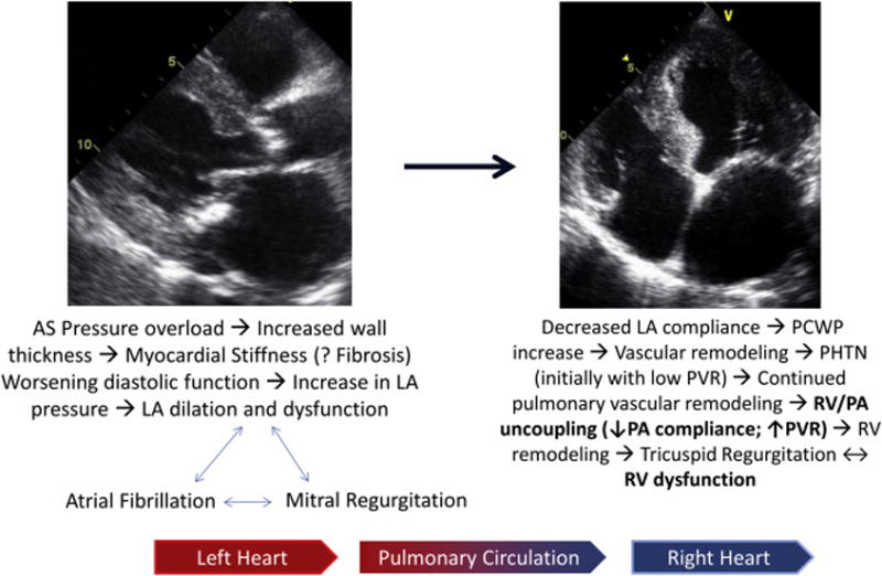
Pathophysiology of right-heart involvement in AS backward pressure transmission. LA, Left atrial; PCWP, pulmonary capillary wedge pressure; PHTN, pulmonary hypertension; PVR, pulmonary vascular resistance.
The prevalence of RV dysfunction in AS will vary according to the disease stage and patient comorbidities. Galli et al.26 found a prevalence of 24% in a population of patients with normal or high-gradient AS with preserved LVEFs. In our own single-center cohort of patients with low-flow, low-gradient AS and reduced LVEFs,27 we found that the presence of RV dysfunction (defined as tricuspid annular plane systolic excursion [TAPSE] < 16 mm) was very common (57% of patients). Although guidelines generally recommend proceeding with dobutamine stress echocardiography for these patients to determine the true AS severity and presence of flow reserve,23 little was known about the prognostic impact of RV function in these patients.
We found that RV dysfunction in these patients was by far the most important prognostic marker of all-cause mortality. More so than baseline LV global longitudinal strain, the presence of flow/contractile reserve, severity of TR, and PH.27 Our findings were confirmed by the Truly or Pseudo-Severe Aortic Stenosis group, which further demonstrated the prognostic value of RV longitudinal strain at both rest and stress28 in these patients with reduced LV systolic function.
HOW TO QUANTIFY RV SYSTOLIC FUNCTION?
Given the peculiar anatomy and shape of the right ventricle, its evaluation using 2D echocardiography is complex. The American Society of Echocardiography guidelines recommend the use of several 2D views that will need to be integrated for the complete assessment of RV function.29 Although the common qualitative evaluation using the “eyeball method” can be used to assess RV systolic function subjectively, quantitative evaluation is necessary and should be implemented in the routine echocardiographic evaluation of these patients. Parameters such as TAPSE describe longitudinal displacement, which although angle dependent, is a simple and reproducible quantitative method to assess RV systolic function and prognostically important marker in patients with PH.29 This measurement is based on the observation that RV stroke volume is largely related to shortening in the longitudinal axis rather than reduction in the cavity diameter in the radial direction; the latter is more important in the case of the left ventricle. Yet there are many other markers of RV function by echocardiography, including fractional area change, tissue Doppler (of both the tricuspid valve annulus and myocardium), strain imaging by speckle-tracking, and measures of function such as the myocardial performance index. No one measure is perfect for assessing systolic function of this complex chamber.
TAVR does not seem to immediately affect RV systolic function in those with normal baseline function.30,31 However, changes in RV systolic function do not seem to be uniform, as worsening of RV systolic function after TAVR, assessed by cardiac magnetic resonance, also has been reported.32 Advanced imaging methods such as speckle-tracking strain imaging,28,33 three-dimensional echocardiography,34 computed tomography, and cardiac magnetic resonance35 complement 2D echocardiography and invasive hemodynamic testing by providing useful surrogate end points for disease progression and may help track response to interventions for future clinical trials in PH.
Another approach has been the integration of RV-PA coupling (RV stroke volume/RV end-systolic volume), which takes into account RV systolic performance at a given degree of afterload. It has greater sensitivity to subtle changes and potentially may be a better metric than the traditional RV ejection fraction.36 In fact, RV-PA coupling, but not RV ejection fraction, was an independent predictor of outcomes in patients with pulmonary arterial hypertension.37 Noninvasive surrogates for RV-PA coupling are emerging, such as tricuspid annular plane systolic excursion to PA systolic pressure ratio, but have yet to be evaluated in the TAVR population.38
PUTTING EVERYTHING TOGETHER: THE INTERPLAY AMONG RV FUNCTION, PH, AND TR AND THE CHANGES PRODUCED BY TAVR TREATMENT
In this issue of JASE, Schwartz et al.39 retrospectively evaluated 519 patients treated with TAVR (access route, valve type, and procedural complications are not mentioned). They quantified RV systolic function, TR, and PH using 2D echocardiography at two time points (baseline and 6 months after TAVR). For RV function assessment, they used both subjective qualitative evaluation and several quantitative 2D methods, of which TAPSE emerged as the best parameter. The investigators observed that TAVR patients with moderate or greater TR (11.3%) had a more advanced stage of the disease with a higher burden of comorbidities (i.e., more RV remodeling with larger tricuspid annular diameter, worse PH, atrial fibrillation, etc.). They conclude that RV systolic function, but not TR severity, is an independent predictor of adverse outcomes, which replicates our findings27 and those of other groups.40 There was also a suggestion that persistence of moderate or greater TR at 6 months after TAVR was associated with lower survival; however, their follow-up had a limited number of patients with severe TR and a high attrition rate.
The work by Schwartz et al.39 highlights the importance of considering TR severity in the context of other relevant comorbidities commonly seen in TAVR patients, in particular the presence of RV dysfunction, which might be the main driver of outcomes. It is important to highlight, however, that only 15 patients (3%) had severe TR, and the majority of patients had preserved RV systolic function at baseline. In addition, gold-standard invasive hemodynamic assessment of PH was not available, which could have added further information about the hemodynamic presentation and outcomes. Last, this study is not suited to answer the question of whether baseline moderate or severe TR should be a clinical target for ancillary treatment beyond TAVR; larger prospective studies will be needed to address this question with a proper study design.
FUTURE DIRECTIONS
Schwartz et al.39 should be commended for evaluating and quantitating these parameters before and after TAVR intervention. Their work certainly builds on the current evidence that RV function, rather than TR severity, dictates outcomes in TAVR patients. This work produces several other questions. For example, what happens to RV function after TAVR treatment in patients with baseline RV dysfunction? What drives the improvement or lack thereof? Should indices of RV function and pulmonary vascular indices be included in preoperative risk assessment scoring models? What should be the thresholds?
The importance of the right heart in patients with AS being considered for TAVR is a topic about which much remains to be learned. The multifactorial pathogenesis of PH and RV dysfunction in these patients will require our continued research efforts to better understand the complex left-right interplay and pulmonary vascular biology. Schwartz et al. should be congratulated for their work emphasizing the need to perform and integrate the comprehensive assessment on both ventricles and hemodynamics in the decision making process of AS patients.
Identification of specific hemodynamic, molecular, and genetic pathways are critically need in patients with group 2 PH, in particular as it relates to valvular heart disease. A better understanding of the importance of right heart function will hopefully guide further strategies beyond TAVR to improve the risk stratification and ultimately long-term outcomes of these patients.
Contributor Information
Jôao L. Cavalcante, Division of Cardiology, Department of Medicine, Pittsburgh Heart, Lung, Blood, and Vascular Medicine Institute, University of Pittsburgh/UPMC, Pittsburgh, Pennsylvania.
Marc A. Simon, Division of Cardiology, Department of Medicine, Heart and Vascular Institute, University of Pittsburgh, Pittsburgh, Pennsylvania; Pittsburgh Heart, Lung, Blood and Vascular Medicine Institute, Department of Bioengineering, University of Pittsburgh, Pittsburgh, Pennsylvania.
Stephen Y. Chan, Center for Pulmonary Vascular Biology and Medicine, Pittsburgh Heart, Lung, Blood, and Vascular Medicine Institute, Division of Cardiology, Department of Medicine, University of Pittsburgh School of Medicine, University of Pittsburgh Medical Center, Pittsburgh, Pennsylvania.
References
- 1.Douglas PS, Hahn RT, Pibarot P, Weissman NJ, Stewart WJ, Xu K, et al. Hemodynamic outcomes of transcatheter aortic valve replacement and medical management in severe, inoperable aortic stenosis: a longitudinal echocardiographic study of cohort B of the PARTNER trial. J Am Soc Echocardiogr. 2015;28:210–7. e1–9. doi: 10.1016/j.echo.2014.10.009. [DOI] [PubMed] [Google Scholar]
- 2.Hutter A, Bleiziffer S, Richter V, Opitz A, Hettich I, Mazzitelli D, et al. Transcatheter aortic valve implantation in patients with concomitant mitral and tricuspid regurgitation. Ann Thorac Surg. 2013;95:77–84. doi: 10.1016/j.athoracsur.2012.08.030. [DOI] [PubMed] [Google Scholar]
- 3.Barbanti M, Binder RK, Dvir D, Tan J, Freeman M, Thompson CR, et al. Prevalence and impact of preoperative moderate/severe tricuspid regurgitation on patients undergoing transcatheter aortic valve replacement. Catheter Cardiovasc Interv. 2015;85:677–84. doi: 10.1002/ccd.25512. [DOI] [PubMed] [Google Scholar]
- 4.Lindman BR, Maniar HS, Jaber WA, Lerakis S, Mack MJ, Suri RM, et al. Effect of tricuspid regurgitation and the right heart on survival after transcatheter aortic valve replacement: insights from the Placement of Aortic Transcatheter Valves II inoperable cohort. Circ Cardiovasc Interv. 2015;8:e002073. doi: 10.1161/CIRCINTERVENTIONS.114.002073. [DOI] [PMC free article] [PubMed] [Google Scholar]
- 5.Quader N, Lindman BR. Shifting the spotlight onto the forgotten ventricle: role of the right ventricle in low-flow, low-gradient aortic stenosis. J Am Soc Echocardiogr. 2016;29:334–6. doi: 10.1016/j.echo.2016.02.006. [DOI] [PubMed] [Google Scholar]
- 6.Dreyfus GD, Martin RP, Chan KM, Dulguerov F, Alexandrescu C. Functional tricuspid regurgitation: a need to revise our understanding. J Am Coll Cardiol. 2015;65:2331–6. doi: 10.1016/j.jacc.2015.04.011. [DOI] [PubMed] [Google Scholar]
- 7.Al-Mohaissen MA, Chan KL. Prevalence and mechanism of tricuspid regurgitation following implantation of endocardial leads for pacemaker or cardioverter-defibrillator. J Am Soc Echocardiogr. 2012;25:245–52. doi: 10.1016/j.echo.2011.11.020. [DOI] [PubMed] [Google Scholar]
- 8.Miglioranza MH, Mihaila S, Muraru D, Cucchini U, Iliceto S, Badano LP. Variability of tricuspid annulus diameter measurement in healthy volunteers. JACC Cardiovasc Imaging. 2015;8:864–6. doi: 10.1016/j.jcmg.2014.09.010. [DOI] [PubMed] [Google Scholar]
- 9.Spinner EM, Lerakis S, Higginson J, Pernetz M, Howell S, Veledar E, et al. Correlates of tricuspid regurgitation as determined by 3D echocardiography: pulmonary arterial pressure, ventricle geometry, annular dilatation, and papillary muscle displacement. Circ Cardiovasc Imaging. 2012;5:43–50. doi: 10.1161/CIRCIMAGING.111.965707. [DOI] [PubMed] [Google Scholar]
- 10.Chen TE, Kwon SH, Enriquez-Sarano M, Wong BF, Mankad SV. Three-dimensional color Doppler echocardiographic quantification of tricuspid regurgitation orifice area: comparison with conventional two-dimensional measures. J Am Soc Echocardiogr. 2013;26:1143–52. doi: 10.1016/j.echo.2013.07.020. [DOI] [PubMed] [Google Scholar]
- 11.van Rosendael PJ, Joyce E, Katsanos S, Debonnaire P, Kamperidis V, van der Kley F, et al. Tricuspid valve remodelling in functional tricuspid regurgitation: multidetector row computed tomography insights. Eur Heart J Cardiovasc Imaging. 2016;17:96–105. doi: 10.1093/ehjci/jev140. [DOI] [PubMed] [Google Scholar]
- 12.Anwar AM, Soliman OI, Nemes A, van Geuns RJ, Geleijnse ML, Ten Cate FJ. Value of assessment of tricuspid annulus: real-time three-dimensional echocardiography and magnetic resonance imaging. Int J Cardiovasc Imaging. 2007;23:701–5. doi: 10.1007/s10554-006-9206-4. [DOI] [PMC free article] [PubMed] [Google Scholar]
- 13.Jeong DS, Sung K, Kim WS, Lee YT, Yang JH, Jun TG, et al. Fate of functional tricuspid regurgitation in aortic stenosis after aortic valve replacement. J Thorac Cardiovasc Surg. 2014;148:1328–13331. doi: 10.1016/j.jtcvs.2013.10.056. [DOI] [PubMed] [Google Scholar]
- 14.Rodes-Cabau J, Hahn RT, Latib A, Laule M, Lauten A, Maisano F, et al. Transcatheter therapies for treating tricuspid regurgitation. J Am Coll Cardiol. 2016;67:1829–45. doi: 10.1016/j.jacc.2016.01.063. [DOI] [PubMed] [Google Scholar]
- 15.Silver K, Aurigemma G, Krendel S, Barry N, Ockene I, Alpert J. Pulmonary artery hypertension in severe aortic stenosis: incidence and mechanism. Am Heart J. 1993;125:146–50. doi: 10.1016/0002-8703(93)90067-j. [DOI] [PubMed] [Google Scholar]
- 16.O’Sullivan CJ, Wenaweser P, Ceylan O, Rat-Wirtzler J, Stortecky S, Heg D, et al. Effect of pulmonary hypertension hemodynamic presentation on clinical outcomes in patients with severe symptomatic aortic valve stenosis undergoing transcatheter aortic valve implantation: insights from the new proposed pulmonary hypertension classification. Circ Cardiovasc Interv. 2015;8:e002358. doi: 10.1161/CIRCINTERVENTIONS.114.002358. [DOI] [PubMed] [Google Scholar]
- 17.Tang M, Liu X, Lin C, He Y, Cai X, Xu Q, et al. Meta-analysis of outcomes and evolution of pulmonary hypertension before and after transcatheter aortic valve implantation [published online September 29, 2016] Am J Cardiol. doi: 10.1016/j.amjcard.2016.09.015. http://dx.doi.org/10.1016/j.amjcard.2016.09.015. [DOI] [PubMed]
- 18.Ben-Dor I, Goldstein SA, Pichard AD, Satler LF, Maluenda G, Li Y, et al. Clinical profile, prognostic implication, and response to treatment of pulmonary hypertension in patients with severe aortic stenosis. Am J Cardiol. 2011;107:1046–51. doi: 10.1016/j.amjcard.2010.11.031. [DOI] [PubMed] [Google Scholar]
- 19.Sinning JM, Hammerstingl C, Chin D, Ghanem A, Schueler R, Sedaghat A, et al. Decrease of pulmonary hypertension impacts on prognosis after transcatheter aortic valve replacement. EuroIntervention. 2014;9:1042–9. doi: 10.4244/EIJV9I9A177. [DOI] [PubMed] [Google Scholar]
- 20.Testa L, Latib A, De Marco F, De Carlo M, Fiorina C, Montone R, et al. Persistence of severe pulmonary hypertension after transcatheter aortic valve replacement: incidence and prognostic impact. Circ Cardiovasc Interv. 2016;9:e003563. doi: 10.1161/CIRCINTERVENTIONS.115.003563. [DOI] [PubMed] [Google Scholar]
- 21.Gerges C, Gerges M, Lang MB, Zhang Y, Jakowitsch J, Probst P, et al. Diastolic pulmonary vascular pressure gradient: a predictor of prognosis in “out-of-proportion” pulmonary hypertension. Chest. 2013;143:758–66. doi: 10.1378/chest.12-1653. [DOI] [PubMed] [Google Scholar]
- 22.Champion HC, Michelakis ED, Hassoun PM. Comprehensive invasive and noninvasive approach to the right ventricle-pulmonary circulation unit: state of the art and clinical and research implications. Circulation. 2009;120:992–1007. doi: 10.1161/CIRCULATIONAHA.106.674028. [DOI] [PubMed] [Google Scholar]
- 23.Nishimura RA, Otto CM, Bonow RO, Carabello BA, Erwin JP, 3rd, Guyton RA, et al. 2014 AHA/ACC guideline for the management of patients with valvular heart disease: a report of the American College of Cardiology/American Heart Association Task Force on Practice Guidelines. J Am Coll Cardiol. 2014;63:e57–185. doi: 10.1016/j.jacc.2014.02.536. [DOI] [PubMed] [Google Scholar]
- 24.Lee N, Taylor MD, Banerjee RK. Right ventricle-pulmonary circulation dysfunction: a review of energy-based approach. Biomed Eng Online. 2015;14(Suppl 1):S8. doi: 10.1186/1475-925X-14-S1-S8. [DOI] [PMC free article] [PubMed] [Google Scholar]
- 25.Chun HJ, Bonnet S, Chan SY. Translating microRNA biology in pulmonary hypertension: it will take more than “miR” words [published online September 20, 2016] Am J Respir Crit Care Med. doi: 10.1164/rccm.201604-0886PP. http://dx.doi.org/10.1164/rccm.201604-0886PP. [DOI] [PMC free article] [PubMed]
- 26.Galli E, Guirette Y, Feneon D, Daudin M, Fournet M, Leguerrier A, et al. Prevalence and prognostic value of right ventricular dysfunction in severe aortic stenosis. Eur Heart J Cardiovasc Imaging. 2015;16:531–8. doi: 10.1093/ehjci/jeu290. [DOI] [PubMed] [Google Scholar]
- 27.Cavalcante JL, Rijal S, Althouse AD, Delgado-Montero A, Katz WE, Schindler JT, et al. Right ventricular function and prognosis in patients with low-flow, low-gradient severe aortic stenosis. J Am Soc Echocardiogr. 2016;29:325–33. doi: 10.1016/j.echo.2015.12.001. [DOI] [PubMed] [Google Scholar]
- 28.Dahou A, Clavel MA, Capoulade R, Bartko PE, Magne J, Mundigler G, et al. Right ventricular longitudinal strain for risk stratification in low-flow, low-gradient aortic stenosis with low ejection fraction. Heart. 2016;102:548–54. doi: 10.1136/heartjnl-2015-308309. [DOI] [PubMed] [Google Scholar]
- 29.Rudski LG, Lai WW, Afilalo J, Hua L, Handschumacher MD, Chandrasekaran K, et al. Guidelines for the echocardiographic assessment of the right heart in adults: a report from the American Society of Echocardiography endorsed by the European Association of Echocardiography, a registered branch of the European Society of Cardiology, and the Canadian Society of Echocardiography. J Am Soc Echocardiogr. 2010;23:685–713. doi: 10.1016/j.echo.2010.05.010. quiz 86–8. [DOI] [PubMed] [Google Scholar]
- 30.Kempny A, Diller GP, Kaleschke G, Orwat S, Funke A, Schmidt R, et al. Impact of transcatheter aortic valve implantation or surgical aortic valve replacement on right ventricular function. Heart. 2012;98:1299–304. doi: 10.1136/heartjnl-2011-301203. [DOI] [PubMed] [Google Scholar]
- 31.Fairbairn TA, Steadman CD, Mather AN, Motwani M, Blackman DJ, Plein S, et al. Assessment of valve haemodynamics, reverse ventricular remodelling and myocardial fibrosis following transcatheter aortic valve implantation compared to surgical aortic valve replacement: a cardiovascular magnetic resonance study. Heart. 2013;99:1185–91. doi: 10.1136/heartjnl-2013-303927. [DOI] [PMC free article] [PubMed] [Google Scholar]
- 32.Crouch G, Bennetts J, Sinhal A, Tully PJ, Leong DP, Bradbrook C, et al. Early effects of transcatheter aortic valve implantation and aortic valve replacement on myocardial function and aortic valve hemodynamics: insights from cardiovascular magnetic resonance imaging. J Thorac Cardiovasc Surg. 2015;149:462–70. doi: 10.1016/j.jtcvs.2014.10.064. [DOI] [PubMed] [Google Scholar]
- 33.Fine NM, Chen L, Bastiansen PM, Frantz RP, Pellikka PA, Oh JK, et al. Outcome prediction by quantitative right ventricular function assessment in 575 subjects evaluated for pulmonary hypertension. Circ Cardiovasc Imaging. 2013;6:711–21. doi: 10.1161/CIRCIMAGING.113.000640. [DOI] [PubMed] [Google Scholar]
- 34.Ryo K, Goda A, Onishi T, Delgado-Montero A, Tayal B, Champion HC, et al. Characterization of right ventricular remodeling in pulmonary hypertension associated with patient outcomes by 3-dimensional wall motion tracking echocardiography. Circ Cardiovasc Imaging. 2015;8:e003176. doi: 10.1161/CIRCIMAGING.114.003176. [DOI] [PubMed] [Google Scholar]
- 35.Freed BH, Collins JD, Francois CJ, Barker AJ, Cuttica MJ, Chesler NC, et al. MR and CT imaging for the evaluation of pulmonary hypertension. JACC Cardiovasc Imaging. 2016;9:715–32. doi: 10.1016/j.jcmg.2015.12.015. [DOI] [PMC free article] [PubMed] [Google Scholar]
- 36.Vanderpool RR, Rischard F, Naeije R, Hunter K, Simon MA. Simple functional imaging of the right ventricle in pulmonary hypertension: can right ventricular ejection fraction be improved? Int J Cardiol. 2016;223:93–4. doi: 10.1016/j.ijcard.2016.08.138. [DOI] [PMC free article] [PubMed] [Google Scholar]
- 37.Vanderpool RR, Pinsky MR, Naeije R, Deible C, Kosaraju V, Bunner C, et al. RV-pulmonary arterial coupling predicts outcome in patients referred for pulmonary hypertension. Heart. 2015;101:37–43. doi: 10.1136/heartjnl-2014-306142. [DOI] [PMC free article] [PubMed] [Google Scholar]
- 38.Hussain I, Mohammed SF, Forfia PR, Lewis GD, Borlaug BA, Gallup DS, et al. Impaired right ventricular-pulmonary arterial coupling and effect of sildenafil in heart failure with preserved ejection fraction: an ancillary analysis from the Phosphodiesterase-5 Inhibition to Improve Clinical Status And Exercise Capacity in Diastolic Heart Failure (RELAX) Trial. Circ Heart Fail. 2016;9:e002729. doi: 10.1161/CIRCHEARTFAILURE.115.002729. [DOI] [PMC free article] [PubMed] [Google Scholar]
- 39.Schwartz LA, Rozenbaum Z, Ghantous E, Kramarz J, Biner S, Ghermezi M, et al. Impact of right ventricular dysfunction and tricuspid regurgitation on outcomes in patients undergoing transcatheter aortic valve replacement. J Am Soc Echocardiogr. 2017;30:36–46. doi: 10.1016/j.echo.2016.08.016. [DOI] [PubMed] [Google Scholar]
- 40.Kammerlander AA, Marzluf BA, Graf A, Bachmann A, Kocher A, Bonderman D, et al. Right ventricular dysfunction, but not tricuspid regurgitation, is associated with outcome late after left heart valve procedure. J Am Coll Cardiol. 2014;64:2633–42. doi: 10.1016/j.jacc.2014.09.062. [DOI] [PubMed] [Google Scholar]