

Abstract
Background
Bibliometric and scientometric methods can be applied to the study of a research field.
Objective
We hypothesized that a bibliometric and scientometric analysis of the Alzheimer’s disease (AD) research field could render trends that provide researchers and funding agencies valuable insight into the history of the field, current tendencies, and potential future directions.
Methods
We performed searches in publicly available databases including PubMed, Scopus, Web of Science, and Alzheimer’s Funding Analyzer for the period 1975–2014, and conducted a curve fitting analysis with non-linear regression.
Results
While the rate and impact of publications continue to increase, the number of patents per year is currently declining after peaking in the late 2000s, and the funding budget has plateaued in the last 5–10 years analyzed. Genetics is the area growing at a fastest pace, whereas pathophysiology and therapy have not grown further in the last decade. Among the targets of pathophysiology research, amyloid-β continues to be the focus of greatest interest, with tau and apolipoprotein E stagnant after a surge in the 1990s. The role of inflammation, microglia, and the synapse are other research topics with growing interest. Regarding preventative strategies, education attainment, diet, and exercise are recently gaining some momentum, whereas NSAIDs and statins have lost the spotlight they once had.
Conclusion
Our bibliometric and scientometric analysis provides distinct trends in AD research in the last four decades, including publication and patent output, funding, impact, and topics. Our findings could inform the decision-making of research funding agencies in the near future.
Keywords: Alzheimer’s disease, amyloid-β peptides, bibliometrics, h-index, neurofibrillary tangle, scientometrics, tau proteins
INTRODUCTION
Alzheimer’s disease (AD) was first described by the German psychiatrist and neuropathologist Alois Alzheimer in 1906, but its existence was then neglected for many decades. In 1976, Robert Katzman published an editorial calling the attention of the research community to both the high prevalence of AD—the most common cause of “senile dementia”—and its fatal nature [1]. In retrospect, Dr. Katzman’s two-page editorial turned out to be premonitory, as indicated by current epidemiological data. According to the US Alzheimer’s Association, in 2016 there were 5.4 million Americans with AD, and 700,000 people age 65 years and older died with AD in the United States, many of them from complications caused by AD [2]. Fortunately, governments are currently responding to this threatening epidemics with specific plans and policies [3–5]. Furthermore, the last four decades have witnessed an unparalleled growth of Neuroscience and AD research.
We sought to examine the body of research in the field of AD over the last 40 years, taking advantage of publicly available databases and resources on scientific literature and research funding. We hypothesized that a bibliometric and scientometric analysis could yield research trends and provide valuable insights into the future directions of the field for both researchers and funding agencies. Bibliometric analyses have been previously conducted on other neurological diseases such as multiple sclerosis [6], Parkinson’s disease [7], frontotemporal dementia [8], and cerebral amyloid angiopathy [9, 10], and rendered interesting clues with regards to the changing landscape of research on these diseases.
METHODS
PubMed search and data collection
To obtain the body of AD literature, a search was performed in the website of the US National Library of Medicine of the National Institutes of Health (http://www.ncbi.nlm.nih.gov/pubmed/). The search was conducted using the Medical Subject Heading (MeSH) terms found in the MeSH website (http://www.ncbi.nlm.nih.gov/mesh/) and its PubMed Search Builder tool [11, 12]. The MeSH search (i.e., adding “Alzheimer disease”, MeSH Unique ID: D000544, to the search builder box) was preferred over the free text search (i.e., typing “Alzheimer disease” in the PubMed Search box) because precision (number of articles relevant to research / number of articles retrieved) is higher with the use of MeSH terms as compared to free text, although at the expense of a lower yield (number of articles retrieved / number of total articles archived) [13, 14]. The MeSH terms used in this study are listed in Supplementary Table 1 together with their unique ID. The PubMed search strategies used are listed in Supplementary Table 2.
A publication date range filter from 01/01/1975 to 12/31/2014 was applied to all searches. The search was limited to the articles after 1974 because prior to 1975 the number of publications was scarce, so that the publication record was intermittent (i.e., no articles were found in 1967, 1968, 1970, and 1972). The years 2015 and 2016 were excluded because, at the time of this search, the process of PubMed indexation for articles published in these years was still ongoing. The results shown here correspond to a search done on March 11, 2016, except for those regarding preventative factors, which were searched for on April 21, 2017.
To characterize this body of literature, PubMed filters were applied that categorize publications by the following criteria: 1) type of publication: journal articles, reviews, clinical trials, meta-analyses, systematic reviews, case reports, and editorials; 2) human versus animal studies; 3) age group targeted by human studies: child (birth to 18 years), adults 18–44 years, adults 45–64 years, aged 65–79 years, and aged 80 + years.
Next, the MeSH subheadings relevant to the field of AD were selected to identify research trends by subfield. These included: therapy, prevention and control, diagnosis, genetics, epidemiology, mortality, psychology, pathology, physiopathology, etiology, economics, blood, cerebrospinal fluid, and radionuclide imaging. The following subheadings were combined: “therapy”, “diet therapy”, and “drug therapy” were combined under “therapy”; “anatomy and histology” and “pathology” were combined under “pathology”; “etiology” and “physiopathology” under “pathophysiology”; “epidemiology” and “mortality” under “epidemiology”; and “economics” and “organization and administration” under “economics”. Because the MeSH subheading “radiography” only yielded 342 articles and did not capture the literature on MRI, it was not included in this analysis. Instead, a search was performed with the combination of MeSH terms “Alzheimer disease” (MeSH Unique ID: D000544) AND “magnetic resonance imaging” (MeSH Unique ID: D008279), which yielded 3,833 articles.
Next, to investigate the research trend on the pathophysiology of AD, combined searches were conducted with the MeSH term “Alzheimer disease” AND the following relevant MeSH terms: “Amyloid beta-peptides” (MeSH Unique ID: D016229), “Plaque, Amyloid” (MeSH Unique ID: D058225), “tau Proteins” (MeSH Unique ID: D016875), “Neurofibrillary Tangles” (MeSH Unique ID: D016874), “Microglia” (MeSH Unique ID: D017628), “Astrocytes” (MeSH Unique ID: D001253), “Inflammation” (MeSH Unique ID: D007249), “Oxidative Stress” (MeSH Unique ID: D018384), “Mitochondria” (MeSH Unique ID: D008928), “Acetylcholine” (MeSH Unique ID: D000109), and “Cerebrovascular Circulation” (MeSH Unique ID: D002560) (see Supplementary Table 1).
Last, to investigate research trends in AD prevention, the MeSH term “Alzheimer disease” was combined with the MeSH terms “Educational Status” (MeSH Unique ID: D004522), “Diet” (MeSH Unique ID: D004032), “Exercise” (MeSH Unique ID: D015444), “Hydroxymethylglutaryl-CoA Reductase Inhibitors” (MeSH Unique ID: D019161), and “Anti-inflammatory Agents, Non-Steroidal” (MeSH Unique ID: D000894) (see Supplementary Table 1).
Web of Science search
The Thomson Scientific Web of Science (WoS) (http://www.webofknowledge.com/WOS) was interrogated on April 19, 2016 to obtain the Hirsh or h-index for the body of AD literature on a yearly basis. This search was conducted entering the keyword “Alzheimer disease” in the title box and each of the 40 years investigated in the time range filter box. When referred to an author —its original and most common use— the h-index represents the number of articles by that author that have received h or more citations [15]. However, a cumulative or average h-index can also be applied to determine the impact of scholarly journals [16], scientific disciplines, and groups of researchers (i.e., university, department, country) [17]. In all cases, the h-index is modified by the year of publication (that is, the seniority of the research work) because older papers have more time to accumulate citations. To account for the effect of time, the raw h-index for each year of AD research was divided by the number of years that those papers were available for citation, as below:
where h(t)y is the time-corrected yearly h-index, hy is the global h-index of each year AD body of literature, and (2014-y) is the number of years that that body of literature has been available to citation. This correction of the h-index has been previously applied to Chemistry research topics and chemical compounds [18] and is analogous to the m-quotient developed by Hirsh to account for the seniority of the individual researcher [19].
Because the h-index for a discipline or a research topic is also influenced by the number of articles available to be cited, herein we also corrected the yearly h-index for the number of AD papers published each year as noted below:
where h(o)y is the output-corrected yearly h-index, hy is again the global h-index of each year AD body of literature, and ny is the number of AD papers published that year. A similar correction of the h-index has been previously used to compare the evolution of impact of Neurology journals with wide differences in the number of articles published per year [20].
Last, it follows that the yearly h-index corrected for both the year of publication and the number of AD papers published each year is:
Scopus search
The website Scopus (http://www.scopus.es) was interrogated on March 19, 2016 to obtain the patents related with AD. The keyword “Alzheimer disease” was entered in the title box and the patents rendered were ordered by year.
Alzheimer’s Funding Analyzer (AFA) search
To obtain data about trends in budget for AD research, we inquired the Alzheimer’s Funding Analyzer (http://www.j-alz.uberresearch.com). This search tool was set up by the Journal of Alzheimer’s Disease and includes information from 96 funding agencies from 33 countries including all the countries in the European Union, Australia, Canada, Ireland, Qatar, the United Kingdom, and the United States. Data for annual US dollars (USD) spent and number of projects funded per year was available since 1976.
Statistical analyses
To investigate research trends, the percent of articles on each theme (MeSH subheading or keyword) published every year was calculated by dividing the number of articles on that theme that appeared that year by the total number of articles under the MeSH keyword “Alzheimer disease” published that year.
The association between each of the research topics, or USD spent in AD research, and time was analyzed using nonlinear regression with the least-square method. XY dot plots were examined and the most appropriate pairs of models, including linear, exponential, Gaussian, and centered polynomial (up to sixth order) were compared with the Akaike’s Informative Criterion (AICc), with no weights or constrains. The results of these comparisons are summarized in Table 1. XY graphs in figures contain the regression line and 95% confidence interval corresponding to the preferred model. Statistical analyses and graphs were performed with GraphPad Prism version 7.0 (GraphPad, La Jolla, CA).
Table 1.
Summary of the statistical results of the present study
| Variable | Comparator model
|
Preferred model
|
ΔAICc | ||
|---|---|---|---|---|---|
| Model | Prob (%) | Model | Prob (%) | ||
| Number of articles | Straight line | <0.01 | Centered 6th order polynomial | >99.99 | 47.79 |
| % of total articles | Straight line | <0.01 | Centered 6th order polynomial | >99.99 | 48.79 |
| Number of patents | Centered 2nd order polynomial (quadratic) | <0.01 | Gaussian | >99.99 | −29.73 |
| Aggregated funding budget | Centered 6th order polynomial | 22.27 | Centered 4th order polynomial | 77.73 | −2.499 |
| Number of starting projects funded | Centered 6th order polynomial | 40.56 | Centered 5th order polynomial | 59.44 | −0.7646 |
|
| |||||
| Raw h-index | Gaussian | <0.01 | Centered 6th order polynomial | >99.99 | 43.04 |
| h-index corrected for number of papers | Centered 5th order polynomial | 36.00 | Centered 6th order polynomial | 64.00 | 1.151 |
| h-index corrected for year of publication | Exponential growth equation | <0.01 | Centered 6th order polynomial | >99.99 | 48.73 |
| h-index corrected for year of publication and number of papers | Centered 6th order polynomial | 17.05 | Centered 5th order polynomial | 82.95 | −3.165 |
|
| |||||
| Journal article | Standard sine wave | <0.01 | Centered 6th order polynomial | >99.99 | 297.7 |
| Reviews | Centered 5th order polynomial | 22.22 | Centered 4th order polynomial | 77.78 | −2.505 |
| Systematic reviews and meta-analyses | Centered 4th order polynomial | 41.25 | Centered 3rd order polynomial (cubic) | 58.75 | −0.7077 |
| Clinical trials | Centered 6th order polynomial | 20.21 | Centered 5th order polynomial | 79.79 | −2.746 |
| Case reports | Centered 3rd order polynomial (cubic) | 11.94 | One phase decay | 88.06 | −3.997 |
| Editorials, news, letters, comments | One phase decay | 14.66 | Centered 5th order polynomial | 85.34 | 3.523 |
|
| |||||
| Human studies | Straight line | 0.61 | Centered 2nd order polynomial (quadratic) | 99.39 | 10.19 |
| Animal studies | Exponential growth equation | 2.51 | Centered 4th order polynomial | 97.49 | 7.316 |
| Birth-18 years | Centered 5th order polynomial | 2.11 | One phase decay | 97.89 | −7.671 |
| Adults 18–44 years | One phase decay | 27.02 | Exponential growth equation | 72.98 | −1.987 |
| Adults 45–64 years | Centered 2nd order polynomial (quadratic) | 23.56 | One phase decay | 76.44 | 2.354 |
| Adults 65–79 years | Centered 4th order polynomial | 48.18 | Centered 6th order polynomial | 51.82 | 0.1455 |
| Adults 80 + years old | Centered 4th order polynomial | 5.79 | Centered 6th order polynomial | 94.21 | 5.578 |
|
| |||||
| Diagnosis | One phase decay | 0.04 | Centered 6th order polynomial | 99.96 | 15.44 |
| Pathology | One phase decay | <0.01 | Centered 6th order polynomial | >99.99 | 28.57 |
| Pathophysiology | Centered 6th order polynomial | 4.36 | Sigmoidal, 4PL, X is log (concentration) | 95.64 | −6.177 |
| Therapy | Centered 6th order polynomial | 6.04 | Sigmoidal, 4PL, X is log (concentration) | 93.96 | −5.487 |
| Genetics | Straight line | <0.01 | Centered 6th order polynomial | >99.99 | 21.14 |
| Epidemiology | Centered 3rd order polynomial (cubic) | 2.28 | Centered 6th order polynomial | 97.72 | 7.518 |
| Prevention | Sigmoidal, 4PL, X is log (concentration) | 0.03 | Centered 6th order polynomial | 99.97 | 16.37 |
| Economics | Centered 3rd order polynomial (cubic) | 1.15 | Centered 6th order polynomial | 98.85 | 8.913 |
|
| |||||
| Psychology | Centered 2nd order polynomial (quadratic) | <0.01 | Centered 3rd order polynomial (cubic) | 29.99 | 24.16 |
| MRI | Exponential growth equation | 2.38 | Straight line | 97.62 | −7.424 |
| Blood | Straight line | 1.67 | Centered 3rd order polynomial (cubic) | 98.33 | 8.146 |
| CSF | Centered 6th order polynomial | 2.39 | Centered 3rd order polynomial (cubic) | 97.61 | −7.423 |
| Radionuclide | Straight line | 0.03 | Centered 6th order polynomial | 99.97 | 16.09 |
|
| |||||
| Amyloid ß peptides & plaques | Centered 6th order polynomial | 7.01 | Straight line | 92.99 | −5.171 |
| Tau protein & NFTs | Centered 6th order polynomial | 31.20 | Centered 5th order polynomial | 68.80 | −1.582 |
| Apolipoprotein E | Centered 2nd order polynomial (quadratic) | <0.01 | Centered 6th order polynomial | >99.99 | 39.87 |
| Inflammation | Straight line | 5.78 | Centered 4th order polynomial | 94.22 | 5.584 |
| Microglia | Straight line | 35.83 | Centered 3rd order polynomial (cubic) | 64.17 | 1.166 |
| Oxidative stress | Centered 3rd order polynomial (cubic) | 41.28 | One phase association | 58.72 | −0.7048 |
| Synapse | Centered 6th order polynomial | 2.02 | Centered 2nd order polynomial (quadratic) | 97.98 | −7.767 |
| Mitochondria | Centered 2nd order polynomial (quadratic) | 0.36 | Centered 6th order polynomial | 99.64 | 11.23 |
| Astrocytes | One phase decay | 25.88 | Centered 4th order polynomial | 74.12 | 2.104 |
| Calcium | Centered 6th order polynomial | 20.78 | Centered 5th order polynomial | 79.22 | −2.676 |
| Acetylcholine | Straight line | 7.39 | Exponential growth equation | 92.61 | −5.055 |
| Cerebrovascular disease | Centered 2nd order polynomial (quadratic) | <0.01 | One phase decay | >99.99 | −30.76 |
|
| |||||
| Education | Straight line | 48.80 | Centered 2nd order polynomial (quadratic) | 51.20 | 0.096 |
| Diet | Centered 5th order polynomial | 28.99 | Centered 6th order polynomial | 71.01 | 1.792 |
| Exercise | Straight line | 2.91 | Centered 3rd order polynomial (cubic) | 97.09 | 7.017 |
| NSAIDs | Centered 2nd order polynomial (quadratic) | 15.67 | Gaussian | 84.33 | 3.366 |
| Statins | Gaussian | 41.67 | Centered 3rd order polynomial (cubic) | 58.33 | 0.6731 |
ΔAICc, difference in Akaike’s Informative Criteria; CSF, cerebrospinal fluid; NFTs, neurofibrillary tangles; Prob, probability that the model is correct.
RESULTS
AD research output and funding in the last four decades (1975–2014)
Figure 1 shows the evolution of AD research output in the last four decades. The frequency in Fig. 1A refers to the percentage of publications in each year with respect to the total number of publications in the whole period of four decades. After a hesitant beginning in the first 7 years of this period, the publication output increased virtually linearly for 25 years. It appears that in the last 5-6 years, the annual rate of publication has increased further as judged by the slope of the fitting curve. Of note, in sharp contrast with the scientific publication output, the number of patents per year as recorded in the database Scopus followed a Gaussian distribution with a peak in the late 2000s (Fig. 1B). Similarly, both the available budget to fund AD research (Fig. 1C) and the number of starting projects funded (Fig. 1D) appear to have plateaued in the last decade after an almost exponential increase for three decades.
Fig. 1.

Evolution of AD research output and funding. A) Total number (left y axis) and relative frequency (right y axis) of AD publications per year between 1975 and 2014, based on PubMed database. B) Number of patents on AD per year based on Scopus database. C) Aggregated funding amount per year in millions of US dollars, based on Alzheimer’s Funding Analyzer database (www.j-alz/uberresearch.com). D) Number of starting projects funded per year, based on Alzheimer’s Funding Analyzer database.
Impact of AD research in the last four decades (1975–2014)
Figure 2 shows the evolution of the impact of AD research across the four decades as indicated by the h-index of the WoS. Because the h-index applied to a research topic is influenced by both the age of the publication (older papers tend to have a higher h-index as they accumulate citations) and the number of publications (the most prolific years have a higher h-index), we present the yearly evolution of the raw h-index (Fig. 2A) and the h-index corrected for: the number of papers published each year according to the WoS (Fig. 2B), the age of publication (Fig. 2C), and both parameters (Fig. 2D). The computations of these corrected versions of the h-index are explained in the method section. The cumulative yearly h-index exhibits an expected increase over time (Fig. 2A) followed by a plateau around 2000, and a decline after 2005. Correcting for the number of papers published each year shows that the spectacular increase in impact between 1975 and 1995 is mostly due to an increase in publication output (Fig. 2B), since the corrected h-index drops dramatically in that period. Correcting for year of publication demonstrates that the decline in impact observed after 2005 is just due to the youth of these most recent papers (Fig. 2C), since the corrected h-index increases exponentially after that year. Remarkably, the evolution of the “doubly-corrected” h-index depicts a U-shape, indicating that the individual publications with highest impact date from late 1970s and early 1980s, but also from the last 5–10 years (Fig. 2D).
Fig. 2.

Evolution of AD research impact. A) h-index of the AD field per year based on Web of Science database. B) Yearly h-index of the AD field corrected for the number of papers published each year. C) Yearly h-index of the AD field corrected for the year (age) of publication. D) Yearly h-index of the AD field corrected by the number of papers and the year (age) of publication.
Trends in publication subtypes
Figure 3 shows the AD research publications split by publication subtype as yielded by PubMed filters: journal article, reviews, clinical trials, systematic reviews and meta-analyses, case reports, and others (editorials, news, letters, comments, and introductory journal articles). The number and percent of publications under each article subtype is listed in Supplementary Table 3.
Fig. 3.

Trends in AD publication subtypes. Graphs represent the relative frequency of publications of each subtype per year with respect to the total number of papers published each year. Classification of articles as journal articles (A), reviews (B), systematic reviews and meta-analyses (C), clinical trials (D), case reports (E), and editorial notes, news, comments, letters, and introductory journal articles (F) was achieved using available PubMed filters. Note that the sum of the frequencies for different publication subtypes in the same year can exceed 100% because some publications are categorized in more than one subtype.
The relative frequency of journal articles, the most common type of publication, dropped in the late 1970s in favor of case reports and short papers (i.e., editorials, letters, etc.), then increased and remained stable for 20 years between 1980 and 2000, and has been increasing slightly again thereafter. The rate of publication of review articles increased more than 2-fold between 1980 and 2000 and has stabilized thereafter, whereas the publication of systematic reviews and meta-analyses has steadily increased since the early 1990s. In contrast, the relative frequency of clinical trial papers peaked between 2000 and 2005, and the publication of case reports, once relatively usual, has continuously declined since the late 1970s.
Human versus animal studies
Figure 4A shows the publication output for human and animal research over the period 1975–2014 and Fig. 4B-F depict the human studies split by target age group (that is, birth-18 years, adults 18–44 years, adults 45–64 years, aged 65–79 years, and aged 80+ years) as yielded by the corresponding PubMed filters.
Fig. 4.

Trends in human versus animal studies and of human studies by age group. Graphs represent the relative frequency of papers within each target group per year with respect to the total number of papers published each year. Classification of studies in human versus animal (A) and of human studies in different age group (B-F) was achieved using available PubMed filters. Note that the sum of the relative frequencies of human and animal publications and of the different human age groups for any given year exceeds 100% because PubMed may classify some publications as both animal and human studies, and because some human studies may include more than one age group.
Since the early 1990s, the publication of animal studies of AD has steadily increased every year. This increase in animal research parallels a slight decline in the rate of publication of human studies. The reduction in human research publications affects all age groups except for the oldest (80+ years), which exhibits a dramatic surge in the late 1990s and a slow gradual increase thereafter.
Trends in research themes
Figure 5 shows the breakdown of AD research publications by major themes as defined by MeSH subheadings: diagnosis, therapy, prevention, epidemiology, pathology, pathophysiology, genetics, and economics. Note that theme graphs are arranged in descending order of frequency of publication, that is, from the highest (A) to the lowest (H) percentage of papers published on that topic each year with respect to the total number of publications published that year. The number and percent of articles under each MeSH subheading is listed in Supplementary Table 4.
Fig. 5.

Trends in AD research themes. Graphs represent the relative frequency of papers about each research theme per year with respect to the total number of papers published each year. Classification of papers under diagnosis (A), pathology (B), pathophysiology (C), therapy (D), genetics (E), epidemiology (F), prevention (G), and economics (H) was based on the MeSH subheadings available under the MeSH term “Alzheimer disease” in PubMed database, as explained in the methods section. Note that the sum of the frequencies of different research themes for the same year exceeds 100% in many years because the same article may be categorized under more than one theme.
Diagnosis, pathophysiology, therapy, genetics, and pathology represent the bulk of AD research, while prevention, epidemiology, and economics remain marginal themes. A closer look reveals that research in AD diagnosis (Fig. 5A) and pathology (Fig. 5B), which were the main themes of publication in the first few years analyzed, then reached a plateau that essentially has lasted until now, although there seems to be some renovated interest in pathology research after 2005. By contrast, AD pathophysiology (Fig. 5C) and therapy (Fig. 5D), which were already prominent research themes between 1975 and 1990, experienced a significant boost after 1990 and 1995, respectively, followed by a plateau after 2000 and 2005, respectively. The evolution of genetics research (Fig. 5E) is remarkably different from the other themes, with a significant increase over the last three decades in two distinct phases: first, between late 1980s and 2000, and again after 2010. Finally, epidemiology (Fig. 5F) and prevention (Fig. 5G) research have increased in parallel between 1985 and 2005 and are declining in the last 5–10 years. Research on economics and organization (Fig. 5H) has been negligible throughout the last 30 years.
Research trends in AD diagnosis
Figure 6 shows the breakdown of AD research publications by major diagnostic tools as defined by the MeSH subheadings: psychology, magnetic resonance imaging, blood, CSF, and radionuclide imaging. Note that again graphs are arranged in descending order of frequency of publication, that is, from the highest (A) to the lowest (E) percentage of papers published on that topic each year with respect to the total number of publications published that year.
Fig. 6.
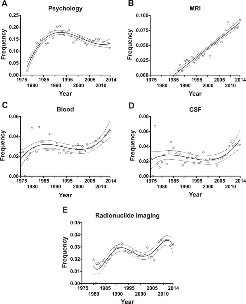
Research trends in AD diagnosis. Graphs represent the relative frequency of each research theme per year with respect to the total number of papers published each year. Classification of papers under psychology (A), blood (C), CSF (D), and radionuclide (E) was based on the MeSH subheadings available under the MeSH term “Alzheimer disease” in PubMed database, as explained in the methods section. MRI papers (B) were obtained by a search combining the MeSH terms “Alzheimer disease” and “Magnetic resonance imaging” because the “radiography” MeSH subheading yielded few papers.
Visual inspection of these graphs reveals that psychology (Fig. 6A) flourished in the decade of 1980s and plateaued in the early 1990s, but continues to be the main diagnostic theme, followed very closely in recent years by MRI. MRI research (Fig. 6B) has followed a linear increase since its application to dementia diagnosis in the late 1980s. Blood (Fig. 6C) and CSF (Fig. 6D) biomarker research has been pursued since 1975 but began receiving more attention starting in 2005. Research on radionuclide imaging (Fig. 6E) exhibits two distinct phases of acceleration: between 1985 and early 1990s, and after 2005.
Research trends in AD pathophysiology
Figure 7 shows the breakdown of research publications in AD pathophysiology using the following MeSH terms: amyloid beta peptides and amyloid plaques, tau proteins and neurofibrillary tangles, apolipoprotein E, astrocytes, microglia, inflammation, acetylcholine, oxidative stress, mitochondria, calcium, synapses, and cerebrovascular circulation.
Fig. 7.

Research trends in AD pathophysiology. Graphs represent the relative frequency of papers on each research topic per year with respect to the total number of papers published each year. Searches were conducted in PubMed database by combining the “Alzheimer disease” MeSH term with MeSH terms of interest. NFTs, neurofibrillary tangles.
Aβ peptide and plaques (Fig. 7A) represent the main research topic within AD pathophysiology and follow a linear increase starting in 1985. By contrast, tau research (Fig. 7B) has been proportionally behind Aβ research. Tau literature experienced a dramatic impulse in the early 1990s with a 4-fold increase in its relative frequency, then declined between 1995 and 2005, but is seemingly regaining momentum in the last decade. Apolipoprotein E research (Fig. 7C) peaked in late 1990s and dropped thereafter, although remains a significant research subject.
Of note, research into inflammation (Fig. 7D), microglia (Fig. 7E), oxidative stress (Fig. 7F), synapses (Fig. 7G), and mitochondria (Fig. 7H) are overall trending up, at least since 1995. By contrast, research on astrocytes (Fig. 7I) and calcium dyshomeostasis (Fig. 7J) seems to have plateaued since early 1990s, whereas acetylcholine (Fig. 7K) and cerebrovascular circulation research (Fig. 7L) were very prominent in the late 1970s and early 1980s, but have experienced a steady downtrend.
Research trends in AD prevention
Figure 8 shows the research publications in AD prevention broken down by individual preventative factors and strategies as defined by the following MeSH terms: educational status (MeSH term for educational attainment), diet, exercise, anti-inflammatory agents, non-steroidal (MeSH term for NSAIDs), and hydroxymethylglutaryl-co A reductase inhibitors (MeSH term for statins).
Fig. 8.

Research trends in AD prevention. Graphs represent the relative frequency of literature on each prevention strategy or factor per year with respect to the total number of papers published each year. Searches were conducted in PubMed database by combining the “Alzheimer disease” MeSH term with MeSH terms “Educational Status” (A), “Diet” (B), “Exercise” (C), “Anti-Inflammatory Agents, Non-Steroidal” (NSAIDs) (D), and “Hydroxymethylglutaryl-CoA Reductase Inhibitors” (statins) (E), as explained in the methods section.
Regression analyses of the relative frequency of publications in each of these prevention topics against time showed that education, diet and exercise are trending up, whereas NSAIDs and statins peaked in the 2000–2005 period and dropped thereafter.
DISCUSSION
Our bibliometric and scientometric analyses of AD research reveal several interesting trends between 1975 and 2014. Based on our PubMed search, the volume of papers has been continuously growing, with an even higher pace in the last 5 years analyzed. However, based on our Scopus search, this ever-increasing rate of publications has not been paralleled by an increase in patents, because the number of patents per year peaked in 2007 and declined thereafter. Moreover, based on our Alzheimer’s Funding Analyzer search, the growth of AD literature in the last 5 years cannot be explained by an increase in funding, as both annual aggregated budget and number of starting projects funded have plateaued. The apparently ever-increasing publication rate also contrasts with the fact that only four drugs (donepezil, galantamine, rivastigmine, and memantine) have been approved by the Food and Drug Administration in this four-decade period, and none of them are disease-modifying drugs.
The impact of AD research as measured by the cumulative h-index grew in the first three decades mostly due to the increasing number of publications and, to a lesser extent, due to the seniority of the earliest publications. However, the impact appears to be increasing in the last decade independently of the volume and age of the publications. Since the h-index is based on number of citations, this observation may be reflecting several parallel developments, such as the current proliferation of scholarly journals (i.e., open access journals) resulting in more citations available for reference, the lengthening of reference lists in publications, a more collaborative research process with more authors per article [21, 22], and the expanding practice of self-citation by both authors and journals [23–25].
We took advantage of PubMed filters and MeSH terms to break down the body of AD research by different criteria. The analysis by publication subtype showed that the relevance of case reports and other short publications has diminished in favor of clinical trials, reviews, systematic reviews, and meta-analysis. The relative proportion represented by clinical trials peaked between 2000 and 2005, when most randomized double-blind placebo-controlled clinical trials of the current FDA-approved drugs donepezil [26–29], galantamine [30–35], rivastigmine [36, 37], and memantine [38, 39], and the first attempt with anti-Aβ immunotherapy [40, 41], were carried out. The relative proportion of systematic reviews and meta-analysis is expectedly increasing, as knowledge is produced and accumulated. Another interesting observation is that publication of human studies is decreasing in favor of animal studies, particularly since 1995. This trend is probably explained by the incessant development of transgenic mouse models that recapitulate the pathological hallmarks of the disease since that year [42–45], and suggests a growing interest in understanding the mechanisms of the disease. Of note, the downtrend in human studies affects all age groups analyzed except for the subgroup of the oldest (80+ years), which exhibits a slight increase, perhaps reflecting the ongoing demographic changes derived from the lengthening of life expectancy in developed countries.
Diagnosis and pathology were the main themes in the early years but have been receiving less attention over time in favor of research on therapeutics, etiology/pathophysiology, genetics and, to a lesser extent, prevention and epidemiology. These findings seem to align with the shift from human to animal studies described above. Importantly, research on organization and economics has not yet flourished, despite the enormous societal and economic threats that AD poses in Western countries. During the past four decades, we have witnessed an evolution of the pathological diagnostic criteria for AD. The first set of criteria described by Khachaturian in 1985 [46] were followed by the Consortium to Establish a Registry for Alzheimer’s Disease (CERAD) criteria [47] and the Braak and Braak criteria from 1991 [48], both unified in the NIA-Reagan Institute criteria of 1997 [49], and most recently updated in 2012 with the National Institute on Aging (NIA)-Alzheimer’s Association criteria [50, 51]. However, the apparently renovated interest in pathology research in the last decade could be related to the birth in 2005 of the National Alzheimer’s Coordinating Center (NACC) (which coordinates a longitudinal cohort study of cognitive aging among all the US NIA-funded AD Centers) [52], and to the inauguration in 2006 of the BrainNet Europe Consortium (a network of European brain banks) [53].
Among the imaging diagnostic studies, MRI publications have increased linearly since the development and clinical application of MRI in 1986, whereas radionuclide imaging studies have increased in a stepwise fashion, which is probably related to the expansion of the use of the FDG-PET since the 1980s, and the development of fibrillar Aβ specific PET radiotracers since 2004 [54–57]. This uptrend is expected to accelerate with the recent development of tau-specific radiotracers [58–60] and the refinement of radiotracers specific for reactive glia [61, 62]. Both blood and CSF biomarkers have been object of interest throughout the 40-year period investigated but are experiencing a growing interest in the last decade, likely thanks to the development of proteomic techniques and multiplex platforms (v.gr. [63]). Besides these technological advances, research consortia like the Alzheimer Disease Neuroimaging Initiative (ADNI) [64], which started recruiting subjects in 2005, also help to explain this increase in research on imaging and biochemical biomarkers. In sharp contrast, the interest in psychology bloomed in the 1980s and early 1990s but declined thereafter.
The first wave of genetic studies in the 1990s is likely related to the introduction and expansion of linkage analysis in the late 1980s. The second wave observed after 2005 can be explained by the introduction and expansion of the GWAS methodology in the late 2000s. The technological advances in the field of genetics have streamlined and reduced the cost of genetic techniques and, consequently, have made them more accessible to researchers. Research collaborations like the Alzheimer’s Disease Genetics Consortium (ADGC), the Cohorts for Heart and Aging Research in Genomic Epidemiology (CHARGE) consortium [65], the Genetic and Environmental Risk for Alzheimer’s Disease (GERAD) consortium, and the European Alzheimer’s Disease Initiative (EADI), have enabled large scale GWAS studies that are rendering new susceptibility loci for AD (v.gr. [66]), and have consequently impacted the number of genetic publications.
Among the pathophysiological targets of AD research, Aβ has been the number one theme since the seminal paper by Glenner and Wong in 1984 [67], with a linear increase trend ever since their publication, surely fueled by the influential amyloid cascade hypothesis in both its original (1991) [68] and refined (2002) [69] versions. Tau research blossomed after the discovery that this microtubule-associated protein is a major component of the neurofibrillary tangles in 1986 [70–72], but plateaued after 1995 as indicated by the relative frequencies of tau-related papers per year. The decade of 1990s witnessed one of the most passionate debates in the history of modern neuroscience, with equally ardent advocates of Aβ (so called “baptists”) and tau (or “tauists”) as central players of AD pathophysiology [73, 74]. Although there is currently a sense in the AD research community that the research efforts should be shifted from Aβ to tau, in part motivated by the failure of anti-Aβ directed clinical trials to substantially impact the rate of cognitive decline [75, 76], this shift is not yet reflected in PubMed. Similarly to tau, apolipoprotein E research also flourished after the seminal papers describing the relevance of its ε4 allele as a genetic risk factor for AD in 1993 [77, 78], but has received proportionally much less attention since 2000. Of note, the inflammation literature has experienced a steady increase since early 1990s paralleled by an increase in microglia literature. The GWAS discoveries of multiple susceptibility loci for AD that are related to microglial function such as CR1 [79], CD33 [80, 81], and TREM2 [82, 83], anticipate a further growth of this subfield in the next decade. Remarkably, our analysis was able to show the downtrends in the literature on the cholinergic and vascular hypotheses of AD.
The literature dealing with the prevention of AD is relatively recent within the 40-year period analyzed, and still scarce if we compare with that on therapeutics, etiology/pathophysiology, and genetics. This is despite between one third and half of AD cases worldwide might be attributable to potentially modifiable risk factors [84, 85]. We examined the main protective factors described in epidemiological studies. Interestingly, the role of educational attainment (well captured by the MeSH term “educational status”), diet, and exercise are gaining some momentum. By contrast, after the significant excitement ignited by the initial epidemiological studies [86–90], NSAIDs and statins appear to be losing the interest of research community, almost certainly due to the failure of clinical trials conducted in AD patients [91–95], mild cognitive impairment patients [96, 97], and individuals at risk of developing AD [98]. Because of the huge benefit in societal and economic cost that a delay in AD onset would entail, clearly more emphasis needs to be placed on prevention research.
In summary, our bibliometric and scientometric analysis of the AD literature in the last four decades provides distinct trends in AD research including publication and patent output, funding, impact, study type, and topics. Our findings could inform future decisions of research funding agencies by highlighting past advances, as well as demonstrating the breath of unresolved and under-addressed questions.
Supplementary Material
Acknowledgments
We want to thank Drs. Brad Hyman and Tim Clark, from the Massachusetts Alzheimer Disease Research Center and Harvard Medical School, for their critical review of our manuscript and valuable insight. This work was funded by the NINDS R25 Educational Research Program for Neurology Residents and Fellows of the University of Iowa (R25NS079173 to AS-P, GA, and QZ); the Roy J. Carver College of Medicine Physician Scientist Training Pathway (PSTP) of the University of Iowa (to AS-P, GA, and QZ), and a Pilot Project Grant from the University of Iowa Aging Mind and Brain Initiative (AMBI) (to QZ).
Authors’ disclosures available online (http://j-alz.com/manuscript-disclosures/17-0184r1).
Footnotes
Handling Associate Editor: Aaron Sorensen
SUPPLEMENTARY MATERIAL
The supplementary material is available in the electronic version of this article: http://dx.doi.org/10.3233/JAD-170184.
References
- 1.Katzman R. Editorial: The prevalence and malignancy of Alzheimer disease. A major killer. Arch Neurol. 1976;33:217–218. doi: 10.1001/archneur.1976.00500040001001. [DOI] [PubMed] [Google Scholar]
- 2.Alzheimer’s Association. 2015 Alzheimer’s disease facts and figures. Alzheimers Dement. 2015;11:332–384. doi: 10.1016/j.jalz.2015.02.003. [DOI] [PubMed] [Google Scholar]
- 3.Department of Health, United Kingdom. The Ministerial Advisory Group on Dementia Research: Headline Report 2011 [Google Scholar]
- 4.Le Duff F, Develay AE, Quetel J, Lafay P, Schück S, Pradier C, Robert P, French National Alzheimer dataBank (BNA) The 2008-2012 French Alzheimer plan: Description of the national Alzheimer information system. J Alzheimers Dis. 2012;29:891–902. doi: 10.3233/JAD-2012-111943. [DOI] [PubMed] [Google Scholar]
- 5.Khachaturian ZS, Khachaturian AS, Thies W. The draft “National Plan” to address Alzheimer’s disease -National Alzheimer’s Project Act (NAPA) Alzheimers Dement. 2012;8:234–236. doi: 10.1016/j.jalz.2012.04.004. [DOI] [PubMed] [Google Scholar]
- 6.Aleixandre-Benavent R, Alonso-Arroyo A, González de Dios J, Vidal-Infer A, González-Muñoz M, Sempere ÁP. Bibliometric profile of the global scientific research on multiple sclerosis (2003-2012) Mult Scler Houndmills Basingstoke Engl. 2015;21:235–245. doi: 10.1177/1352458514540357. [DOI] [PubMed] [Google Scholar]
- 7.Li T, Ho Y-S, Li C-Y. Bibliometric analysis on global Parkinson’s disease research trends during 1991-2006. Neurosci Lett. 2008;441:248–252. doi: 10.1016/j.neulet.2008.06.044. [DOI] [PubMed] [Google Scholar]
- 8.Guido D, Morandi G, Palluzzi F, Borroni B. Telling the story of frontotemporal dementia by bibliometric analysis. J Alzheimers Dis. 2015;48:703–709. doi: 10.3233/JAD-150275. [DOI] [PubMed] [Google Scholar]
- 9.Charidimou A, Song M. Evolving trends in cerebral amyloid angiopathy research themes: Insights from medical subject heading analysis. J Neurol Sci. 2015;357:341–342. doi: 10.1016/j.jns.2015.08.001. [DOI] [PubMed] [Google Scholar]
- 10.Charidimou A, Fox Z, Werring DJ, Song M. Mapping the landscape of cerebral amyloid angiopathy research: An informetric analysis perspective. J Neurol Neurosurg Psychiatry. 2016;87:252–259. doi: 10.1136/jnnp-2015-310690. [DOI] [PubMed] [Google Scholar]
- 11.Lowe HJ, Barnett GO. Understanding and using the medical subject headings (MeSH) vocabulary to perform literature searches. JAMA. 1994;271:1103–1108. [PubMed] [Google Scholar]
- 12.Coletti MH, Bleich HL. Medical subject headings used to search the biomedical literature. J Am Med Inform Assoc. 2001;8:317–323. doi: 10.1136/jamia.2001.0080317. [DOI] [PMC free article] [PubMed] [Google Scholar]
- 13.Jenuwine ES, Floyd JA. Comparison of Medical Subject Headings and text-word searches in MEDLINE to retrieve studies on sleep in healthy individuals. J Med Libr Assoc. 2004;92:349–353. [PMC free article] [PubMed] [Google Scholar]
- 14.Chang AA, Heskett KM, Davidson TM. Searching the literature using medical subject headings versus text word with PubMed. Laryngoscope. 2006;116:336–340. doi: 10.1097/01.mlg.0000195371.72887.a2. [DOI] [PubMed] [Google Scholar]
- 15.Hirsch JE. An index to quantify an individual’s scientific research output. Proc Natl Acad Sci U S A. 2005;102:16569–16572. doi: 10.1073/pnas.0507655102. [DOI] [PMC free article] [PubMed] [Google Scholar]
- 16.Saper CB. Weighed and measured. Ann Neurol. 2014;76:319–320. doi: 10.1002/ana.24258. [DOI] [PubMed] [Google Scholar]
- 17.Khan NR, Thompson CJ, Taylor DR, Venable GT, Wham RM, Michael LM, Klimo P. An analysis of publication productivity for 1225 academic neurosurgeons and 99 departments in the United States. J Neurosurg. 2014;120:746–755. doi: 10.3171/2013.11.JNS131708. [DOI] [PubMed] [Google Scholar]
- 18.Banks MG. An extension of the Hirsch index: Indexing scientific topics and compounds. Scientometrics. 2006;69:161–168. [Google Scholar]
- 19.Hirsch JE. An index to quantify an individual’s scientific research output. Proc Natl Acad Sci U S A. 2005;102:16569–16572. doi: 10.1073/pnas.0507655102. [DOI] [PMC free article] [PubMed] [Google Scholar]
- 20.Saper CB. Weighed and measured. Ann Neurol. 2014;76:319–320. doi: 10.1002/ana.24258. [DOI] [PubMed] [Google Scholar]
- 21.Falagas ME, Zarkali A, Karageorgopoulos DE, Bardakas V, Mavros MN. The impact of article length on the number of future citations: A bibliometric analysis of general medicine journals. PLoS One. 2013;8:e49476. doi: 10.1371/journal.pone.0049476. [DOI] [PMC free article] [PubMed] [Google Scholar]
- 22.Hughes ME, Peeler J, Hogenesch JB, Trojanowski JQ. The growth and impact of Alzheimer disease centers as measured by social network analysis. JAMA Neurol. 2014;71:412–420. doi: 10.1001/jamaneurol.2013.6225. [DOI] [PMC free article] [PubMed] [Google Scholar]
- 23.Bartneck C, Kokkelmans S. Detecting h-index manipulation through self-citation analysis. Scientometrics. 2011;87:85–98. doi: 10.1007/s11192-010-0306-5. [DOI] [PMC free article] [PubMed] [Google Scholar]
- 24.Chorus C, Waltman L. A large-scale analysis of impact factor biased journal self-citations. PLoS One. 2016;11:e0161021. doi: 10.1371/journal.pone.0161021. [DOI] [PMC free article] [PubMed] [Google Scholar]
- 25.Heneberg P. From excessive journal self-cites to citation stacking: Analysis of journal self-citation kinetics in search for journals, which boost their scientometric indicators. PLoS One. 2016;11:e0153730. doi: 10.1371/journal.pone.0153730. [DOI] [PMC free article] [PubMed] [Google Scholar]
- 26.Greenberg SM, Tennis MK, Brown LB, Gomez-Isla T, Hayden DL, Schoenfeld DA, Walsh KL, Corwin C, Daffner KR, Friedman P, Meadows ME, Sperling RA, Growdon JH. Donepezil therapy in clinical practice: A randomized crossover study. Arch Neurol. 2000;57:94–99. doi: 10.1001/archneur.57.1.94. [DOI] [PubMed] [Google Scholar]
- 27.Homma A, Takeda M, Imai Y, Udaka F, Hasegawa K, Kameyama M, Nishimura T. Clinical efficacy and safety of donepezil on cognitive and global function in patients with Alzheimer’s disease. A 24-week, multicenter, double-blind, placebo-controlled study in Japan. E2020 Study Group. Dement Geriatr Cogn Disord. 2000;11:299–313. doi: 10.1159/000017259. [DOI] [PubMed] [Google Scholar]
- 28.Feldman H, Gauthier S, Hecker J, Vellas B, Subbiah P, Whalen E. Donepezil MSAD Study Investigators Group, A 24-week, randomized, double-blind study of donepezil in moderate to severe Alzheimer’s disease. Neurology. 2001;57:613–620. doi: 10.1212/wnl.57.4.613. [DOI] [PubMed] [Google Scholar]
- 29.Winblad B, Engedal K, Soininen H, Verhey F, Waldemar G, Wimo A, Wetterholm AL, Zhang R, Haglund A, Subbiah P, Donepezil Nordic Study, Group A 1-year, randomized, placebo-controlled study of donepezil in patients with mild to moderate AD. Neurology. 2001;57:489–495. doi: 10.1212/wnl.57.3.489. [DOI] [PubMed] [Google Scholar]
- 30.Raskind MA, Peskind ER, Wessel T, Yuan W. Galantamine in AD: A 6-month randomized, placebo-controlled trial with a 6-month extension. The Galantamine USA-1 Study Group. Neurology. 2000;54:2261–2268. doi: 10.1212/wnl.54.12.2261. [DOI] [PubMed] [Google Scholar]
- 31.Tariot PN, Solomon PR, Morris JC, Kershaw P, Lilienfeld S, Ding C. A 5-month, randomized, placebo-controlled trial of galantamine in AD. The Galantamine USA-10 Study Group. Neurology. 2000;54:2269–2276. doi: 10.1212/wnl.54.12.2269. [DOI] [PubMed] [Google Scholar]
- 32.Wilcock GK, Lilienfeld S, Gaens E. Efficacy and safety of galantamine in patients with mild to moderate Alzheimer’s disease: Multicentre randomised controlled trial. Galantamine International-1 Study Group. BMJ. 2000;321:1445–1449. doi: 10.1136/bmj.321.7274.1445. [DOI] [PMC free article] [PubMed] [Google Scholar]
- 33.Wilkinson D, Murray J. Galantamine: A randomized, double-blind, dose comparison in patients with Alzheimer’s disease. Int J Geriatr Psychiatry. 2001;16:852–857. doi: 10.1002/gps.409. [DOI] [PubMed] [Google Scholar]
- 34.Erkinjuntti T, Kurz A, Gauthier S, Bullock R, Lilienfeld S, Damaraju CV. Efficacy of galantamine in probable vascular dementia and Alzheimer’s disease combined with cerebrovascular disease: A randomised trial. Lancet. 2002;359:1283–1290. doi: 10.1016/S0140-6736(02)08267-3. [DOI] [PubMed] [Google Scholar]
- 35.Raskind MA, Peskind ER, Truyen L, Kershaw P, Damaraju CV. The cognitive benefits of galantamine are sustained for at least 36 months: A long-term extension trial. Arch Neurol. 2004;61:252–256. doi: 10.1001/archneur.61.2.252. [DOI] [PubMed] [Google Scholar]
- 36.Farlow M, Anand R, Messina J, Hartman R, Veach J. A 52-week study of the efficacy of rivastigmine in patients with mild to moderately severe Alzheimer’s disease. Eur Neurol. 2000;44:236–241. doi: 10.1159/000008243. [DOI] [PubMed] [Google Scholar]
- 37.Ballard C, Margallo-Lana M, Juszczak E, Douglas S, Swann A, Thomas A, O’Brien J, Everratt A, Sadler S, Maddison C, Lee L, Bannister C, Elvish R, Jacoby R. Quetiapine and rivastigmine and cognitive decline in Alzheimer’s disease: Randomised double blind placebo controlled trial. BMJ. 2005;330:874. doi: 10.1136/bmj.38369.459988.8F. [DOI] [PMC free article] [PubMed] [Google Scholar]
- 38.Reisberg B, Doody R, Stöffler A, Schmitt F, Ferris S, Möbius HJ, Memantine Study, Group Memantine in moderate-to-severe Alzheimer’s disease. N Engl J Med. 2003;348:1333–1341. doi: 10.1056/NEJMoa013128. [DOI] [PubMed] [Google Scholar]
- 39.Tariot PN, Farlow MR, Grossberg GT, Graham SM, McDonald S, Gergel I, Memantine Study, Group Memantine treatment in patients with moderate to severe Alzheimer disease already receiving donepezil: A randomized controlled trial. JAMA. 2004;291:317–324. doi: 10.1001/jama.291.3.317. [DOI] [PubMed] [Google Scholar]
- 40.Orgogozo J-M, Gilman S, Dartigues J-F, Laurent B, Puel M, Kirby LC, Jouanny P, Dubois B, Eisner L, Flitman S, Michel BF, Boada M, Frank A, Hock C. Subacute meningoencephalitis in a subset of patients with AD after Abeta42 immunization. Neurology. 2003;61:46–54. doi: 10.1212/01.wnl.0000073623.84147.a8. [DOI] [PubMed] [Google Scholar]
- 41.Gilman S, Koller M, Black RS, Jenkins L, Griffith SG, Fox NC, Eisner L, Kirby L, Rovira MB, Forette F, Orgogozo J-M, AN1792(QS-21)-201 Study Team Clinical effects of Abeta immunization (AN1792) in patients with AD in an interrupted trial. Neurology. 2005;64:1553–1562. doi: 10.1212/01.WNL.0000159740.16984.3C. [DOI] [PubMed] [Google Scholar]
- 42.Games D, Adams D, Alessandrini R, Barbour R, Berthelette P, Blackwell C, Carr T, Clemens J, Donaldson T, Gillespie F. Alzheimer-type neuropathology in transgenic mice overexpressing V717F beta-amyloid precursor protein. Nature. 1995;373:523–527. doi: 10.1038/373523a0. [DOI] [PubMed] [Google Scholar]
- 43.Moran PM, Higgins LS, Cordell B, Moser PC. Age-related learning deficits in transgenic mice expressing the 751-amino acid isoform of human beta-amyloid precursor protein. Proc Natl Acad Sci U S A. 1995;92:5341–5345. doi: 10.1073/pnas.92.12.5341. [DOI] [PMC free article] [PubMed] [Google Scholar]
- 44.Borchelt DR, Ratovitski T, van Lare J, Lee MK, Gonzales V, Jenkins NA, Copeland NG, Price DL, Sisodia SS. Accelerated amyloid deposition in the brains of transgenic mice coexpressing mutant presenilin 1 and amyloid precursor proteins. Neuron. 1997;19:939–945. doi: 10.1016/s0896-6273(00)80974-5. [DOI] [PubMed] [Google Scholar]
- 45.Holcomb L, Gordon MN, McGowan E, Yu X, Benkovic S, Jantzen P, Wright K, Saad I, Mueller R, Morgan D, Sanders S, Zehr C, O’Campo K, Hardy J, Prada CM, Eckman C, Younkin S, Hsiao K, Duff K. Accelerated Alzheimer-type phenotype in transgenic mice carrying both mutant amyloid precursor protein and presenilin 1 transgenes. Nat Med. 1998;4:97–100. doi: 10.1038/nm0198-097. [DOI] [PubMed] [Google Scholar]
- 46.Khachaturian ZS. Diagnosis of Alzheimer’s disease. Arch Neurol. 1985;42:1097–1105. doi: 10.1001/archneur.1985.04060100083029. [DOI] [PubMed] [Google Scholar]
- 47.Mirra SS, Heyman A, McKeel D, Sumi SM, Crain BJ, Brownlee LM, Vogel FS, Hughes JP, van Belle G, Berg L. The Consortium to Establish a Registry for Alzheimer’s Disease (CERAD). Part II. Standardization of the neuropathologic assessment of Alzheimer’s disease. Neurology. 1991;41:479–486. doi: 10.1212/wnl.41.4.479. [DOI] [PubMed] [Google Scholar]
- 48.Braak H, Braak E. Neuropathological stageing of Alzheimer-related changes. Acta Neuropathol (Berl) 1991;82:239–259. doi: 10.1007/BF00308809. [DOI] [PubMed] [Google Scholar]
- 49.Consensus recommendations for the postmortem diagnosis of Alzheimer’s disease. The National Institute on Aging, and Reagan Institute Working Group on Diagnostic Criteria for the Neuropathological Assessment of Alzheimer’s Disease. Neurobiol Aging. 1997;18:S1–2. [PubMed] [Google Scholar]
- 50.Hyman BT, Phelps CH, Beach TG, Bigio EH, Cairns NJ, Carrillo MC, Dickson DW, Duyckaerts C, Frosch MP, Masliah E, Mirra SS, Nelson PT, Schneider JA, Thal DR, Thies B, Trojanowski JQ, Vinters HV, Montine TJ. National Institute on Aging-Alzheimer’s Association guidelines for the neuropathologic assessment of Alzheimer’s disease. Alzheimers Dement. 2012;8:1–13. doi: 10.1016/j.jalz.2011.10.007. [DOI] [PMC free article] [PubMed] [Google Scholar]
- 51.Montine TJ, Phelps CH, Beach TG, Bigio EH, Cairns NJ, Dickson DW, Duyckaerts C, Frosch MP, Masliah E, Mirra SS, Nelson PT, Schneider JA, Thal DR, Trojanowski JQ, Vinters HV, Hyman BT, National Institute on Aging, Alzheimer’s Association National Institute on Aging-Alzheimer’s Association guidelines for the neuropathologic assessment of Alzheimer’s disease: A practical approach. Acta Neuropathol (Berl) 2012;123:1–11. doi: 10.1007/s00401-011-0910-3. [DOI] [PMC free article] [PubMed] [Google Scholar]
- 52.Beekly DL, Ramos EM, van Belle G, Deitrich W, Clark AD, Jacka ME, Kukull WA, NIA-Alzheimer’s Disease, Centers The National Alzheimer’s Coordinating Center (NACC) Database: An Alzheimer disease database. Alzheimer Dis Assoc Disord. 2004;18:270–277. [PubMed] [Google Scholar]
- 53.Alafuzoff I, Pikkarainen M, Al-Sarraj S, Arzberger T, Bell J, Bodi I, Bogdanovic N, Budka H, Bugiani O, Ferrer I, Gelpi E, Giaccone G, Graeber MB, Hauw J-J, Kamphorst W, King A, Kopp N, Korkolopoulou P, Kovács GG, Meyronet D, Parchi P, Patsouris E, Preusser M, Ravid R, Roggendorf W, Seilhean D, Streichenberger N, Thal DR, Kretzschmar H. Interlaboratory comparison of assessments of Alzheimer disease-related lesions: A study of the BrainNet Europe Consortium. J Neuropathol Exp Neurol. 2006;65:740–757. doi: 10.1097/01.jnen.0000229986.17548.27. [DOI] [PubMed] [Google Scholar]
- 54.Klunk WE, Engler H, Nordberg A, Wang Y, Blomqvist G, Holt DP, Bergström M, Savitcheva I, Huang G, Estrada S, Ausén B, Debnath ML, Barletta J, Price JC, Sandell J, Lopresti BJ, Wall A, Koivisto P, Antoni G, Mathis CA, Långström B. Imaging brain amyloid in Alzheimer’s disease with Pittsburgh Compound-B. Ann Neurol. 2004;55:306–319. doi: 10.1002/ana.20009. [DOI] [PubMed] [Google Scholar]
- 55.Clark CM, Schneider JA, Bedell BJ, Beach TG, Bilker WB, Mintun MA, Pontecorvo MJ, Hefti F, Carpenter AP, Flitter ML, Krautkramer MJ, Kung HF, Coleman RE, Doraiswamy PM, Fleisher AS, Sabbagh MN, Sadowsky CH, Reiman EP, Reiman PEM, Zehntner SP, Skovronsky DM, AV45-A07 Study, Group Use of florbetapir-PET for imaging beta-amyloid pathology. JAMA. 2011;305:275–283. doi: 10.1001/jama.2010.2008. [DOI] [PMC free article] [PubMed] [Google Scholar]
- 56.Vandenberghe R, Van Laere K, Ivanoiu A, Salmon E, Bastin C, Triau E, Hasselbalch S, Law I, Andersen A, Korner A, Minthon L, Garraux G, Nelissen N, Bormans G, Buckley C, Owenius R, Thurfjell L, Farrar G, Brooks DJ. 18F-flutemetamol amyloid imaging in Alzheimer disease and mild cognitive impairment: A phase 2 trial. Ann Neurol. 2010;68:319–329. doi: 10.1002/ana.22068. [DOI] [PubMed] [Google Scholar]
- 57.Barthel H, Gertz H-J, Dresel S, Peters O, Bartenstein P, Buerger K, Hiemeyer F, Wittemer-Rump SM, Seibyl J, Reininger C, Sabri O, Florbetaben Study, Group Cerebral amyloid-β PET with florbetaben (18F) in patients with Alzheimer’s disease and healthy controls: A multicentre phase 2 diagnostic study. Lancet Neurol. 2011;10:424–435. doi: 10.1016/S1474-4422(11)70077-1. [DOI] [PubMed] [Google Scholar]
- 58.Maruyama M, Shimada H, Suhara T, Shinotoh H, Ji B, Maeda J, Zhang M-R, Trojanowski JQ, Lee VM-Y, Ono M, Masamoto K, Takano H, Sahara N, Iwata N, Okamura N, Furumoto S, Kudo Y, Chang Q, Saido TC, Takashima A, Lewis J, Jang M-K, Aoki I, Ito H, Higuchi M. Imaging of tau pathology in a tauopathy mouse model and in Alzheimer patients compared to normal controls. Neuron. 2013;79:1094–1108. doi: 10.1016/j.neuron.2013.07.037. [DOI] [PMC free article] [PubMed] [Google Scholar]
- 59.Schöll M, Lockhart SN, Schonhaut DR, O’Neil JP, Janabi M, Ossenkoppele R, Baker SL, Vogel JW, Faria J, Schwimmer HD, Rabinovici GD, Jagust WJ. PET imaging of tau deposition in the aging human brain. Neuron. 2016;89:971–982. doi: 10.1016/j.neuron.2016.01.028. [DOI] [PMC free article] [PubMed] [Google Scholar]
- 60.Marquié M, Normandin MD, Vanderburg CR, Costantino IM, Bien EA, Rycyna LG, Klunk WE, Mathis CA, Ikonomovic MD, Debnath ML, Vasdev N, Dickerson BC, Gomperts SN, Growdon JH, Johnson KA, Frosch MP, Hyman BT, Gómez-Isla T. Validating novel tau positron emission tomography tracer [F-18]-AV-1451 (T807) on postmortem brain tissue. Ann Neurol. 2015;78:787–800. doi: 10.1002/ana.24517. [DOI] [PMC free article] [PubMed] [Google Scholar]
- 61.Cagnin A, Brooks DJ, Kennedy AM, Gunn RN, Myers R, Turkheimer FE, Jones T, Banati RB. In-vivo measurement of activated microglia in dementia. Lancet. 2001;358:461–467. doi: 10.1016/S0140-6736(01)05625-2. [DOI] [PubMed] [Google Scholar]
- 62.Kreisl WC, Lyoo CH, McGwier M, Snow J, Jenko KJ, Kimura N, Corona W, Morse CL, Zoghbi SS, Pike VW, McMahon FJ, Turner RS, Innis RB, Biomarkers Consortium PET, Radioligand Project Team In vivo radioligand binding to translocator protein correlates with severity of Alzheimer’s disease. Brain. 2013;136:2228–2238. doi: 10.1093/brain/awt145. [DOI] [PMC free article] [PubMed] [Google Scholar]
- 63.Ray S, Britschgi M, Herbert C, Takeda-Uchimura Y, Boxer A, Blennow K, Friedman LF, Galasko DR, Jutel M, Karydas A, Kaye JA, Leszek J, Miller BL, Minthon L, Quinn JF, Rabinovici GD, Robinson WH, Sabbagh MN, So YT, Sparks DL, Tabaton M, Tinklenberg J, Yesavage JA, Tibshirani R, Wyss-Coray T. Classification and prediction of clinical Alzheimer’s diagnosis based on plasma signaling proteins. Nat Med. 2007;13:1359–1362. doi: 10.1038/nm1653. [DOI] [PubMed] [Google Scholar]
- 64.Mueller SG, Weiner MW, Thal LJ, Petersen RC, Jack CR, Jagust W, Trojanowski JQ, Toga AW, Beckett L. Ways toward an early diagnosis in Alzheimer’s disease: The Alzheimer’s Disease Neuroimaging Initiative (ADNI) Alzheimers Dement. 2005;1:55–66. doi: 10.1016/j.jalz.2005.06.003. [DOI] [PMC free article] [PubMed] [Google Scholar]
- 65.Psaty BM, O’Donnell CJ, Gudnason V, Lunetta KL, Folsom AR, Rotter JI, Uitterlinden AG, Harris TB, Witteman JCM, Boerwinkle E, Consortium CHARGE Cohorts for Heart and Aging Research in Genomic Epidemiology (CHARGE) Consortium: Design of prospective meta-analyses of genome-wide association studies from 5 cohorts. Circ Cardiovasc Genet. 2009;2:73–80. doi: 10.1161/CIRCGENETICS.108.829747. [DOI] [PMC free article] [PubMed] [Google Scholar]
- 66.Lambert JC, Ibrahim-Verbaas CA, Harold D, Naj AC, Sims R, Bellenguez C, DeStafano AL, Bis JC, Beecham GW, Grenier-Boley B, Russo G, Thorton-Wells TA, Jones N, Smith AV, Chouraki V, Thomas C, Ikram MA, Zelenika D, Vardarajan BN, Kamatani Y, Lin CF, Gerrish A, Schmidt H, Kunkle B, Dunstan ML, Ruiz A, Bihoreau MT, Choi SH, Reitz C, Pasquier F, Cruchaga C, Craig D, Amin N, Berr C, Lopez OL, De Jager PL, Deramecourt V, Johnston JA, Evans D, Lovestone S, Letenneur L, Morón FJ, Rubinsztein DC, Eiriksdottir G, Sleegers K, Goate AM, Fiévet N, Huentelman MW, Gill M, Brown K, Kamboh MI, Keller L, Barberger-Gateau P, McGuiness B, Larson EB, Green R, Myers AJ, Dufouil C, Todd S, Wallon D, Love S, Rogaeva E, Gallacher J, St George-Hyslop P, Clarimon J, Lleo A, Bayer A, Tsuang DW, Yu L, Tsolaki M, Bossú P, Spalletta G, Proitsi P, Collinge J, Sorbi S, Sanchez-Garcia F, Fox NC, Hardy J, Deniz Naranjo MC, Bosco P, Clarke R, Brayne C, Galimberti D, Mancuso M, Matthews F, European Alzheimer’s Disease Initiative (EADI), Genetic Environmental Risk in Alzheimer’s Disease, Alzheimer’s Disease Genetic Consortium, Cohorts for Heart, Aging Research in Genomic Epidemiology. Moebus S, Mecocci P, Del Zompo M, Maier W, Hampel H, Pilotto A, Bullido M, Panza F, Caffarra P, Nacmias B, Gilbert JR, Mayhaus M, Lannefelt L, Hakonarson H, Pichler S, Carrasquillo MM, Ingelsson M, Beekly D, Alvarez V, Zou F, Valladares O, Younkin SG, Coto E, Hamilton-Nelson KL, Gu W, Razquin C, Pastor P, Mateo I, Owen MJ, Faber KM, Jonsson PV, Combarros O, O’Donovan MC, Cantwell LB, Soininen H, Blacker D, Mead S, Mosley TH, Bennett DA, Harris TB, Fratiglioni L, Holmes C, de Bruijn RF, Passmore P, Montine TJ, Bettens K, Rotter JI, Brice A, Morgan K, Foroud TM, Kukull WA, Hannequin D, Powell JF, Nalls MA, Ritchie K, Lunetta KL, Kauwe JS, Boerwinkle E, Riemenschneider M, Boada M, Hiltuenen M, Martin ER, Schmidt R, Rujescu D, Wang LS, Dartigues JF, Mayeux R, Tzourio C, Hofman A, Nöthen MM, Graff C, Psaty BM, Jones L, Haines JL, Holmans PA, Lathrop M, Pericak-Vance MA, Launer LJ, Farrer LA, van Duijn CM, Van Broeckhoven C, Moskvina V, Seshadri S, Williams J, Schellenberg GD, Amouyel P. Meta-analysis of 74,046 individuals identifies 11 new susceptibility loci for Alzheimer’s disease. Nat Genet. 2013;45:1452–1458. doi: 10.1038/ng.2802. [DOI] [PMC free article] [PubMed] [Google Scholar]
- 67.Glenner GG, Wong CW. Alzheimer’s disease: Initial report of the purification and characterization of a novel cerebrovascular amyloid protein. Biochem Biophys Res Commun. 1984;120:885–890. doi: 10.1016/s0006-291x(84)80190-4. [DOI] [PubMed] [Google Scholar]
- 68.Selkoe DJ. The molecular pathology of Alzheimer’s disease. Neuron. 1991;6:487–498. doi: 10.1016/0896-6273(91)90052-2. [DOI] [PubMed] [Google Scholar]
- 69.Hardy J, Selkoe DJ. The amyloid hypothesis of Alzheimer’s disease: Progress and problems on the road to therapeutics. Science. 2002;297:353–356. doi: 10.1126/science.1072994. [DOI] [PubMed] [Google Scholar]
- 70.Grundke-Iqbal I, Iqbal K, Quinlan M, Tung YC, Zaidi MS, Wisniewski HM. Microtubule-associated protein tau. A component of Alzheimer paired helical filaments. J Biol Chem. 1986;261:6084–6089. [PubMed] [Google Scholar]
- 71.Wood JG, Mirra SS, Pollock NJ, Binder LI. Neurofibrillary tangles of Alzheimer disease share antigenic determinants with the axonal microtubule-associated protein tau (tau) Proc Natl Acad Sci U S A. 1986;83:4040–4043. doi: 10.1073/pnas.83.11.4040. [DOI] [PMC free article] [PubMed] [Google Scholar]
- 72.Ihara Y, Nukina N, Miura R, Ogawara M. Phosphorylated tau protein is integrated into paired helical filaments in Alzheimer’s disease. J Biochem (Tokyo) 1986;99:1807–1810. doi: 10.1093/oxfordjournals.jbchem.a135662. [DOI] [PubMed] [Google Scholar]
- 73.Trojanowski JQ. Tauists, Baptists, Syners, Apostates, and new data. Ann Neurol. 2002;52:263–265. doi: 10.1002/ana.10281. [DOI] [PubMed] [Google Scholar]
- 74.Mudher A, Lovestone S. Alzheimer’s disease-do tauists and baptists finally shake hands? Trends Neurosci. 2002;25:22–26. doi: 10.1016/s0166-2236(00)02031-2. [DOI] [PubMed] [Google Scholar]
- 75.Karran E, Mercken M, De Strooper B. The amyloid cascade hypothesis for Alzheimer’s disease: An appraisal for the development of therapeutics. Nat Rev Drug Discov. 2011;10:698–712. doi: 10.1038/nrd3505. [DOI] [PubMed] [Google Scholar]
- 76.Karran E, Hardy J. A critique of the drug discovery and phase 3 clinical programs targeting the amyloid hypothesis for Alzheimer disease. Ann Neurol. 2014;76:185–205. doi: 10.1002/ana.24188. [DOI] [PMC free article] [PubMed] [Google Scholar]
- 77.Corder EH, Saunders AM, Strittmatter WJ, Schmechel DE, Gaskell PC, Small GW, Roses AD, Haines JL, Pericak-Vance MA. Gene dose of apolipoprotein E type 4 allele and the risk of Alzheimer’s disease in late onset families. Science. 1993;261:921–923. doi: 10.1126/science.8346443. [DOI] [PubMed] [Google Scholar]
- 78.Rebeck GW, Reiter JS, Strickland DK, Hyman BT. Apolipoprotein E in sporadic Alzheimer’s disease: Allelic variation and receptor interactions. Neuron. 1993;11:575–580. doi: 10.1016/0896-6273(93)90070-8. [DOI] [PubMed] [Google Scholar]
- 79.Lambert J-C, Heath S, Even G, Campion D, Sleegers K, Hiltunen M, Combarros O, Zelenika D, Bullido MJ, Tavernier B, Letenneur L, Bettens K, Berr C, Pasquier F, Fiévet N, Barberger-Gateau P, Engelborghs S, De Deyn P, Mateo I, Franck A, Helisalmi S, Porcellini E, Hanon O, European Alzheimer’s Disease Initiative Investigators. de Pancorbo MM, Lendon C, Dufouil C, Jaillard C, Leveillard T, Alvarez V, Bosco P, Mancuso M, Panza F, Nacmias B, Bossú P, Piccardi P, Annoni G, Seripa D, Galimberti D, Hannequin D, Licastro F, Soininen H, Ritchie K, Blanché H, Dartigues J-F, Tzourio C, Gut I, Van Broeckhoven C, Alpérovitch A, Lathrop M, Amouyel P. Genome-wide association study identifies variants at CLU and CR1 associated with Alzheimer’s disease. Nat Genet. 2009;41:1094–1099. doi: 10.1038/ng.439. [DOI] [PubMed] [Google Scholar]
- 80.Hollingworth P, Harold D, Sims R, Gerrish A, Lambert J-C, Carrasquillo MM, Abraham R, Hamshere ML, Pahwa JS, Moskvina V, Dowzell K, Jones N, Stretton A, Thomas C, Richards A, Ivanov D, Widdowson C, Chapman J, Lovestone S, Powell J, Proitsi P, Lupton MK, Brayne C, Rubinsztein DC, Gill M, Lawlor B, Lynch A, Brown KS, Passmore PA, Craig D, McGuinness B, Todd S, Holmes C, Mann D, Smith AD, Beaumont H, Warden D, Wilcock G, Love S, Kehoe PG, Hooper NM, Vardy ERLC, Hardy J, Mead S, Fox NC, Rossor M, Collinge J, Maier W, Jessen F, Rüther E, Schürmann B, Heun R, Kölsch H, van den Bussche H, Heuser I, Kornhuber J, Wiltfang J, Dichgans M, Frölich L, Hampel H, Gallacher J, Hüll M, Rujescu D, Giegling I, Goate AM, Kauwe JSK, Cruchaga C, Nowotny P, Morris JC, Mayo K, Sleegers K, Bettens K, Engelborghs S, De Deyn PP, Van Broeckhoven C, Livingston G, Bass NJ, Gurling H, McQuillin A, Gwilliam R, Deloukas P, Al-Chalabi A, Shaw CE, Tsolaki M, Singleton AB, Guerreiro R, Mühleisen TW, Nöthen MM, Moebus S, Jöckel K-H, Klopp N, Wichmann H-E, Pankratz VS, Sando SB, Aasly JO, Barcikowska M, Wszolek ZK, Dickson DW, Graff-Radford NR, Petersen RC, Alzheimer’s Disease Neuroimaging Initiative. van Duijn CM, Breteler MMB, Ikram MA, DeStefano AL, Fitzpatrick AL, Lopez O, Launer LJ, Seshadri S, consortium CHARGE. Berr C, Campion D, Epelbaum J, Dartigues J-F, Tzourio C, Alpérovitch A, Lathrop M, EADI1 consortium. Feulner TM, Friedrich P, Riehle C, Krawczak M, Schreiber S, Mayhaus M, Nicolhaus S, Wagenpfeil S, Steinberg S, Stefansson H, Stefansson K, Snaedal J, Björnsson S, Jonsson PV, Chouraki V, Genier-Boley B, Hiltunen M, Soininen H, Combarros O, Zelenika D, Delepine M, Bullido MJ, Pasquier F, Mateo I, Frank-Garcia A, Porcellini E, Hanon O, Coto E, Alvarez V, Bosco P, Siciliano G, Mancuso M, Panza F, Solfrizzi V, Nacmias B, Sorbi S, Bossú P, Piccardi P, Arosio B, Annoni G, Seripa D, Pilotto A, Scarpini E, Galimberti D, Brice A, Hannequin D, Licastro F, Jones L, Holmans PA, Jonsson T, Riemenschneider M, Morgan K, Younkin SG, Owen MJ, O’Donovan M, Amouyel P, Williams J. Common variants at ABCA7, MS4A6A/MS4A4E, EPHA1, CD33 and CD2AP are associated with Alzheimer’s disease. Nat Genet. 2011;43:429–435. doi: 10.1038/ng.803. [DOI] [PMC free article] [PubMed] [Google Scholar]
- 81.Naj AC, Jun G, Beecham GW, Wang L-S, Vardarajan BN, Buros J, Gallins PJ, Buxbaum JD, Jarvik GP, Crane PK, Larson EB, Bird TD, Boeve BF, Graff-Radford NR, De Jager PL, Evans D, Schneider JA, Carrasquillo MM, Ertekin-Taner N, Younkin SG, Cruchaga C, Kauwe JSK, Nowotny P, Kramer P, Hardy J, Huentelman MJ, Myers AJ, Barmada MM, Demirci FY, Baldwin CT, Green RC, Rogaeva E, St George-Hyslop P, Arnold SE, Barber R, Beach T, Bigio EH, Bowen JD, Boxer A, Burke JR, Cairns NJ, Carlson CS, Carney RM, Carroll SL, Chui HC, Clark DG, Corneveaux J, Cotman CW, Cummings JL, DeCarli C, DeKosky ST, Diaz-Arrastia R, Dick M, Dickson DW, Ellis WG, Faber KM, Fallon KB, Farlow MR, Ferris S, Frosch MP, Galasko DR, Ganguli M, Gearing M, Geschwind DH, Ghetti B, Gilbert JR, Gilman S, Giordani B, Glass JD, Growdon JH, Hamilton RL, Harrell LE, Head E, Honig LS, Hulette CM, Hyman BT, Jicha GA, Jin L-W, Johnson N, Karlawish J, Karydas A, Kaye JA, Kim R, Koo EH, Kowall NW, Lah JJ, Levey AI, Lieberman AP, Lopez OL, Mack WJ, Marson DC, Martiniuk F, Mash DC, Masliah E, McCormick WC, McCurry SM, McDavid AN, McKee AC, Mesulam M, Miller BL, Miller CA, Miller JW, Parisi JE, Perl DP, Peskind E, Petersen RC, Poon WW, Quinn JF, Rajbhandary RA, Raskind M, Reisberg B, Ringman JM, Roberson ED, Rosenberg RN, Sano M, Schneider LS, Seeley W, Shelanski ML, Slifer MA, Smith CD, Sonnen JA, Spina S, Stern RA, Tanzi RE, Trojanowski JQ, Troncoso JC, Van Deerlin VM, Vinters HV, Vonsattel JP, Weintraub S, Welsh-Bohmer KA, Williamson J, Woltjer RL, Cantwell LB, Dombroski BA, Beekly D, Lunetta KL, Martin ER, Kamboh MI, Saykin AJ, Reiman EM, Bennett DA, Morris JC, Montine TJ, Goate AM, Blacker D, Tsuang DW, Hakonarson H, Kukull WA, Foroud TM, Haines JL, Mayeux R, Pericak-Vance MA, Farrer LA, Schellenberg GD. Common variants at MS4A4/MS4A6E, CD2AP, CD33 and EPHA1 are associated with late-onset Alzheimer’s disease. Nat Genet. 2011;43:436–441. doi: 10.1038/ng.801. [DOI] [PMC free article] [PubMed] [Google Scholar]
- 82.Jonsson T, Stefansson H, Steinberg S, Jonsdottir I, Jonsson PV, Snaedal J, Bjornsson S, Huttenlocher J, Levey AI, Lah JJ, Rujescu D, Hampel H, Giegling I, Andreassen OA, Engedal K, Ulstein I, Djurovic S, Ibrahim-Verbaas C, Hofman A, Ikram MA, van Duijn CM, Thorsteinsdottir U, Kong A, Stefansson K. Variant of TREM2 associated with the risk of Alzheimer’s disease. N Engl J Med. 2013;368:107–116. doi: 10.1056/NEJMoa1211103. [DOI] [PMC free article] [PubMed] [Google Scholar]
- 83.Guerreiro R, Wojtas A, Bras J, Carrasquillo M, Rogaeva E, Majounie E, Cruchaga C, Sassi C, Kauwe JSK, Younkin S, Hazrati L, Collinge J, Pocock J, Lashley T, Williams J, Lambert J-C, Amouyel P, Goate A, Rademakers R, Morgan K, Powell J, St George-Hyslop P, Singleton A, Hardy J, Alzheimer Genetic Analysis, Group TREM2 variants in Alzheimer’s disease. N Engl J Med. 2013;368:117–127. doi: 10.1056/NEJMoa1211851. [DOI] [PMC free article] [PubMed] [Google Scholar]
- 84.Barnes DE, Yaffe K. The projected effect of risk factor reduction on Alzheimer’s disease prevalence. Lancet Neurol. 2011;10:819–828. doi: 10.1016/S1474-4422(11)70072-2. [DOI] [PMC free article] [PubMed] [Google Scholar]
- 85.Norton S, Matthews FE, Barnes DE, Yaffe K, Brayne C. Potential for primary prevention of Alzheimer’s disease: An analysis of population-based data. Lancet Neurol. 2014;13:788–794. doi: 10.1016/S1474-4422(14)70136-X. [DOI] [PubMed] [Google Scholar]
- 86.in t’ Veld BA, Ruitenberg A, Hofman A, Launer LJ, van Duijn CM, Stijnen T, Breteler MM, Stricker BH. Non-steroidal antiinflammatory drugs and the risk of Alzheimer’s disease. N Engl J Med. 2001;345:1515–1521. doi: 10.1056/NEJMoa010178. [DOI] [PubMed] [Google Scholar]
- 87.Stewart WF, Kawas C, Corrada M, Metter EJ. Risk of Alzheimer’s disease and duration of NSAID use. Neurology. 1997;48:626–632. doi: 10.1212/wnl.48.3.626. [DOI] [PubMed] [Google Scholar]
- 88.Anthony JC, Breitner JC, Zandi PP, Meyer MR, Jurasova I, Norton MC, Stone SV. Reduced prevalence of AD in users of NSAIDs and H2 receptor antagonists: The Cache County study. Neurology. 2000;54:2066–2071. doi: 10.1212/wnl.54.11.2066. [DOI] [PubMed] [Google Scholar]
- 89.Wolozin B, Kellman W, Ruosseau P, Celesia GG, Siegel G. Decreased prevalence of Alzheimer disease associated with 3-hydroxy-3-methyglutaryl coenzyme A reductase inhibitors. Arch Neurol. 2000;57:1439–1443. doi: 10.1001/archneur.57.10.1439. [DOI] [PubMed] [Google Scholar]
- 90.Jick H, Zornberg GL, Jick SS, Seshadri S, Drachman DA. Statins and the risk of dementia. Lancet. 2000;356:1627–1631. doi: 10.1016/s0140-6736(00)03155-x. [DOI] [PubMed] [Google Scholar]
- 91.Reines SA, Block GA, Morris JC, Liu G, Nessly ML, Lines CR, Norman BA, Baranak CC, Rofecoxib Protocol 091 Study, Group Rofecoxib: No effect on Alzheimer’s disease in a 1-year, randomized, blinded, controlled study. Neurology. 2004;62:66–71. doi: 10.1212/wnl.62.1.66. [DOI] [PubMed] [Google Scholar]
- 92.Aisen PS, Schafer KA, Grundman M, Pfeiffer E, Sano M, Davis KL, Farlow MR, Jin S, Thomas RG, Thal LJ, Alzheimer’s Disease Cooperative, Study Effects of rofecoxib or naproxen vs placebo on Alzheimer disease progression: A randomized controlled trial. JAMA. 2003;289:2819–2826. doi: 10.1001/jama.289.21.2819. [DOI] [PubMed] [Google Scholar]
- 93.Green RC, Schneider LS, Amato DA, Beelen AP, Wilcock G, Swabb EA, Zavitz KH, Tarenflurbil Phase 3 Study, Group Effect of tarenflurbil on cognitive decline and activities of daily living in patients with mild Alzheimer disease: A randomized controlled trial. JAMA. 2009;302:2557–2564. doi: 10.1001/jama.2009.1866. [DOI] [PMC free article] [PubMed] [Google Scholar]
- 94.Feldman HH, Doody RS, Kivipelto M, Sparks DL, Waters DD, Jones RW, Schwam E, Schindler R, Hey-Hadavi J, DeMicco DA, Breazna A, LEADe Investigators Randomized controlled trial of atorvastatin in mild to moderate Alzheimer disease: LEADe. Neurology. 2010;74:956–964. doi: 10.1212/WNL.0b013e3181d6476a. [DOI] [PubMed] [Google Scholar]
- 95.Sano M, Bell KL, Galasko D, Galvin JE, Thomas RG, van Dyck CH, Aisen PS. A randomized, double-blind, placebo-controlled trial of simvastatin to treat Alzheimer disease. Neurology. 2011;77:556–563. doi: 10.1212/WNL.0b013e318228bf11. [DOI] [PMC free article] [PubMed] [Google Scholar]
- 96.Thal LJ, Ferris SH, Kirby L, Block GA, Lines CR, Yuen E, Assaid C, Nessly ML, Norman BA, Baranak CC, Reines SA, Rofecoxib Protocol 078 study group A randomized, double-blind, study of rofecoxib in patients with mild cognitive impairment. Neuropsychopharmacology. 2005;30:1204–1215. doi: 10.1038/sj.npp.1300690. [DOI] [PubMed] [Google Scholar]
- 97.Gómez-Isla T, Blesa R, Boada M, Clarimón J, Del Ser T, Domenech G, Ferro JM, Gómez-Ansón B, Manubens JM, Martínez-Lage JM, Muñoz D, Peña-Casanova J, Torres F, Study TRIMCI, Group A randomized, double-blind, placebo controlled-trial of triflusal in mild cognitive impairment: The TRIMCI study. Alzheimer Dis Assoc Disord. 2008;22:21–29. doi: 10.1097/WAD.0b013e3181611024. [DOI] [PubMed] [Google Scholar]
- 98.ADAPT Research Group. Martin BK, Szekely C, Brandt J, Piantadosi S, Breitner JCS, Craft S, Evans D, Green R, Mullan M. Cognitive function over time in the Alzheimer’s Disease Anti-inflammatory Prevention Trial (ADAPT): Results of a randomized, controlled trial of naproxen and celecoxib. Arch Neurol. 2008;65:896–905. doi: 10.1001/archneur.2008.65.7.nct70006. [DOI] [PMC free article] [PubMed] [Google Scholar]
Associated Data
This section collects any data citations, data availability statements, or supplementary materials included in this article.