

Abstract
Cutaneous metastasis from colon cancer is rare, occurs in less than 6% of patients and its associated with poor prognosis. Most often it presents in the abdomen, inguinal or perineal regions, supraclavicular area, and less commonly on the face, neck, scalp, and prior surgical sites. We present a case of a 41-year-old female with colon cancer who developed cutaneous metastases to the scalp, and was treated with topical 5-FU and radiation therapy. Treatment options for cutaneous metastases usually include systemic therapy, topical chemotherapy, surgical excision, or radiation. Our case is probably the first report who was treated with topical 5-FU in addition to radiation therapy. This treatment modality is easy to use and we would recommend clinical trials to be conducted to further study the use of topical 5-FU.
Keywords: Adenocarcinoma, cutaneous metastasis, colon cancer, fluorouracil imiquimod, skin-directed treatment, synergy, synergy of topical and systemic treatments, topical treatment, 5-FU
Cutaneous metastasis from colon cancer is rare, occurs in less than 6% of patients and its associated with poor prognosis.[1] Most often it presents in the abdomen, inguinal or perineal regions, supraclavicular area, and less commonly on the face, neck, scalp, and prior surgical sites.[2] Treatment options include systemic therapy, topical chemotherapy, surgical excision, or radiation. We present a case of a 41-year-old female with colon cancer who developed cutaneous metastases to the scalp, and was treated with topical 5-FU and radiation therapy.
Case Report
The patient is a 42-year-old Caucasian woman who presented with significant weight loss, progressive right-side abdominal pain and distention for close to a year. Family history of a maternal aunt with ovarian cancer diagnosed at the age of 44 as well as another maternal aunt with breast cancer. Initial imaging, CT scan revealed a 30 cm mass within the distal small bowel, an 11 cm complex cystic mass posterior to the uterus, a 16 ×7 cm liver mass and numerous small nodules throughout the liver and lungs. Her liver biopsy revealed adenocarcinoma of colonic origin. Immunohistochemistry was positive for pankeratin, CK20, and CDX-2, and negative for TTF-1, ER, PR, CK7 and CA-125. Tumor markers were elevated with CEA of 409.7 ng/mL (normal range 0.0–4.9 ng/mL), CA-125 of 70.6 U/mL (normal range 0–34 U/mL) and CA 19–9 of 1726 U/mL (normal range <34 U/mL). She underwent diagnostic laparoscopic transverse colostomy placement to relieve bowel obstruction. Intraoperative peritoneal fluid pathology was negative for malignant cells. Further imaging with MRI abdomen showed a complex cystic right adnexal lesion most consistent with primary ovarian cancer. She was evaluated by gynecologic oncology who recommended against biopsy due to high risk of rupture because of its cystic nature. Genetic analysis of the colon cancer showed KRAS and TP53 mutations. No loss of nuclear expression of mismatch repair proteins. She had homozygous wild-type polymorphism for UGT1A1 (UDP glucuronosyltransferase family 1 member A1).
In the setting of stage IV colorectal cancer and suspicion of ovarian cancer, she was started on chemotherapy with FOLFIRINOX (5-FU without bolus, leucovorin, irinotecan and oxaliplatin) and completed 12 cycles. Following that she underwent total abdominal hysterectomy and bilateral salpingo-oophorectomy for suspected ovarian cancer. The pathology was consistent with metastatic adenocarcinoma of colonic origin. Her follow up CT scan showed progressive metastatic disease. She initiated second line treatment with FOLFIRI (Irinotecan with 5-FU and folinic acid) with bevacizumab.
During this treatment, she noticed a non-healing cyst-like lesion on the posterior right parietal scalp, which was slowly growing over one year (Fig. 1). The lesion was sometimes painful with spontaneous serosanguineous discharge. She was evaluated by dermatology followed by resection of her right parietal scalp 2.5×2.2 cm lesion. An MRI of the head (Fig. 2) was done to assess the depth of the lesion and deep tissue involvement. The pathology was consistent with adenocarcinoma of colonic origin with positive positive. Palliative radiation therapy (PRT) was recommended in case of worsening symptoms such as bleeding or pain. Patient received 24 Gy in 3 fractions.
Figure 1.
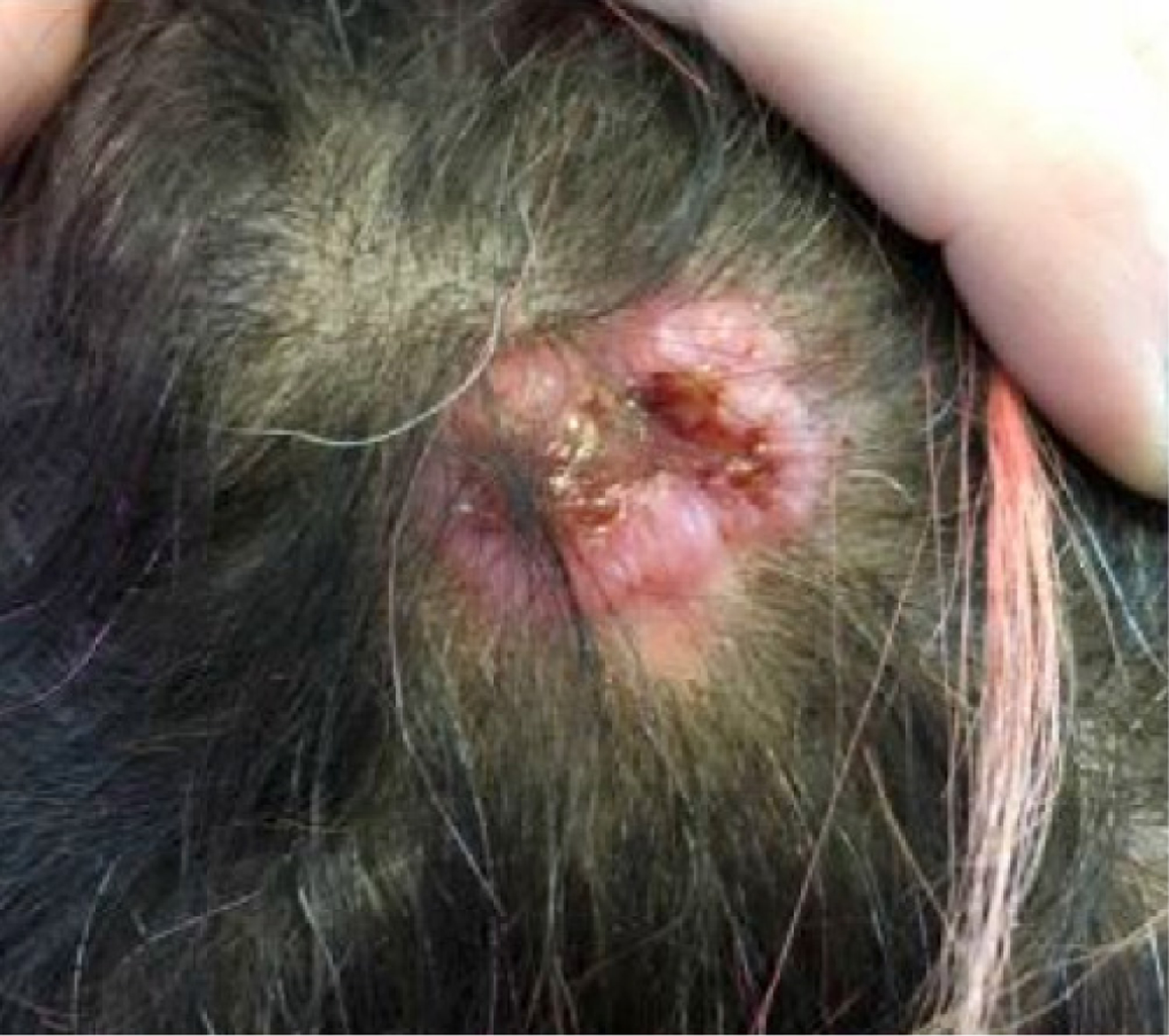
Initial presentation of the scalp lesion.
Figure 2.

MRI scan of headshowing the location and depth of scalp mets.
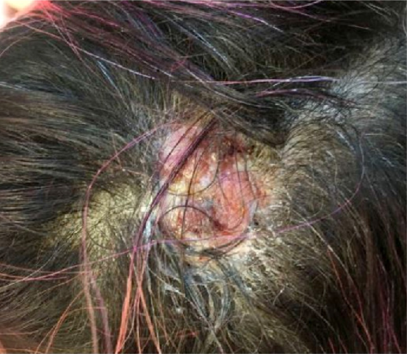
Due to progression of the scalp lesion, topical 5-FU twice daily was started. Her scalp lesion started showing signs of healing with scab formation starting at week 4 with decreased in size and dramatically improved in 8 weeks (Fig. 3).
Figure 3.
MRI scan of headshowing the location and depth of scalp mets.
Five months after initiation of topical 5-FU the patient reported increasing frequency of local symptoms including a sharp stabbing pain, increased headache and tender to touch with deep aching. She also developed a palpable small nodule in the right sub-occipital region with concern for a new metastatic lesion. MRI head revealed a soft tissue lesion in the right parietal scalp, measuring 2.4 cm (previously 1.6 cm) with a new 1 cm enhancing soft tissue lesion in the right sub-occipital scalp.
Despite the numerous systemic treatments including FOLFIRI with bevacizumab, Regorafenib, MIRI (mitomycin-C, irinotecan), FOLFOXIRI (folinic acid, 5-fluorouracil, oxaliplatin and irinotecan) and bevacizumab and topical 5-FU her scalp lesion progressed. Palliative radiation therapy was started. Following radiation therapy with concurrent 5-FU her scalp lesions remained stable and her symptoms improved.
Her overall systemic disease continued to progress while on therapy and after significant decline in functional status, the patient was transitioned to comfort measures.
Discussion
Colon cancer rarely metastasizes to the skin and occurs in less than 6% of patients.[1,2] Cutaneous metastases occur concurrently with widespread metastatic disease to other sites such as liver, peritoneum and lung.[3,4] Most frequent sites of cutaneous metastasis are the abdomen, inguinal or perineal regions, and prior surgical sites, and occur with less frequency on the face, neck and scalp.[5,6] Adenocarcinoma has the highest rate of cutaneous metastasis compared to other histologic subtypes. The skin metastasis can appear as sessile, pedunculated, single or multiple nodules, as a mass with ulceration, or as a cyst.
The mechanism is thought to be secondary to lymphatic or hematogenous spread, local extension of the tumor, and surgical implantation during resection of the primary lesion.
Cutaneous metastases have been associated with a poor prognosis, with overall survival approximately 12–18 months.[2–10] Aravind et al.[10] in their article have divided cutaneous metastasis cases in to two groups:
Group 1: One who primarily present with cutaneous manifestations with primary not identified
Group 2:This group is the one which has been treated with resection of the primary tumor and is being followed up by medical oncology.
The investigators pointed out that the former group is usually associated with more poor outcomes and other visceral involvement.
No clear guidelines exist for treatment of cutaneous metastasis with primary go to treatment being wide excision of the lesion. Systemic chemotherapies and local radiation have also been used but no topical treatment options have been ever reported. Radiation is mostly used in palliation for painful or bleeding lesions but with modest results.[3–9] Table 1 summarizes all case reports published on PubMed with English abstracts since 2013 and treatment options utilized. Overall, the goal of treatment is symptomatic relief. No topical chemotherapy has been previously reported. Our patient was prescribed topical 5-FU (5%) once daily and initially her lesion responded as shown above in the picture, but eventually her overall disease progressed though being on systemic chemotherapy and eventually passed away after a total treatment time of 18 months.
Table 1.
Summary of previously published Case Reports with cutaneous metastases associated with CRC
| Author et. al | Age | Gender | Site of cancer treatment | Phase of Mets | Site of skin systemic Mets | Presence of | Treatment | Outcome (survival after cut Mets diagnosed) |
|---|---|---|---|---|---|---|---|---|
| Ha JY et. al; 2016[6] | 78 | Male | Ascending Colon | Follow up | Right Parietal Scalp | Lung | Systemic Capecitabine | Poor |
| Góes HF et. al; 2016[11] | 76 | Female | Descending Colon | Initial | Right Parietal Scalp | None | Surgical Excision and FOLFOX | Good |
| Udkoff J et. al; 2016[12] | 56 | Male | Colon | Follow up | Scrotum | None | FOLFIRI | Fair |
| Dehal A et. al; 2016[13] | 47 | Male | Rectal | Follow up | Genital area and perineum | Local lymph nodes and vessels | Local Radiation | Good |
| Reusser NM et. al; 2015[14] | 58 | Male | Rectal | Follow up | Right Flank | NA | Palliative | Poor |
| Abt NB et. al; 2015[15] | 67 | Male | Sigmoid Colon | Initial | Pelvis and Scrotum | Local Lymph nodes and skin | Patient declined treatment | Poor |
| Sheets N et. al; 2014[16] | 78 | Male | Ileocecal Valve | Initial | Left Scapula | None | Surgical Excision | Good |
| de Miguel Valencia MJ et. al; 2013[17] |
55 | Male | Rectal | Follow up | Multiple subcutaneous lesions on face, axilla, chest, flank and lower extremities | Lung and Liver | None | Poor |
| Nesseris 1 et. al; 2013[4] | 80 | Male | Ascending Colon | Follow up | Abdominal Surgical Scar | None | Surgical Excision | Good |
| Hashimi Y et. al; 2013[3] | 70 | Male | Rectal | Follow up | Right Cheek | Lung | Surgical Excision | Fair |
| Aravind Bet.al; 2013[10] | 61 | Female | Rectal | Follow up | Scalp (recurrent) | Lung | Surgical resection | Good |
| Balta AZ et.al; 2013[18] | 84 | Female | Rectal | Initial | Left Occiput | None | Chemotherapy and Radiation | Poor |
| Relies Det.al; 2012[19] | 55 | Male | Sigmoid | Follow up | Right upper lip | Liver | Excision | Poor |
| Balta 1 et.al; 2012[20] | 46 | Male | Rectal | Follow up | Anogenital region | Local | Not resectable. Patient denied chemo | Unknown |
| Nguyen VX et. al; 2012[21] | 65 | Male | Caecum | Initial | Right Flank | None | Systemic Capecitabine and cyclophosphamide, progressed then on FOLFOX | Poor |
We searched the medical literature with diligence and finally found two more cases who received topical 5-FU for skin metastases in patients with breast cancer.[22] Among a case series, two patients used topical 5-FU. When used alone, 5-FU reduced bleeding and drainage of lesions and when combined with cryotherapy and systemic therapy, rapidly decreased tumor burden. The authors concluded that combined treatment with cryotherapy and topical 5FU is superior to cryotherapy alone, suggesting that 5-FU induces an antitumor activity independent of cryotherapy. Similar to imiquimod, the authors also suggested that the dramatic response in both patients is in part owing to a favorable immune milieu induced by 5-FU that synergizes with systemic therapies.
Our case showed that topical 5-FU can result in partial regression or local control when used as monotherapy or in combination with other treatment modalities like in our patient along with radiation therapy. Side effects include irritation to the applied area and burning sensation. This treatment modality is feasible with a favorable side effect profile; however, the role of topical 5-FU needs to be further investigated, including tests to characterize the antitumor responses elicited by 5-FU.
Conclusions
Cutaneous metastasis is of rare occurrence. In literature they occur either as primary presentation or develop while he patient has been treated and is being followed up appropriately. No specific guidelines exist for treatment options of cutaneous metastasis with wide surgical excision being the most reported treatment. Topical 5-FU can be used as a potential treatment but clinical trials need to be conducted.
Footnotes
Informed consent: Written informed consent was obtained from the patient for the publication of the case report and the accompanying images.
Peer-review: Externally peer-reviewed.
Conflict of Interest: None declared.
References
- 1.Qiu M, Hu J, Yang D, et al. Pattern of distant metastases in colorectal cancer: a SEER based study. Oncotarget 2015;17:38658–66. [DOI] [PMC free article] [PubMed] [Google Scholar]
- 2.Amal A, Krishnaprasad K, Praveen GP, Shankar V, Keerthi N N: A rare case of carcinoma rectum with scalp metastasis: an unusual presentation. Int Surg J 2018;5:1574–6. [Google Scholar]
- 3.Hashimi Y, Dholakia S. Facial cutaneous metastasis of colorectal adenocarcinoma. BMJ Case Rep 2013;1–3. [DOI] [PMC free article] [PubMed] [Google Scholar]
- 4.Nesseris I, Tsamakis C, Gregoriou S: Cutaneous metastasis of colon adenocarcinoma: case report and review of the literature. An Bras Dermatol 2013;88:56–8. [DOI] [PMC free article] [PubMed] [Google Scholar]
- 5.Wang DY, Ye F, Lin JJ, Xu X. Cutaneous metastasis: a rare phenomenon of colorectal cancer. Ann Surg Treat Res 2017;93:277–280. [DOI] [PMC free article] [PubMed] [Google Scholar]
- 6.Ha JY, Oh EH, Jung MK, Park SE, Kim JT, Hwang IG. Choroidal and skin metastases from colorectal cancer. World J Gastroenterol 2016;22:9650–3. [DOI] [PMC free article] [PubMed] [Google Scholar]
- 7.Horiuchi A, Nozawa K, Akahane T, Shimada R, Shibuya H, Aoyagi Y, et al. Skin metastasis from sigmoid colon cancer. Int Surg 2011;96:135–8. [DOI] [PubMed] [Google Scholar]
- 8.Shah SR, Applebaum DS, Potenziani S, Huttenbach YT, Wolf J, Orengo IF. Cutaneous metastasis to the scalp as the primary presentation of colorectal adenocarcinoma. Dermatol Online J 2017;23:13030/qt31p698nz. [PubMed] [Google Scholar]
- 9.Reusser NM, Wu W, Mir M. Zosteriform metastasis of rectal adenocarcinoma: a case report. Dermatol Online J 2015;21:pii: 13030/qt0d5455rt. [PubMed] [Google Scholar]
- 10.Aravind B, Kumar R, Basnyat P. Cutaneous metastases secondary to colorectal carcinoma may not be as ominous as previously thought: a case report and review of the literature. BMJ Case Rep 2013;pii: bcr2013008556. [DOI] [PMC free article] [PubMed] [Google Scholar]
- 11.Góes HF, Lima Cdos S, Souza MB, Estrella RR, Faria MA, Rochael MC. Single cutaneous metastasis of colon adenocarcinoma -Case report. An Bras Dermatol 2016;91:517–9. [DOI] [PMC free article] [PubMed] [Google Scholar]
- 12.Udkoff J, Cohen PR. Adenocarcinoma of the colon presenting with scrotal metastasis: case report and review of the literature. Dermatology online journal 2016;22. [PubMed] [Google Scholar]
- 13.Dehal A, Patel S, Kim S, et al. Cutaneous Metastasis of Rectal Cancer: A Case Report and Literature Review. The Permanente journal 2016;20:74–8. [DOI] [PMC free article] [PubMed] [Google Scholar]
- 14.Reusser NM, Wu W, Mir M. Zosteriform metastasis of rectal adenocarcinoma: a case report. Dermatology online journal 2015;21. [PubMed] [Google Scholar]
- 15.Abt NB, Sampah MES, Boonyasai RT, et al. Primary colonic adenocarcinoma diagnosed with cutaneous shave biopsy. BMJ case reports 2015;2015:bcr2015210046. [DOI] [PMC free article] [PubMed] [Google Scholar]
- 16.Sheets N, Powers J, Richmond B. Cutaneous metastasis of colon cancer: case report and literature review. The West Virginia medical journal 2014;110:22–4. [PubMed] [Google Scholar]
- 17.de Miguel Valencia MJ, Fraile Gonzalez M, Yague Hernando A, et al. [Cutaneous metastases of rectal cancer]. Anales del sistema sanitario de Navarra 2013;36:557–61. [DOI] [PubMed] [Google Scholar]
- 18.Balta AZ, Sucullu I, Ozdemir Y, Dandin O. A rare clinical manifestation of rectal adenocarcinoma and synchronous scalp metastasis: A case report. Ulusal cerrahi dergisi 2013;29:197–9. [DOI] [PMC free article] [PubMed] [Google Scholar]
- 19.Relles D, Fong Z, Burkhart R, Maxwell PJ. Facial cutaneous metastasis of colon adenocarcinoma. The American surgeon. 2012;78:E454–6. [PubMed] [Google Scholar]
- 20.Balta I, Vahaboglu G, Karabulut AA, Yetisir F, Astarci M, Gungor E, et al. Cutaneous metastases of rectal mucinous adenocarcinoma mimicking granuloma inguinale. Internal medicine (Tokyo, Japan) 2012;51:2479–81. 10.2169/internalmedicine.51.7802 [DOI] [PubMed] [Google Scholar]
- 21.Nguyen VX, Nguyen BD, Ram PC. Occult colon cancer with initial cutaneous metastatic manifestation: PET/CT detection. Clinical nuclear medicine 2012;37:506–8. 10.1097/RLU.0b013e318238f4dc [DOI] [PubMed] [Google Scholar]
- 22.Krishnasamy SR, Almazan TH, Suero-Abreu GA, Jung JY. Successful treatment of cutaneous metastatic breast cancer with topical treatments that potentially synergize with systemic therapy: A case series. JAAD Case Reports 2018;4:711–5. [DOI] [PMC free article] [PubMed] [Google Scholar]