

Abstract
Atopic dermatitis is a heterogeneous disease and resists classification. In this review, we discuss atopic dermatitis nomenclature and identify morphologic phenotypes, which will facilitate correct diagnoses and development of treatment strategies. We support using the term ‘atopic dermatitis’ rather than eczema, because it describes the allergic background and inflammation (‘itis’) as drivers of the disease. Atopic dermatitis has many morphologic manifestations that vary by topographic area affected, age, or race and require consideration in differential diagnosis. Different phenotypes based on morphology and topographic location, ethnicity, and age are discussed. A better-defined phenotype identification for atopic dermatitis will facilitate earlier and correct diagnosis of this complex condition and inform selection of the most appropriate treatment choice in an era in which targeted therapies may generate more individualized patient care.
Keywords: atopic, dermatitis, eczema, nomenclature, phenotype, treatment
Introduction
Atopic dermatitis (AD) is a chronic inflammatory skin condition affecting both children (15–20%) and adults (2–5%).1–3 Its prevalence is increasing, particularly in lower-income countries, and it occurs in a variety of geographic and ethnic patterns.3,4 Previously classified as either early-onset [i.e. infantile (aged <2 years), childhood (aged 2–12 years), adolescent (aged 12–18 years)], or adult-onset (aged >18 years), a separate subgroup of elderly-onset AD (aged ⩾60 years) has recently been described.5–7 AD can therefore be regarded as a lifelong disease, and clinical and laboratory differences observed in these subtypes contribute to the heterogenous nature of the disease.8
Traditionally, clinical lesions in eczema are classified as ‘acute’ (oozing, oedema, and erythema) or ‘chronic’ (dyspigmentation, xerosis, and lichenification) (Figure 1). However, as AD is a chronic relapsing condition, both types of lesions can coexist during flares. Pruritus is a hallmark of AD, and excoriations secondary to scratching are often present. Diagnosis of AD is largely based on the morphology and distribution of lesions,2,3,9 but also on the patient’s clinical course, comorbid conditions, and family history. Our understanding of the pathogenesis of AD in part reflects the complex interplay between genetic and environmental factors.9
Figure 1.

Clinical signs of atopic dermatitis.
Many genes have been linked to AD across different populations, but the extent of genetic risk for AD is yet to be fully defined. However, candidate gene studies, genome-wide association studies, and genetic sequencing studies have shown that genetic susceptibility is associated with epidermal barrier dysfunction and type 2 dominated immune responses.9–13 Indeed, type 2 inflammation plays a key role in both acute and chronic phases of the disease.14–17 However, an incomplete understanding of the underlying immunologic mechanisms of AD and its heterogeneous manifestations has resulted in a therapeutic approach based more on disease severity than on phenotype or its underlying specific biologic pathways (endotype).
Objectives
First, we discuss the nomenclature associated with AD. Second, we identify and illustrate different morphologic phenotypes seen in patients of different age ranges or races and in different anatomic locations. To guide appropriate treatment decisions, it is critical that the different AD phenotypes are diagnosed efficiently and correctly and in a timely manner.
AD nomenclature
Atopy, which is Greek for ‘out of place’, entered the medical lexicon in 1923, with ‘atopic dermatitis’ following a decade later, soon after the term ‘atopic eczema’ had first been used.18–21 The American Academy of Asthma and Immunology defines atopy as ‘the genetic tendency to develop allergic diseases such as allergic rhinitis, asthma, and AD’ (or ‘eczema’, depending on the terminology used).22 Most individuals with AD are sensitized to allergens and have a coexisting allergic disease, and allergen exposure can have an effect on AD symptoms, severity, and flares.
The term ‘dermatitis’ was excluded from early discussions of hereditary and clinical presentation of allergic sensitization because it was considered difficult to define.19,23 An electronic survey of experts from the International Eczema Council led to a consensus (97.2%) on the need to include the prefix ‘atopic’ in all descriptions of the disease, regardless of a preference for ‘eczema’ or ‘dermatitis’.18 We agree that use of the term ‘atopic’ provides important mechanistic information and helps group AD with other diseases with the same aetiology.
Differences in nomenclature create potential for confusion and for issues with the quality of epidemiologic data24 given that AD and eczema are represented by different codes in the International Classification of Diseases, Tenth Revision (ICD-10), system. Besides the ICD-10 codes for AD (L20.x; Table 1), other ICD-10 codes can be used in diagnosis.
Table 1.
ICD-10 codes that can be used for AD and its different morphologic phenotypes.
| L20 Atopic dermatitis | L29 Pruritus |
| L20.0 Besnier’s prurigo | L29.0 Pruritus ani |
| L20.8 Other atopic dermatitis | L29.1 Pruritus scroti |
| L20.81 Atopic neurodermatitis | L29.2 Pruritus vulvae |
| L20.82 Flexural eczema | L29.3 Anogenital pruritus, unspecified |
| L20.83 Infantile (acute) (chronic) eczema | L29.8 Other pruritus |
| L20.84 Intrinsic (allergic) eczema | L29.9 Pruritus, unspecified |
| L20.89 Other atopic dermatitis | L30 Other and unspecified dermatitis |
| L20.9 Atopic dermatitis, unspecified | L30.0 Nummular dermatitis |
| L26 Exfoliative dermatitis | L30.1 Dyshidrosis [pompholyx] |
| L28 Lichen simplex chronicus and prurigo | L30.2 Cutaneous autosensitization |
| L28.0 Lichen simplex chronicus | L30.8 Other specified dermatitis |
| L28.1 Prurigo nodularis | L30.9 Dermatitis, unspecified |
| L28.2 Other prurigo | L53 Other erythematous conditions |
| L53.8 Other specified erythematous conditions | |
| L53.9 Erythematous condition, unspecified |
AD, atopic dermatitis; ICD, International Classification of Diseases, Tenth Edition.
Thus, the heterogeneity of AD presentation may be a source of the varied terminology used to describe AD. Consensus within the medical community is necessary to avoid confusion, bias, and errors in epidemiologic data. We argue for the use of ‘atopic dermatitis’ over ‘atopic eczema’ because it more fully captures the inflammatory aetiology of the disease, an important feature when considering use of new targeted therapies. Education of the lay community will be a key next step to ensuring use of consistent terminology.
Diagnosis of AD
The diagnostic criteria used for AD have been thoroughly reviewed by Andersen et al.19 Diagnostic guidelines from the UK Working Party25 and the American Academy of Dermatology (AAD)26,27 are established for paediatric and adult diagnoses. These guidelines are refinements of the Hanifin–Rajka criteria, first introduced in 1980 and comprising four major and 23 minor clinical features, of which at least three major and three minor criteria must be present to confirm an AD diagnosis.28 The UK Working Party Criteria mandate that pruritus must be present along with at least three of five major criteria: history of flexural involvement, history of dry skin, onset of AD before 2 years of age, history of any other atopic condition, and flexural dermatitis.25 The AAD guidelines comprise essential, important, and associated clinical features. For a positive diagnosis of AD, essential features (pruritus and eczema) must be present, and important features (early AD onset, history of atopy and xerosis) are usually present, lending further support to the diagnosis; evidence of associated features (e.g. facial pallor, lichenification), while suggestive of AD, are not a mandatory requirement.27 The Millennium Criteria were the first to require the presence of antigen-specific immunoglobulin E (IgE) as a mandatory criterion for the diagnosis of AD.29 The presence of IgE antibodies is also required for some definitions of atopy, such as that of the World Allergy Organization.18 However, this criterion is controversial because it excludes as many as two-thirds of patients with AD who do not display this type of response30 and is generally not used as a diagnostic criterion in practice.
Several challenging clinical phenotypes of AD exist that display peculiar morphologic features or topographic distributions, and some of these phenotypes have difficult-to-discern aetiologies or may mimic or coexist with other dermatologic conditions. These challenging phenotypes can be defined morphologically [nummular dermatitis, prurigo nodularis (PN)-like lesions, erythroderma, lichenified dermatitis, follicular/papular dermatitis and pompholyx (dyshidrosis)]4,31–43 (Figure 2) and topographically (flexural or periorificial or occurring on the face, lips [eczematous cheilitis], eyelid, head and neck, hand and foot, or nipple)34,36,38,39,41–46 (Figure 3). Figures 4 and 5 show histologic samples of AD.
Figure 2.
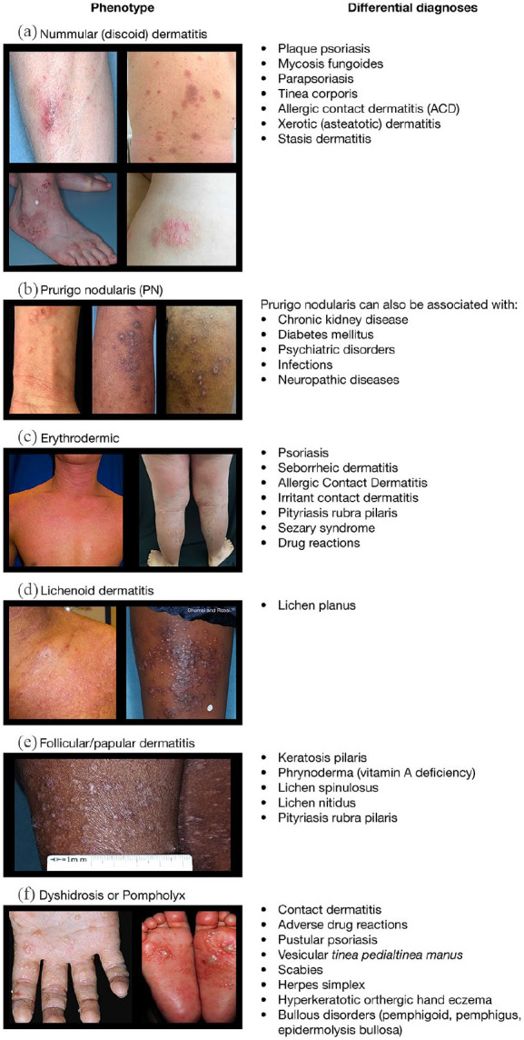
Morphologic phenotypes. (a) Nummular (discoid) dermatitis. (b) Prurigo nodularis. (c) Erythrodermic. (d) Lichenoid dermatitis. (e) Follicular/papular dermatitis. (f) Dyshidrosis or Pompholyx.
Figure 3.

Topographic phenotypes of AD. (a) Typical flexural dermatitis. (b) Face. (c) Eczematous cheilitis. (d) Eyelid dermatitis. (e) Head and neck dermatitis. (f) Hand dermatitis. (g) Nipple dermatitis.
†Inadequate treatment of eyelid dermatitis can lead to severe chronic inflammation with scarring or ‡ocular surface diseases such as atopic keratoconjunctivitis (picture shows sclerosis and symblepharon of conjunctiva).
Figure 4.

Atopic dermatitis: histology. (a) Normal skin. (b) Atopic dermatitis. (c) Dyshidrosis. (d) Acute dermatitis. (e) Chronic dermatitis.
Figure 5.

Histology: subacute chronic dermatitis, psoriasiform dermatitis. (a) Sub-acute-chronic dermatitis. (b) Psoriasiform dermatitis.
Histological images generously provided by Dr Maria Teresa Fernández-Figueras.
No consensus exists on whether some of these challenging phenotypes represent exclusive forms of AD, and this lack of consensus is further complicated by the availability of separate ICD-10 codes for these manifestations (Table 1). There is a need to confirm which of, and when, these conditions should be considered forms of AD and which are independent diagnoses. We propose that nummular dermatitis, PN-like AD, erythroderma, pompholyx, lichenified dermatitis, flexural dermatitis, facial dermatitis, eczematous cheilitis, eyelid dermatitis, head and neck dermatitis (HND), hand and foot dermatitis, and nipple dermatitis should be considered part of the spectrum of AD when other AD features (e.g. typical flexural eczematous lesions), elevated IgE, or atopic comorbidities (history of asthma, rhinoconjunctivitis, food allergy) are present, especially when there is no evidence of other diseases known to cause these phenotypes.
Morphologic AD phenotypes
Nummular or discoid dermatitis
The term ‘nummular’ derives from the coin-like appearance of lesions that form rapidly by the merging of papules and papulovesicles into circinate and ovoid plaques (Figure 2a).34,40 Lesions are typically 1.5–3 cm in diameter and progress from a highly inflammatory acute phase characterized by marked erythema, crusty appearance, and oozing/weeping to a smoother, scaly phase that may become annular because of central clearing and peripheral extension. Lesions are predominantly present on lower extremities but can also appear on upper extremities and the trunk.47 Nummular dermatitis is the most common atypical morphologic variant of AD,48 but no agreement exists as to whether nummular dermatitis is AD in all cases. In most cases, nummular dermatitis is idiopathic, and no allergic or irritant cause can be discerned. It is more common in children (although not infants) and adult-onset AD,49 and coexists in some individuals with typical flexural AD or elevated serum IgE (or both).50,51 As occurs in AD, staphylococcal superantigen-producing Staphylococcus aureus colonization is commonly present in nummular dermatitis.52 Nummular dermatitis may be considered AD when other features of AD (e.g. typical flexural eczematous lesions), elevated IgE, and atopic comorbidities (history of asthma, rhinoconjunctivitis, food allergy) are currently or have been present and when no evidence exists for other diseases (e.g., stasis dermatitis) that are also known to cause nummular dermatitis.31,32
Prurigo nodularis
PN (Figure 2b) is a condition distinct from AD, but PN secondary to AD can occur. PN is characterized by single to multiple excoriated hyperkeratotic and intensely itchy papules and nodules that occur predominantly on the extremities.33,53 Pruriginous lesions are persistent and tend to be symmetrically distributed in areas accessible to scratching, with normal or lichenified skin between the lesions, and a characteristic ‘butterfly’ sign on the back where no lesions are present in areas inaccessible to scratching. PN is commonly located on the extensor surfaces of the extremities and rarely affects the face.54 Pruritus may be accompanied by burning, stinging, pain, and other symptoms. There is often neuronal sensitization, demonstrated by allokinesis (light touch-evoked itch) and hyperkinesis (exaggerated itch response to a pruritic stimulus).55 The key immune mediators and mechanisms behind atopic itch in AD have been reviewed and include histamine, TSLP and type-2 cytokines.56 The key role of type 2 cytokines in PN is emphasized by the very good therapeutic response to dupilumab.57
AD has been identified as an underlying or contributing cause in nearly one-half of PN cases.58,59 PN secondary to AD is more common in adults and in individuals of South-East Asian or African origin.4,59,60 In an AD registry study performed in Japan, the prevalence of prurigo nodules in 300 patients with AD was high: 30.9% in patients with moderate AD and 56.3% in patients with severe AD.61 Itch is a cardinal symptom in AD, and the itch–scratch cycle could lead to secondary PN lesions. Accordingly, PN can coexist with AD or persist after cessation of AD.33
Erythroderma
Erythroderma (Figure 2c), also known as exfoliative dermatitis, is the presence of erythema on >90% of the body surface area. Erythroderma typically begins with the appearance of erythemato-pruritic lesions of varied primary morphology, most often on the head, trunk, and genital region, and rapidly spreads to all or most of the body within days or a few weeks. The palms of the hands and soles of the feet tend to be spared, along with the nose (nose sign) in some cases.62,63 Scaling of the skin follows, with large scales in acute cases and small scales in chronic cases.62 Erythrodermic AD is more common in adolescents and adults (aged 12–60 years) in East Asia, particularly those with a longer disease course.4,64,65 Erythroderma is not specific to AD and a differential diagnosis must consider numerous causes, but AD has been reported to be the underlying cause of erythroderma in 5%–24% of cases.66 Erythrodermic AD is a serious condition because it is associated with a high rate of hospitalization, skin infections, and potential life-threatening complications.67
Lichenified dermatitis
Lichenified dermatitis (Figure 2d) refers to a thickening of the skin, which appears elevated, with accentuated creases and a leathery appearance due to prolonged scratching and rubbing. In an analysis of AD clinical trial data, lichenification was found to be more common in patients from South-East Asia or Africa than in Caucasian patients.4 The condition is most common in young people (median age 7 years).35 Along with pronounced greater lichenification, individuals from Africa or of African descent can present with perifollicular accentuation and post-inflammatory depigmentation, which can complicate the diagnosis.60 People from Asia or of Asian descent with AD exhibit increased epidermal hyperplasia (increased epidermal thickness and Ki67 counts) on lesional biopsy specimens, which is associated with increased gene expression of T helper (Th)17- and Th22-related markers.60,65
Follicular/papular dermatitis
Follicular and papular dermatitis is a morphological subtype more commonly identified in darker skin phototypes (Figure 2e). An analysis of AD feature prevalence reported among clinical studies by region found that studies from Africa reported a higher prevalence of papular lichenoid lesions (22%) compared with the overall prevalence from all studies (22%).4
Topographical phenotypes
AD typically affects the face, neck, and flexural zones.27 Many of the morphologic variants show predisposition for certain areas of the body. Some topographical phenotypes of AD are described as follows.
Typical flexural dermatitis
A tendency for AD to affect flexural areas was first noted in Roman Emperor Augustus by Suetonius,68–70 and remains a prominent feature of both US and UK diagnostic guidelines.12,25,27,64,71 The signs most frequently seen with flexural involvement are erythema, oedema or papulation, excoriation, lichenification, and oozing or crusting (Figure 1). Flexural areas typically affected by AD include the neck, cubital and popliteal fossae, wrists, and ankles (Figure 3a).
The extent of flexural involvement appears to vary with disease course and differs by racial origin. Flexural involvement is more common in patients with early-onset AD and a chronic persisting course.72 There may be sparing of flexural involvement in patients aged >60 years.5 Whereas flexural involvement is common in adolescent and adult Caucasians, extensor involvement appears to be more common in infants and Asians and can occur in those of African descent35,43,73,74 (Figure 3a, right two panels).
Face
Facial involvement (Figure 3b) is common in AD, not only in children and infants,27 but also in adolescents and adults. In many individuals, AD may involve the face alone; in infants aged <1 year, AD is typically restricted to the face, where it is characterized by erythematous and highly pruritic lesions alongside moist, oozing papulovesicles that sometime form crusts or scales45,73 and can be concentrated in the perioral region,5,75 on the cheeks, or, in some cases, the scalp.73 Periorificial lesions of the head tend to occur in AD during childhood.5
Eczematous cheilitis
Eczematous cheilitis (Figure 3c) can be a manifestation of AD and presents as red, dry lips, and sometimes with a median fissure of the lower lip, alongside lateral fissures (angular cheilitis).76 In nearly one-fifth of cases, cheilitis is a manifestation of AD.77 Chronic cheilitis can be the result of exposure to irritants or allergic contact dermatitis to sensitizers such as nickel, fragrances, Balsam of Peru, chromium salts and manganese salts present in cosmetics, dental materials, and oral hygiene products.77 In contrast, angular cheilitis (Figure 3c) is usually infectious in nature.
Eyelid dermatitis
Substantial eyelid involvement was highlighted as a key feature in the first description of AD, but eyelid involvement is common in several forms of dermatitis (Figure 3d).19,73 Itching, burning, and crusting of the eyelids is common, and scaling may occur at the base of the eyelashes, along with depigmentation, lichenification, and loss of lashes.39,78 AD is associated with ocular diseases, including conjunctivitis and cataracts.79,80 Inadequate treatment of eyelid dermatitis, blepharitis, or periocular eczema can lead to severe chronic inflammation with scarring and subsequent complications like ectropion or entropion as well as to ocular surface diseases such as keratoconjunctivitis (Figure 3d).
Similar to eczematous cheilitis, eyelid dermatitis is a common manifestation of AD.45,64 Eyelid dermatitis is more common in childhood and may also be more common in people of Asian descent, particularly females.4,81 Onset after 6 months of age and a personal history of atopy are the main risk factors for eyelid dermatitis in AD.82 Of note, long-term use of potent topical steroids to treat eyelid dermatitis can cause eyelid skin atrophy due to the thin skin of the eyelid, as well as serious adverse events such as glaucoma and cataracts, which are also associated with use of systemic corticosteroids.83–85
Head and neck dermatitis
In adolescents and adults, facial AD, often seen as xerotic, scaly, erythematous, and sometimes lichenified plaques affecting the face and neck and eventually the scalp, is one of the causative factors of HND (Figure 3e).
There are many other potential causes for HND, such as aeroallergen sensitization, allergic contact dermatitis to topical products, rosacea, seborrheic dermatitis, topical corticosteroid (TCS) withdrawal syndrome, and sensitization to Malassezia furfur,37,38,76,81,86 and these may coexist with AD in the same person.
Allergic contact dermatitis (ACD) commonly involves the lips, eyelids, and neck, among other regions, and may be mistaken for or complicate AD.87 The delayed hypersensitivity reaction observed with ACD following prior sensitization to an allergen results in highly pruritic erythematous plaques. AD treatments may be implicated in ACD, and allergens with higher rates of sensitization among patients with AD include formaldehyde releasers, metal allergens, cocamidopropyl betaine, and fragrances. ACD should also be investigated in paediatric patients; although rates of sensitization are comparable with the general population, ACD is a highly relevant diagnosis in children, and paediatric skin with an immature barrier function is more susceptible to toxicity.88 In a UK retrospective case study of 114 children aged 3–15 years who were patch-tested over a 3-year period, findings on patch test were positive in 33% of patients with facial dermatitis, 40% with perioral dermatitis, and 86% with eyelid dermatitis.89 Paediatric patients with moderate-to-severe and early-onset AD have a higher incidence of sensitization to topical products, most commonly to emollients and antiseptics.90 Sunscreens can also be a cause of ACD on the face and neck, and both sensitization and photosensitization should be investigated when suspected.91,92
HND can be induced by TCS withdrawal. In particular, overuse of potent TCS on the face can lead to many adverse events upon withdrawal, including burning, itching, and facial redness.93,94 A retrospective analysis of 55 people in Australia suspected to be undergoing TCS withdrawal found that 100% of patients experienced redness and itch. Of these patients, TCS were initially prescribed for AD (76%), contact dermatitis (15%), or other rashes (9%).93
In addition, recent reports have suggested an association between treatment of AD with the biologic dupilumab and development of new regional dermatoses, with a predilection for the face, head, and neck regions.95–97 This rare phenomenon has yet to be fully clinically characterized and a unifying phenotype has not been identified. These reports may represent disparate rashes with multiple underlying aetiologies. It is possible that dupilumab treatment potentially unmasks previously undiagnosed ACD or seborrheic like-dermatitis, which had been kept at bay prior to dupilumab initiation. A recent case report suggests that topical ketoconazole is successful in treating facial rashes refractory to dupilumab.98
Elevated serum levels of Malassezia-specific IgE are characteristic of HND in contrast to seborrheic dermatitis or pityriasis versicolor.99–101 In 2000, Devos and Van der Valk investigated whether positive prick tests to P. ovale were associated with a specific localization in the head and neck region in a cohort of 589 patients. They found that serum levels of Malassezia-specific IgE were elevated in all patients with AD and HND but in only 13.6% of patients with AD without HND.100
A positive response to oral itraconazole may support the clinical diagnosis of HND. Randomized, placebo-controlled trials have shown significant clinical improvement of suspected HND in patients with AD treated with systemic antifungals.102–104
Hand and foot dermatitis
AD of the hand (Figure 3f) presents usually as xerotic, scaly, lichenified, and fissured skin, mainly on the dorsal hands.105 This phenotype predominates in adulthood, although specific manifestations do occur in children (juvenile palmoplantar dermatitis or dermatitis plantaris sicca).4,73 In adults, the lifetime prevalence of hand dermatitis is 15%,106 and it is twice as common in females as in males.106,107 In a study of Swedish children, approximately 70% of those with hand dermatitis at age 16 years had a history of AD, and the risk of hand dermatitis was greater in children with persistent or more severe AD.108 In a French study that evaluated the benefit of therapeutic patient education in 72 adults with chronic hand eczema, 43.7% had a personal history of AD.109 A retrospective review and clinical examination of 725 German adolescent and adults with AD found that two-thirds of patients whose dermatitis developed after infancy and then persisted into adulthood had hand dermatitis.75 A systematic review and meta-analysis of 35 studies in 12 countries showed that patients with AD have a 3- to 4-fold increased incidence of hand dermatitis when compared with controls,110 and that hand dermatitis in schoolchildren with AD frequently affects only the dorsa.111
Hand dermatitis is often associated with occupational or domestic exposure to irritants and can be aggravated by daily tasks that necessitate contact with water, such as dishwashing, laundry, and childcare. However, a history of childhood AD appears to be the main risk factor for development of hand dermatitis.105 Approximately 29% of people with hand dermatitis have a history of AD.112 A Norwegian study found that 90% of school children with hand eczema also reported AD, which suggests that hand eczema in children is closely associated with AD.111 In most cases, distinguishing between contact dermatitis and AD of the hand caused by endogenous factors is impossible by clinical presentation alone, and patch testing is warranted to determine whether any concomitant contact dermatitis is present. Nevertheless, the presence of lesions at the wrist is indicative of an underlying endogenous cause.105 A 7-year follow-up study in patients with hand dermatitis found that 45% of patients also had concomitant AD lesions on other parts of their body during the follow-up period.112
Unlike hand dermatitis, foot dermatitis is predominantly caused by allergens in footwear.113 Foot dermatitis may also be associated with AD.113 In most cases, the condition is found concomitantly with hand dermatitis.114 A dorsal pattern similar to that found in hand dermatitis suggests an allergic aetiology.115 The prevalence of foot dermatitis in patients with AD is approximately 30%.116
Juvenile palmoplantar dermatitis is a distinct subtype of AD. As the name suggests, it affects the palms and plantar surfaces of the hands and feet, rather than the dorsal surfaces. In affected children, symptoms are most common in winter and tend to resolve between the ages of 12 and 16 years.64,117
Dyshidrosis, also called dyshidrotic eczema or pompholyx, may be a clinical phenotype of AD that manifests as vesicles and blisters on the palms and soles; it accounts for <10% of all cases of hand and foot dermatitis35 (Figures 2f, 6). This condition is very itchy and disabling. Complications can lead to or include secondary infection.118,119
Figure 6.

Most common phenotypes of hand eczema: dyshidrotic and hyperkeratotic.
(a) Dyshidrotic eczema. The main component of dyshidrotic eczema is the presence of multiple vesicles containing clear fluid; the hands are usually wet. Sometimes due to the thick stratum corneum, vesicles do not come out of the skin surface and are only visible on close examination. (b) Hyperkeratotic eczema. Features include skin thickening (hyperkeratosis), fissures and bleeding, desquamation, and scaling. The hands are usually very dry.
Hyperkeratotic, dry fissured hand eczema, pulpitis, and nummular hand eczema are other clinical manifestations of hand dermatitis (Figure 6).120
Nipple dermatitis
AD-related nipple dermatitis presents symmetrically, typically involving both the nipples and areolas (Figure 3g).64 Nipple dermatitis can be aggravated or triggered by breastfeeding121,122 and is found in 11–23% of individuals with AD, most commonly in postpubertal girls and young adults.4,114 Despite being described as a minor diagnostic feature of the Hanifin–Rajka criteria,30 the specificity of nipple eczema as a symptom of AD remains unclear.27,29,123,124
AD in the elderly: a new phenotype
Phenotyping and stratification of AD type by both age-related clinical picture and age at onset have been reviewed by Bieber and colleagues.5 Elderly-type AD (AD in patients aged ⩾60 years) has been suggested to be a distinct age-related phenotype alongside previously recognized infantile, childhood, and adult phenotypes.5–7,125
As in adult-type AD, lichenified lesions are common, but tend to be localized to the cubital and popliteal extensor areas rather than the flexural areas, as is typical of adult-type AD.5,126,127 Three forms of elderly-type AD are apparent – elderly onset, relapsing, and continuous – and are subdivided by age at onset.126–132 A comparison of studies carried out in various geographic locations indicates that the prevalence of AD in those aged >60 years is relatively stable at 2–5%.127,130,133 Immune system changes due to aging (immunosenescence), such as increased type 2 cytokine production [mainly interleukin (IL)-4 and IL-13], may play a key role in the pathogenesis of elderly-onset AD,127,134 as they do in chronic idiopathic pruritus of the elderly.135 Additional considerations in the elderly include changes in the composition of the cornified envelope with aging that may lead to impaired barrier function,136 and the effects of medications, including calcium channel blocker-induced dermatitis137 and statin-exacerbated xerotic/asteatotic dermatitis.138
Guidelines are lacking on the differential diagnosis of AD versus other skin conditions in the elderly, and diagnosis relies on persistence of symptoms for 6 months after excluding other causes such as comorbidities, treatment-related adverse events, cutaneous T-cell lymphoma, and nonbullous variants of bullous pemphigoid.51,126,127,139
Treatment of atopic dermatitis
Preventive skin hygiene, including barrier-stabilizing topical treatment, hydration, and monitoring and treatment for superinfections are critical in the care of patients with AD. Guidance exists on the treatment of AD in adults and children,140 and typical treatment strategies for AD include treating visible skin lesions with topical anti-inflammatory medications such as corticosteroids and calcineurin inhibitors in combination with frequently applied emollients to minimize the impact of skin barrier dysfunction.140,141 TCS are an effective first line of anti-inflammatory treatment when applied to skin lesions of patients with AD, and they and are associated with minor side effects.99,140 Steroid-sparing topical agents that are licensed for the treatment of AD include the calcineurin inhibitors tacrolimus ointment and pimecrolimus cream and the phosphodiesterase 4 inhibitor crisaborole; these are preferred for sensitive locations and are used commonly for HND,142 with narrowband UVB phototherapy as another approved option. Patients with severe AD can also be treated with traditional systemic immunosuppressive medication, such as cyclosporine A or oral glucocorticoids, with off-label use of azathioprine, methotrexate or mycophenolate mofetil sometimes considered.140 However, use of these systemic immunosuppressants can be limited by their adverse effects and tolerability, particularly for long-term treatment.27,142,143 Novel biologic therapies include the approved therapy dupilumab, which targets the underlying inflammatory mechanism of AD by selectively blocking type 2 inflammation.144–147 Many novel systemic and topical treatments are also under investigation, including the JAK inhibitors baricitinib, upadacitinib, and abrocitinib, the dual JAK-SYK inhibitor ASN002, as well as antagonists of histamine and TSLP, which are each implicated in the pathogenesis of AD.148 The implementation and success of these agents in treating AD relies, however, on further elucidation of the various phenotypes and appropriate diagnosis of the disease.
Conclusions
Lack of consensus on AD terminology may lead to confusion and result in erroneous data and flawed epidemiologic assumptions. Efforts continue to address this issue, and the increasing use of the consensus term ‘atopic dermatitis’ in the literature and in medical records, may be a sign of progress. Wider use of the term ‘atopic dermatitis’ for diagnosis should help generate more accurate epidemiologic data and improve evaluation of disease burden, outcome measures, and reimbursement decisions, as well as development of new treatments.
AD is a heterogeneous condition that continues to frustrate attempts at classification based on age, morphology, and topographic location. This review considers the clinical characteristics of morphologically (nummular dermatitis, PN-like lesions, erythroderma, pompholyx, papular/follicular and lichenified dermatitis) and topographically defined (flexural, facial, including eczematous cheilitis and eyelid dermatitis, head and neck, hand and feet, or nipple) phenotypes of AD, which we consider to be a practical guide to discerning the distinct manifestations of this heterogenous disease. The subtyping of AD by phenotype may be important to identify the more appropriate treatment for patients, and a protocol has been developed for a systematic review of phenotypes in AD.149 Given the diversity of AD phenotypes, this reference will facilitate earlier and correct diagnoses of AD, leading to better treatment decisions and management of the disease.
Acknowledgments
Microphotographs in Figure 5 have been generously provided by Dr. Maria Teresa Fernández-Figueras, Department of Pathology, Hospital Universitari General de Catalunya, Universitat Internacional de Catalunya, Sant Cugat del Vallès (Barcelona), Spain. The authors especially thank the patients who provided written agreement for the use of their photographs in scientific publications, and Noah Levit (Regeneron Pharmaceuticals, Inc.) for critical feedback on the manuscript.
Footnotes
Author contributions: GG conceived the manuscript, MdB, ABR, KK, and AB performed literature review references, ABR analyzed data, ABR, GG, MdB, VA, MD, and LP provided clinical photos, MdB and ABR contributed to writing. All authors reviewed and approved all versions of the manuscript.
Conflict of interest statement: Dr. Giampiero Girolomoni has been principal investigator in sponsored clinical trials and received personal fees from AbbVie, Abiogen, Almirall, Amgen, Biogen, Boehringer Ingelheim, Bristol-Myers Squibb, Celgene, Celltrion, LEO Pharma, Lilly, Menlo Therapeutics, Merck, Merck Sharp & Dohme, Novartis, OM Pharma, Pfizer, Regeneron, Samsung, Sandoz, Sanofi Genzyme and UCB Pharma.
Dr. Marjolein De Bruin-Weller has been principal investigator, advisory board member, and consultant for Regeneron, Sanofi Genzyme; principal investigator, and advisory board member for AbbVie, Pfizer, and Leo Pharma; and advisory board member for Lilly, UCB, Galderma, and Janssen.
Dr. Valeria Aoki has received honoraria for lecturing from Sanofi.
Dr. Kenji Kabashima has received consulting fees, honoraria, grant support, and lecturing fees from Japan Tobacco, LEO Pharma, Maruho, Mitsubishi Tanabe, Ono, Procter & Gamble, Sanofi, Taiho, and Torii.
Dr. Mette Deleuran has received research support, honoraria for lecturing and is on consulting/advisory board agreements with AbbVie, Almirall, Galapagos, LEO Pharma, Lilly, Meda Pharma, Pfizer, Pierre Fabre, Regeneron, and Sanofi Genzyme.
Dr. Luis Puig has received research support, honoraria for lecturing, and is on consulting/advisory board agreements with AbbVie, Almirall, Amgen, Baxalta, Biogen, Boehringer Ingelheim, Celgene, Gebro, Janssen, LEO Pharma, Lilly, Merck Serono, Merck Sharp & Dohme, Pfizer, Mylan, Novartis, Roche, Sandoz, Samsung Bioepis, Sanofi, and UCB. Dr Puig is also an Associate Editor of Therapeutic Advances in Chronic Disease and, therefore, the peer review process was managed by alternative members of the Board and the submitting Editor had no involvement in the decision-making process.
Dr. Ashish Bansal is an employee and shareholder of Regeneron Pharmaceuticals, Inc.
Dr. Ana B. Rossi is an employee and may hold stock and/or stock options in Sanofi Genzyme.
Funding: The authors disclosed receipt of the following financial support for the research, authorship, and/or publication of this article: Research sponsored by Sanofi and Regeneron Pharmaceuticals, Inc. Editorial assistance was provided by Lauren D. Van Wassenhove, PhD, and Carolyn Ellenberger, PhD, of Excerpta Medica, funded by Sanofi Genzyme and Regeneron Pharmaceuticals, Inc., according to the Good Publication Practice guideline.
ORCID iD: Giampiero Girolomoni  https://orcid.org/0000-0001-8548-0493
Contributor Information
Giampiero Girolomoni, Department of Medicine, Section of Dermatology and Venereology, University of Verona, Piazzale A. Stefani 1, Verona, 37126, Italy.
Marjolein de Bruin-Weller, National Expertise Center of Atopic Dermatitis, Department of Dermatology and Allergology, University Medical Center, Utrecht, Netherlands.
Valeria Aoki, Department of Dermatology, University of São Paulo School of Medicine, São Paulo, Brazil.
Kenji Kabashima, Department of Dermatology, Kyoto University Graduate School of Medicine, Kyoto, Japan.
Mette Deleuran, Department of Dermatology, Aarhus University Hospital, Aarhus, Denmark.
Luis Puig, Department of Dermatology, Hospital de la Santa Creu i Sant Pau, Universitat Autònoma de Barcelona, Barcelona, Spain.
Ashish Bansal, Regeneron Pharmaceuticals, Inc., Tarrytown, NY, USA.
Ana B. Rossi, Sanofi Genzyme, Cambridge, MA, USA
References
- 1. Barbarot S, Auziere S, Gadkari A, et al. Epidemiology of atopic dermatitis in adults: results from an international survey. Allergy 2018; 73: 1284–1293. [DOI] [PubMed] [Google Scholar]
- 2. Bieber T. Atopic dermatitis. N Engl J Med 2008; 358: 1483–1494. [DOI] [PubMed] [Google Scholar]
- 3. Nutten S. Atopic dermatitis: global epidemiology and risk factors. Ann Nutr Metab 2015; 66(Suppl. 1): 8–16. [DOI] [PubMed] [Google Scholar]
- 4. Yew YW, Thyssen JP, Silverberg JI. A systematic review and meta-analysis of the regional and age-related differences in atopic dermatitis clinical characteristics. J Am Acad Dermatol 2019; 80: 390–401. [DOI] [PubMed] [Google Scholar]
- 5. Bieber T, D’Erme AM, Akdis CA, et al. Clinical phenotypes and endophenotypes of atopic dermatitis: where are we, and where should we go? J Allergy Clin Immunol 2017; 139: S58–S64. [DOI] [PubMed] [Google Scholar]
- 6. Tanei R. Atopic dermatitis in older adults: a review of treatment options. Drugs Aging 2020; 37: 149–160. [DOI] [PMC free article] [PubMed] [Google Scholar]
- 7. Chello C, Carnicelli G, Sernicola A, et al. Atopic dermatitis in the elderly Caucasian population: diagnostic clinical criteria and review of the literature. Int J Dermatol 2020; 59: 716–721. [DOI] [PubMed] [Google Scholar]
- 8. Ha DL, Park GH, Kim HS, et al. Clinical and laboratory differences between early-onset and late-onset adult atopic dermatitis. J Cutan Med Surg 2020; 24: 360–366. [DOI] [PubMed] [Google Scholar]
- 9. Weidinger S, Novak N. Atopic dermatitis. Lancet 2016; 387: 1109–1122. [DOI] [PubMed] [Google Scholar]
- 10. Elias PM. Primary role of barrier dysfunction in the pathogenesis of atopic dermatitis. Exp Dermatol 2018; 27: 847–851. [DOI] [PubMed] [Google Scholar]
- 11. Løset M, Brown SJ, Saunes M, et al. Genetics of atopic dermatitis: from DNA sequence to clinical relevance. Dermatology 2019; 235: 355–364. [DOI] [PubMed] [Google Scholar]
- 12. Novak N, Bieber T, Leung DYM. Immune mechanisms leading to atopic dermatitis. J Allergy Clin Immunol 2003; 112: S128–S139. [DOI] [PubMed] [Google Scholar]
- 13. Klonowska J, Gleń J, Nowicki RJ, et al. New cytokines in the pathogenesis of atopic dermatitis-new therapeutic targets. Int J Mol Sci 2018; 19: 3086. [DOI] [PMC free article] [PubMed] [Google Scholar]
- 14. Cabanillas B, Brehler A-C, Novak N. Atopic dermatitis phenotypes and the need for personalized medicine. Curr Opin Allergy Clin Immunol 2017; 17: 309–315. [DOI] [PMC free article] [PubMed] [Google Scholar]
- 15. Czarnowicki T, He H, Krueger JG, et al. Atopic dermatitis endotypes and implications for targeted therapeutics. J Allergy Clin Immunol 2019; 143: 1–11. [DOI] [PubMed] [Google Scholar]
- 16. Simpson EL, Paller AS, Siegfried EC, et al. Efficacy and safety of dupilumab in adolescents with uncontrolled moderate to severe atopic dermatitis: a phase 3 randomized clinical trial. JAMA Dermatol 2020; 156: 44–56. [DOI] [PMC free article] [PubMed] [Google Scholar]
- 17. Hijnen DJ. Shifting paradigms in the immunology of atopic dermatitis. J Allergy Clin Immunol 2020; 145: 1360–1362. [DOI] [PubMed] [Google Scholar]
- 18. Silverberg JI, Thyssen JP, Drucker AS, et al. What’s in a name? Atopic dermatitis or atopic eczema, but not eczema alone. Allergy 2017; 72: 2026–2030. [DOI] [PubMed] [Google Scholar]
- 19. Andersen RM, Thyssen JP, Maibach HI. Qualitative vs quantitative atopic dermatitis criteria – in historical and present perspectives. J Eur Acad Dermatol Venereol 2016; 30: 604–618. [DOI] [PubMed] [Google Scholar]
- 20. Wise F, Sulzberger MB. (eds). Editor’s remarks. In: Yearbook of dermatology and syphilology. Chicago: Year Book Medical, 1933, pp.1−3. [Google Scholar]
- 21. Rudikoff D, Cohen SR, Scheinfeld N. Atopic dermatitis and eczematous disorders. Boca Raton, FL: CRC Press, Taylor & Francis Group, 2014. [Google Scholar]
- 22. American Academy of Allergy Asthma & Immunology. Atopy definition, www.aaaai.org/conditions-and-treatments/conditions-dictionary/Atopy (2020, accessed 28 September 2020).
- 23. Cooke RA, Vander Veer A., Jr. Human sensitization. J Immunol 1916; 1: 201–305. [Google Scholar]
- 24. Nakamura T, Haider S, Colicino S, et al. Different definitions of atopic dermatitis: impact on prevalence estimates and associated risk factors. Br J Dermatol 2019; 181: 1272–1279. [DOI] [PMC free article] [PubMed] [Google Scholar]
- 25. Williams HC, Burney PG, Pembroke AC, et al. The U.K. Working Party’s diagnostic criteria for atopic dermatitis. I. Derivation of a minimum set of discriminators for atopic dermatitis. Br J Dermatol 1994; 131: 383–396. [DOI] [PubMed] [Google Scholar]
- 26. Eichenfield LF, Hanifin JM, Luger TA, et al. Consensus conference on pediatric atopic dermatitis. J Am Acad Dermatol 2003; 49: 1088–1095. [DOI] [PubMed] [Google Scholar]
- 27. Eichenfield LF, Tom WL, Chamlin SL, et al. Guidelines of care for the management of atopic dermatitis: section 1. Diagnosis and assessment of atopic dermatitis. J Am Acad Dermatol 2014; 70: 338–351. [DOI] [PMC free article] [PubMed] [Google Scholar]
- 28. Hanifin JM, Rajka G. Diagnostic features of atopic dermatitis. Acta Derm Venereol Suppl (Stockh) 1980; 92: 44–47. [Google Scholar]
- 29. Bos JD, Van Leent EJM, Sillevis Smitt JH. The millennium criteria for the diagnosis of atopic dermatitis. Exp Dermatol 1998; 7: 132–138. [DOI] [PubMed] [Google Scholar]
- 30. Flohr C, Johansson SG, Wahlgren CF, et al. How atopic is atopic dermatitis? J Allergy Clin Immunol 2004; 114: 150–158. [DOI] [PubMed] [Google Scholar]
- 31. Fung MA. Inflammatory diseases of the dermis and epidermis. In: Busam KJ. (ed.) Dermatopathology. Philadelphia: Elsevier, 2010, pp.11–81. [Google Scholar]
- 32. Weedon D. (ed.). The spongiotic reaction pattern. In: Weedon’s skin pathology. 3rd ed. London: Churchill Livingstone, 2010, pp.93–122. [Google Scholar]
- 33. Zeidler C, Tsianakas A, Pereira M, et al. Chronic prurigo of nodular type: a review. Acta Derm Venereol 2018; 98: 173–179. [DOI] [PubMed] [Google Scholar]
- 34. Saulite I, Hoetzenecker W, Weidinger S, et al. Sézary syndrome and atopic dermatitis: comparison of immunological aspects and targets. Biomed Res Int 2016; 2016: 9717530. [DOI] [PMC free article] [PubMed] [Google Scholar]
- 35. Agner T, Aalto-Korte K, Andersen KE, et al.; European Environmental and Contact Dermatitis Research Group. Classification of hand eczema. J Eur Acad Dermatol Venereol 2015; 29: 2417–2422. [DOI] [PubMed] [Google Scholar]
- 36. Sterry W, Steinhoff M. Erythroderma. In: Bolognia JL, Jorizzo JL, Schaffer JV. (eds) Dermatology. 3rd ed. Philadelphia: Elsevier Saunders, 2012, pp.171–181. [Google Scholar]
- 37. Wollina U. Pompholyx: a review of clinical features, differential diagnosis, and management. Am J Clin Dermatol 2010; 11: 305–314. [DOI] [PubMed] [Google Scholar]
- 38. Wedi B, Kapp A. Differential diagnosis of atopic eczema. In: Ring J, Przybilla B, Ruzicka T. (eds) Handbook of atopic eczema. New York: Springer, 2006, pp.100–107. [Google Scholar]
- 39. Dusefante A, Mauro M, Belloni Fortina A, et al. Contact allergy to methylchloroisothiazolinone/methylisothiazolinone in north-eastern Italy: a temporal trend from 1996 to 2016. J Eur Acad Derm Venereol 2019; 33: 912–917. [DOI] [PubMed] [Google Scholar]
- 40. Moy AP, Murali M, Kroshinsky D, et al. Immunologic overlap of helper T-cell subtypes 17 and 22 in erythrodermic psoriasis and atopic dermatitis. JAMA Dermatol 2015; 151: 753–760. [DOI] [PubMed] [Google Scholar]
- 41. Shumel B, Rossi AB. Dupilumab treatment provides multidimensional improvement of signs, symptoms, and quality of life in children with severe atopic dermatitis: a pictorial guide. Dermatologist 2020; 28: 42–46. [Google Scholar]
- 42. Mistry N, Gupta A, Alavi A, et al. A review of the diagnosis and management of erythroderma (generalized red skin). Adv Skin Wound Care 2015; 28: 228–238. [DOI] [PubMed] [Google Scholar]
- 43. Allen HB, Jones NP, Bowen SE. Lichenoid and other clinical presentations of atopic dermatitis in an inner city practice. J Am Acad Dermatol 2008; 58: 503–504. [DOI] [PubMed] [Google Scholar]
- 44. Miller JL. What are the histology findings in nummular dermatitis? www.medscape.com/answers/1123605-37375/what-are-the-histology-findings-in-nummular-dermatitis (2019, accessed 28 September 2020).
- 45. Song HS, Jung S-E, Kim YC, et al. Nipple eczema, an indicative manifestation of atopic dermatitis? A clinical, histological, and immunohistochemical study. Am J Dermatopathol 2015; 37: 284–288. [DOI] [PubMed] [Google Scholar]
- 46. Weigelt N, Metze D, Ständer S. Prurigo nodularis: systematic analysis of 58 histological criteria in 136 patients. J Cutan Pathol 2010; 37: 578–586. [DOI] [PubMed] [Google Scholar]
- 47. Jiamton S, Tangjaturonrusamee C, Kulthanan K. Clinical features and aggravating factors in nummular eczema in Thais. Asian Pac J Allergy Immunol 2013; 31: 36–42. [PubMed] [Google Scholar]
- 48. Kulthanan K, Samutrapong P, Jiamtom S, et al. Adult-onset atopic dermatitis: a cross-sectional study of natural history and clinical manifestation. Asian Pacific J Allergy Immunol 2007; 25: 207–214. [PubMed] [Google Scholar]
- 49. Silvestre Salvador JF, Romero-Pérez D, Encabo-Durán B. Atopic dermatitis in adults: a diagnostic challenge. J Investig Allergol Clin Immunol 2017; 27: 78–88. [DOI] [PubMed] [Google Scholar]
- 50. Lange L, Rietschel E, Hunzelmann N, et al. Elevated levels of tryptase in children with nummular eczema. Allergy 2008; 63: 947–949. [DOI] [PubMed] [Google Scholar]
- 51. Siegfried EC, Hebert AA. Diagnosis of atopic dermatitis: mimics, overlaps, and complications. J Clin Med 2015; 4: 884–917. [DOI] [PMC free article] [PubMed] [Google Scholar]
- 52. Kim WJ, Ko HC, Kim MB, et al. Features of Staphylococcus aureus colonization in patients with nummular eczema. Br J Dermatol 2013; 168: 658–660. [DOI] [PubMed] [Google Scholar]
- 53. Pereira MP, Basta S, Moore J, et al. Prurigo nodularis: a physician survey to evaluate current perceptions of its classification, clinical experience and unmet need. J Eur Acad Dermatol Venereol 2018; 32: 2224–2229. [DOI] [PMC free article] [PubMed] [Google Scholar]
- 54. Mullins TB, Sharma P, Riley CA, et al. Prurigo nodularis. In: StatPearls [Internet]. Treasure Island, FL: StatPearls Publishing, https://www.ncbi.nlm.nih.gov/books/NBK459204/ (2020, accessed 16 March 2020). [PubMed] [Google Scholar]
- 55. Ständer HF, Elmariah S, Zeidler C, et al. Diagnostic and treatment algorithm for chronic nodular prurigo. J Am Acad Dermatol 2020; 82: 460–468. [DOI] [PubMed] [Google Scholar]
- 56. Trier AM, Kim BS. Cytokine modulation of atopic itch. Curr Opin Immunol 2018; 54: 7–12. [DOI] [PMC free article] [PubMed] [Google Scholar]
- 57. Chiricozzi A, Maurelli M, Gori N, et al. Dupilumab improves clinical manifestations, symptoms, and quality of life in adult patients with chronic nodular prurigo. J Am Acad Dermatol 2020; 83: 39–45. [DOI] [PubMed] [Google Scholar]
- 58. Iking A, Grundmann S, Chtzigeorgakidis E, et al. Prurigo as a symptom of atopic and non-atopic diseases: aetiological survey in a consecutive cohort of 108 patients. J Eur Acad Derm Venereol 2013; 27: 550–557. [DOI] [PubMed] [Google Scholar]
- 59. Vachiramon V, Tey HL, Thompson AE, et al. Atopic dermatitis in African American children: addressing unmet needs of a common disease. Pediatr Dermatol 2012; 29: 395–402. [DOI] [PubMed] [Google Scholar]
- 60. Kaufman BP, Guttman-Yassky E, Alexis AF. Atopic dermatitis in diverse racial and ethnic groups – variations in epidemiology, genetics, clinical presentation and treatment. Exp Dermatol 2018; 27: 340–357. [DOI] [PubMed] [Google Scholar]
- 61. Katoh N, Saeki H, Kataoka Y, et al. Atopic dermatitis disease registry in Japanese adult patients with moderate to severe atopic dermatitis (ADDRESS-J): baseline characteristics, treatment history and disease burden. J Dermatol 2019; 46: 290–300. [DOI] [PMC free article] [PubMed] [Google Scholar]
- 62. Karakayli G, Beckham G, Orengo I, et al. Exfoliative dermatitis. Am Fam Physician 1999; 59: 625–630. [PubMed] [Google Scholar]
- 63. Agarwal S, Khullar R, Kalla G, et al. Nose sign of exfoliative dermatitis: a possible mechanism. Arch Dermatol 1992; 128: 704. [PubMed] [Google Scholar]
- 64. Jaworek AK, Wojas-Pelc A. Clinical phenotypes of atopic dermatitis. Przegl Dermatol 2018; 105: 273–284. [Google Scholar]
- 65. Noda S, Suárez-Fariñas M, Ungar B, et al. The Asian atopic dermatitis phenotype combines features of atopic dermatitis and psoriasis with increased TH17 polarization. J Allergy Clin Immunol 2015; 136: 1254–1264. [DOI] [PubMed] [Google Scholar]
- 66. Cuellar-Barboza A, Ocampo-Candiani J, Herz-Ruelas ME. A practical approach to the diagnosis and treatment of adult erythroderma. Actas Dermosifiliogr 2018; 109: 777–790. [DOI] [PubMed] [Google Scholar]
- 67. Boh E. Exfoliative dermatitis (erythroderma), www.cancertherapyadvisor.com/home/decision-support-in-medicine/hospital-medicine/exfoliative-dermatitis-erythroderma (2017, accessed 30 September 2020).
- 68. Wallach D, Coste J, Tilles G, et al. The first images of atopic dermatitis: an attempt at retrospective diagnosis in dermatology. J Am Acad Dermatol 2005; 53: 684–689. [DOI] [PubMed] [Google Scholar]
- 69. Cleveland DE. Lichen simplex chronicus. Can Med Assoc J 1933; 29: 368–374. [PMC free article] [PubMed] [Google Scholar]
- 70. Ring J. Erstbeschreibung einer “atopischen Familienanamnese” im Julisch-Claudischen Kaiserhaus: Augustus, Claudius, Britannicus [1st description of an “atopic family anamnesis” in the Julio-Claudian imperial house: Augustus, Claudius, Britannicus]. Hautarzt 1985; 36: 470–471. [PubMed] [Google Scholar]
- 71. Wise F, Sulzberger M. (eds). The 1933 year book of dermatology and syphilology. Chicago: The Year Book Publishers, 1933. [Google Scholar]
- 72. Garmhausen D, Hagemann T, Bieber T, et al. Characterization of different courses of atopic dermatitis in adolescent and adult patients. Allergy 2013; 68: 498–506. [DOI] [PMC free article] [PubMed] [Google Scholar]
- 73. Weidinger S, Ring J. Diagnosis of atopic eczema. In: Ring J, Przybilla B, Ruzicka T. (eds) Handbook of atopic eczema. Berlin: Springer, 2006, pp.84–97. [Google Scholar]
- 74. Sanyal RD, Pavel AB, Glickman J, et al. Atopic dermatitis in African American patients is TH2/TH22-skewed with TH1/TH17 attenuation. Ann Allergy Asthma Immunol 2019; 122: 99–110. [DOI] [PubMed] [Google Scholar]
- 75. Kellen R, Silverberg NB. Pediatric periorifical dermatitis. Cutis 2017; 100: 385–388. [PubMed] [Google Scholar]
- 76. Ring J. Atopic dermatitis: eczema. Cham, Switzerland: Springer International Publishing, 2016. [Google Scholar]
- 77. Shena D, Fantuzzi F, Girolomoni G. Contact allergy in chronic eczematous lip dermatitis. Eur J Dermatol 2008; 18: 688–692. [DOI] [PubMed] [Google Scholar]
- 78. Eberhardt M, Rammohan G. Blepharitis. In: StatPearls. Treasure Island, FL: StatPearls Publishing, 2019. [PubMed] [Google Scholar]
- 79. Govind K, Whang K, Khanna R, et al. Atopic dermatitis is associated with increased prevalence of multiple ocular comorbidities. J Allergy Clin Immunol Pract 2019; 7: 298–299. [DOI] [PubMed] [Google Scholar]
- 80. Thyssen JP, Toft PB, Halling-Overgaard AS, et al. Incidence, prevalence, and risk of selected ocular disease in adults with atopic dermatitis. J Am Acad Dermatol 2017; 77: 280–286.e1. [DOI] [PubMed] [Google Scholar]
- 81. Kiken DA, Silverberg NB. Atopic dermatitis in children, part 1: epidemiology, clinical features, and complications. Cutis 2006; 78: 241–247. [PubMed] [Google Scholar]
- 82. Ayala F, Fabbrocini G, Bacchilega R, et al. Eyelid dermatitis: an evaluation of 447 patients. Am J Contact Dermat 2003; 14: 69–74. [PubMed] [Google Scholar]
- 83. Lam CS, Umi Kalthum MN, Norshamsiah MD, et al. Case series of children with steroid-induced glaucoma. Malays Fam Physician 2018; 13: 32–37. [PMC free article] [PubMed] [Google Scholar]
- 84. Maeng MM, De Moraes CG, Winn BJ, et al. Effect of topical periocular steroid use on intraocular pressure: a retrospective analysis. Ophthalmic Plast Reconstr Surg 2019; 35: 465–468. [DOI] [PubMed] [Google Scholar]
- 85. Caplan A, Fett N, Rosenbach M, et al. Prevention and management of glucocorticoid-induced side effects: a comprehensive review: ocular, cardiovascular, muscular, and psychiatric side effects and issues unique to pediatric patients. J Am Acad Dermatol 2017; 76: 201–207. [DOI] [PubMed] [Google Scholar]
- 86. Pongpairoj K, Morar N, McFadden JP. ‘Seborrhoeic dermatitis’ of the head and neck without scalp involvement—remember nail varnish allergy. Contact Dermatitis 2016; 74: 306–307. [DOI] [PubMed] [Google Scholar]
- 87. Barrett M, Luu M. Differential diagnosis of atopic dermatitis. Immunol Allergy Clin North Am 2017; 37: 11–34. [DOI] [PubMed] [Google Scholar]
- 88. Ribet V, Gurdak M, Ferret PJ, et al. Stepwise approach of development of dermo-cosmetic products in healthy and atopic dermatitis paediatric population: safety evaluation, clinical development and postmarket surveillance. J Eur Acad Dermatol Venereol 2019; 33: 2319–2326. [DOI] [PMC free article] [PubMed] [Google Scholar]
- 89. Beattie PE, Green C, Lowe G, et al. Which children should we patch test? Clin Exp Dermatol 2007; 32: 6–11. [DOI] [PubMed] [Google Scholar]
- 90. Mailhol C, Lauwers-Cances V, Rancé F, et al. Prevalence and risk factors for allergic contact dermatitis to topical treatment in atopic dermatitis: a study in 641 children. Allergy 2009; 64: 801–806. [DOI] [PubMed] [Google Scholar]
- 91. Schauder S, Ippen H. Contact and photocontact sensitivity to sunscreens. Review of a 15-year experience and of the literature. Contact Dermatitis 1997; 37: 221–232. [DOI] [PubMed] [Google Scholar]
- 92. Scheuer E, Warshaw E. Sunscreen allergy: a review of epidemiology, clinical characteristics, and responsible allergens. Dermatitis 2006; 17: 3–11. [DOI] [PubMed] [Google Scholar]
- 93. Sheary B. Steroid withdrawal effects following long-term topical corticosteroid use. Dermatitis 2018; 29: 213–218. [DOI] [PubMed] [Google Scholar]
- 94. Hajar T, Leshem YA, Hanifin JM, et al. A systematic review of topical corticosteroid withdrawal (“steroid addiction”) in patients with atopic dermatitis and other dermatoses. J Am Acad Dermatol 2015; 72: 541–549. [DOI] [PubMed] [Google Scholar]
- 95. Zhu GA, Chen JK, Chiou A, et al. Assessment of the development of new regional dermatoses in patients treated for atopic dermatitis with dupilumab. JAMA Dermatol 2019; 155: 850–852. [DOI] [PMC free article] [PubMed] [Google Scholar]
- 96. Soria A, Du-Thanh A, Seneschal J, et al. Development or exacerbation of head and neck dermatitis in patients treated for atopic dermatitis with dupilumab. JAMA Dermatol 2019; 155: 1312–1315. [DOI] [PMC free article] [PubMed] [Google Scholar]
- 97. Jaros J, Hendricks AJ, Shi VY, et al. A practical approach to recalcitrant face and neck dermatitis in atopic dermatitis. Dermatitis 2020; 31: 169–177. [DOI] [PubMed] [Google Scholar]
- 98. Okiyama N, Nakamura Y, Ishitsuka Y, et al. Successful topical treatment with ketoconazole for facial rashes refractory to dupilumab in patients with atopic dermatitis: case reports. J Eur Acad Dermatol Venereol. Epub ahead of print 9 June 2020. DOI: 10.1111/jdv.16383. [DOI] [PubMed] [Google Scholar]
- 99. Darabi K, Hostetler SG, Bechtel MA, et al. The role of Malassezia in atopic dermatitis affecting the head and neck of adults. J Am Acad Dermatol 2009; 60: 125–136. [DOI] [PubMed] [Google Scholar]
- 100. Devos SA, van der Valk PGM. The relevance of skin prick tests for Pityrosporum ovale in patients with head and neck dermatitis. Allergy 2000; 55: 1056–1058. [DOI] [PubMed] [Google Scholar]
- 101. Nordvall SL, Lindgren L, Johansson SG, et al. IgE antibodies to Pityrosporum orbiculare and Staphylococcus aureus in patients with very high serum total IgE. Clin Exp Allergy 1992; 22: 756–761. [DOI] [PubMed] [Google Scholar]
- 102. Lintu P, Savolainen J, Kortekangas-Savolainen O, et al. Systemic ketoconazole is an effective treatment of atopic dermatitis with IgE-mediated hypersensitivity to yeasts. Allergy 2001; 56: 512–517. [DOI] [PubMed] [Google Scholar]
- 103. Svejgaard E, Larsen PO, Deleuran M, et al. Treatment of head and neck dermatitis comparing itraconazole 200 mg and 400 mg daily for 1 week with placebo. J Eur Acad Dermatol Venereol 2004; 18: 445–449. [DOI] [PubMed] [Google Scholar]
- 104. Kaffenberger BH, Mathis J, Zirwas MJ. A retrospective descriptive study of oral azole antifungal agents in patients with patch test-negative head and neck predominant atopic dermatitis. J Am Acad Dermatol 2014; 71: 480–483. [DOI] [PubMed] [Google Scholar]
- 105. Diepgen TL. Occupational aspects of atopic eczema with emphasis on atopic hand eczema. In: Ring J, Przybilla B, Ruzicka T. (eds) Handbook of atopic eczema. Berlin, Heidelberg: Springer, 2006, pp.166–177. [Google Scholar]
- 106. Thyssen JP, Johansen JD, Linneberg A, et al. The epidemiology of hand eczema in the general population – prevalence and main findings. Contact Dermatitis 2010; 62: 75–87. [DOI] [PubMed] [Google Scholar]
- 107. Johannisson A, Pontén A, Svensson Å. Prevalence, incidence and predictive factors for hand eczema in young adults – a follow-up study. BMC Dermatol 2013; 13: 14. [DOI] [PMC free article] [PubMed] [Google Scholar]
- 108. Grönhagen C, Lidén C, Wahlgreen C-F, et al. Hand eczema and atopic dermatitis in adolescents: a prospective cohort study from the BAMSE project. Br J Dermatol 2015; 173: 1175–1182. [DOI] [PubMed] [Google Scholar]
- 109. Tauber M, Lourari S, Bérard E, et al. Positive change in hand care habits using therapeutic patient education in chronic hand eczema. Contact Dermatitis 2020; 82: 10–17. [DOI] [PubMed] [Google Scholar]
- 110. Ruff SMD, Engebretsen KA, Zachariae C, et al. The association between atopic dermatitis and hand eczema: a systematic review and meta-analysis. Br J Dermatol 2018; 178: 879–888. [DOI] [PubMed] [Google Scholar]
- 111. Dotterud LK, Falk ES. Contact allergy in relation to hand eczema and atopic diseases in north Norwegian schoolchildren. Acta Paediatr 1995; 84: 402–406. [DOI] [PubMed] [Google Scholar]
- 112. Petersen AH, Johansen JD, Hald M. Hand eczema—prognosis and consequences: a 7-year follow-up study. Br J Dermatol 2014; 171: 1428–1433. [DOI] [PubMed] [Google Scholar]
- 113. Ortiz-Salvador J-M, Esteve-Martinez A, Garcia-Rabasco A, et al. Dermatitis of the foot: epidemiologic and clinical features in 389 children. Pediatr Dermatol 2017; 34: 535–539. [DOI] [PubMed] [Google Scholar]
- 114. Rudzki E, Samochocki Z, Rebandel P, et al. Frequency and significance of the major and minor features of Hanifin and Rajka among patients with atopic dermatitis. Dermatology 1994; 189: 41–46. [DOI] [PubMed] [Google Scholar]
- 115. Lazzarini R, Duarte I, Marzagão C. Contact dermatitis of the feet: a study of 53 cases. Dermatitis 2004; 15: 125–130. [DOI] [PubMed] [Google Scholar]
- 116. Holm JG, Agner T, Clausen M-L, et al. Quality of life and disease severity in patients with atopic dermatitis. J Eur Acad Dermatol Venereol 2016; 30: 1760–1767. [DOI] [PubMed] [Google Scholar]
- 117. Jones SK, English JSC, Forsyth A. Juvenile plantar dermatosis – an 8–year follow-up of 102 patients. Clin Exp Dermatol 1987; 12: 5–7. [DOI] [PubMed] [Google Scholar]
- 118. Masuda-Kuroki K, Murakami M, Kishibe M, et al. Diagnostic histopathological features distinguishing palmoplantar pustulosis from pompholyx. J Dermatol 2019; 46: 399–408. [DOI] [PubMed] [Google Scholar]
- 119. Nishizawa A. Dyhidrotic eczema and its relationship to metal allergy. Curr Probl Dermatol 2016; 51: 80–85. [DOI] [PubMed] [Google Scholar]
- 120. Johansen JD, Hald M, Andersen BL, et al. Classification of hand eczema: clinical and aetiological types. Based on the guideline of the Danish Contact Dermatitis Group. Contact Dermatitis 2011; 65: 13–21. [DOI] [PubMed] [Google Scholar]
- 121. Barankin B, Gross MS. Nipple and areolar eczema in the breastfeeding woman. J Cutan Med Surg 2004; 8: 126–130. [DOI] [PubMed] [Google Scholar]
- 122. Waldman RA, Finch J, Grant-Kels JM, et al. Skin diseases of the breast and nipple: inflammatory and infectious diseases. J Am Acad Dermatol 2019; 80: 1483–1494. [DOI] [PubMed] [Google Scholar]
- 123. Nagaraja S, Kanwar AM, Dhar S, et al. Frequency and significance of minor clinical features in various age-related subgroups of atopic dermatitis in children. Pediatr Dermatol 1996; 13: 10–13. [DOI] [PubMed] [Google Scholar]
- 124. Mevorah B, Frenk E, Wietlisbach V, et al. Minor clinical features of atopic dermatitis. Evaluation of their diagnostic significance. Dermatologica 1988; 177: 360–364. [DOI] [PubMed] [Google Scholar]
- 125. Patruno C, Napolitano M, Argenziano G, et al. Dupilumab therapy of atopic dermatitis of the elderly: a multicentre, real-life study. J Eur Acad Dermatol Venereol. Epub ahead of print 17 December 2020. DOI: 10.1111/jdv.17094. [DOI] [PubMed] [Google Scholar]
- 126. Tanei R, Hasegawa Y. Atopic dermatitis in older adults: a viewpoint from geriatric dermatology. Geriatr Gerontol Int 2016; 16(Suppl. 1): 75–86. [DOI] [PubMed] [Google Scholar]
- 127. Williamson S, Merritt J, De Benedetto A. Atopic dermatitis in the elderly: a review of clinical and pathophysiological hallmarks. Br J Dermatol 2020; 182: 47–54. [DOI] [PubMed] [Google Scholar]
- 128. Tanei R, Katsuoka K. Clinical analysis of atopic dermatitis in the aged. J Dermatol 2008; 35: 562–569. [DOI] [PubMed] [Google Scholar]
- 129. Tanei R, Hasegawa Y, Sawabe M. Abundant immunoglobulin E-positive cells in skin lesions support an allergic etiology of atopic dermatitis in the elderly. J Eur Acad Dermatol Venereol 2013; 27: 952–960. [DOI] [PMC free article] [PubMed] [Google Scholar]
- 130. Wolkewitz M, Rothenbacher D, Low M, et al. Lifetime prevalence of self-reported atopic diseases in a population-based sample of elderly subjects: results of the ESTHER study. Br J Dermatol 2007; 156: 693–697. [DOI] [PubMed] [Google Scholar]
- 131. Forsyth EL, Millard TP. Diagnosis and pharmacological treatment of chronic actinic dermatitis in the elderly: an update. Drugs Aging 2010; 27: 451–456. [DOI] [PubMed] [Google Scholar]
- 132. Hello M, Aubert H, Bernier C, et al. Dermatite atopique de l’adulte [Atopic dermatitis of the adult]. Rev Med Interne 2016; 37: 91–99. [DOI] [PubMed] [Google Scholar]
- 133. Muto T, Hsieh SD, Sakurai Y, et al. Prevalence of atopic dermatitis in Japanese adults. Br J Dermatol 2003; 148: 117–121. [DOI] [PubMed] [Google Scholar]
- 134. Hakim FT, Gress RE. Immunosenescence: deficits in adaptive immunity in the elderly. Tissue Antigens 2007; 70: 179–189. [DOI] [PubMed] [Google Scholar]
- 135. Valdez-Rodriquez R, Stull C, Yosipovitch G. Chronic pruritus in the elderly: pathophysiology, diagnosis and management. Drugs Aging 2015; 32: 201–205. [DOI] [PubMed] [Google Scholar]
- 136. Streubel MK, Rinnerthaler M, Bischof J, et al. Changes in the composition of the cornified envelope during skin aging: a calcium centric point of view. In: Farage M, Miller K, Maibach H. (eds) Textbook of aging skin. Berlin, Heidelberg: Springer, 2017, pp.265–284. [Google Scholar]
- 137. Joly P, Benoit-Corven C, Baricault S, et al. Chronic eczematous eruptions of the elderly are associated with chronic exposure to calcium channel blockers: results from a case-control study. J Invest Dermatol 2007; 127: 2766–2771. [DOI] [PubMed] [Google Scholar]
- 138. Cheung K, Powers EM, McKillip J, et al. Effect of statin use on incidence of eczema and atopic dermatitis: a retrospective cohort study. J Am Acad Dermatol. Epub ahead of print 11 May 2020. DOI: 10.1016/j.jaad.2020.05.015. [DOI] [PubMed] [Google Scholar]
- 139. Chinniah N, Gupta M. Pruritus in the elderly – a guide to assessment and management. Aust Fam Physician 2014; 43: 710–713. [PubMed] [Google Scholar]
- 140. Wollenberg A, Christen-Zäch S, Taieb A, et al. ETFAD/EADV Eczema task force 2020 position paper on diagnosis and treatment of atopic dermatitis in adults and children. J Eur Acad Dermatol Venereol 2020; 34: 2717–2744. [DOI] [PubMed] [Google Scholar]
- 141. Leung DYM, Guttman-Yassky E. Deciphering the complexities of atopic dermatitis: shifting paradigms in treatment approaches. J Allergy Clin Immunol 2014; 134: 769–779. [DOI] [PMC free article] [PubMed] [Google Scholar]
- 142. Hoeger PH, Lee KH, Jautova J, et al. The treatment of facial atopic dermatitis in children who are intolerant of, or dependent on, topical corticosteroids: a randomized, controlled clinical trial. Br J Dermatol 2009; 160: 415–422. [DOI] [PubMed] [Google Scholar]
- 143. Simpson EL, Bruin-Weller M, Flohr C, et al. When does atopic dermatitis warrant systemic therapy? Recommendations from an expert panel of the International Eczema Council. J Am Acad Dermatol 2017; 77: 623–633. [DOI] [PubMed] [Google Scholar]
- 144. Gandhi NA, Pirozzi B, Graham NMH. Commonality of the IL-4/IL-13 pathway in atopic diseases. Exp Rev Clin Immunol 2017; 13: 425–437. [DOI] [PubMed] [Google Scholar]
- 145. Simpson EL, Bieber T, Guttman-Yassky E, et al. Two phase 3 trials of dupilumab versus placebo in atopic dermatitis. N Engl J Med 2016; 375: 2335–2348. [DOI] [PubMed] [Google Scholar]
- 146. de Bruin-Weller M, Thaçi D, Smith CH, et al. Dupilumab with concomitant topical corticosteroid treatment in adults with atopic dermatitis with an inadequate response or intolerance to ciclosporin A or when this treatment is medically inadvisable: a placebo-controlled, randomized phase III clinical trial (LIBERTY AD CAFÉ). Br J Dermatol 2018; 178: 1083–1101. [DOI] [PubMed] [Google Scholar]
- 147. Blauvelt A, de Bruin-Weller M, Gooderham M, et al. Long-term management of moderate-to-severe atopic dermatitis with dupilumab and concomitant topical corticosteroids (LIBERTY AD CHRONOS): a 1-year, randomised, double-blinded, placebo-controlled, phase 3 trial. Lancet 2017; 389: 2287–2303. [DOI] [PubMed] [Google Scholar]
- 148. Renert-Yuval Y, Guttman-Yassky E. New treatments for atopic dermatitis targeting beyond IL-4/IL-13 cytokines. Ann Allergy Asthma Immunol 2020; 124: 28–35. [DOI] [PubMed] [Google Scholar]
- 149. Mulick AR, Allen V, Williams HC, et al. Classifying atopic dermatitis: protocol for a systematic review of subtypes (phenotypes) and associated characteristics. BMJ Open 2018; 8: e023097. [DOI] [PMC free article] [PubMed] [Google Scholar]