

Abstract
Objectives:
The study intended to detect the presence and distribution of avian encephalomyelitis virus (AEV)-specific antibodies in Sonali (cross-bred) parent chickens regarding farm location, flock size, and age in Bogura district of Bangladesh, a Sonali chicken belt.
Materials and methods:
A total of 275 Sonali parent chickens’ blood samples were collected randomly from 39 flocks during laying age with a healthy and non-vaccination history against AEV. Blood samples were collected aseptically from the wing veins of chickens using 3-ml syringes and sera were separated. Then, the sera were transferred to the laboratory by maintaining a cool chain. Indirect enzyme-linked immunosorbent assay was used to detect the specific antibodies against AEV present in the sera samples.
Results:
Overall, 70.18% of the chickens were found seropositive for AEV antibodies. Based on the location, the highest seropositivity was recorded in Bogura Sadar [91.30%, confidence intervals (CI) 79.21%–97.58%], and the lowest was in the Adomdighi sub-district (45.45%, CI 29.49%–63.08%). For flock size, AEV seropositivity was significantly (p < 0.05) higher in the large flock (82.22%, CI 72.74%–89.48%). Regarding age groups, the seropositivity of AEV was significantly (p < 0.05) increased with chickens’ age. Higher seropositivity was noted in chickens aged >51 weeks (89.32%, CI 81.69%–94.55%).
Conclusion:
The results indicate that AEV is circulating in the environment, and chickens were exposed to the field strain of AEV. To the best of our knowledge, this is the first report on AEV in chickens in Bangladesh. Proper vaccination and standard farm biosecurity practice could minimize AEV infection in chickens. A detailed epidemiology study, detection, and characterization of the AEV would be essential for effective AEV infection control.
Keywords: Avian encephalomyelitis virus, indirect ELISA, Sonali chickens, seropositivity
Introduction
Avian encephalomyelitis is a viral infectious disease caused by the avian encephalomyelitis virus (AEV). The disease widely occurs in chickens, turkeys, quails, pigeons, and pheasant [1]. Structurally, it is a positive sense, single-stranded RNA virus with 7-kb nucleotides in the genome that belongs to the newly created genus Tremovirus under the family Picornaviridae [2,3]. AEV has also been recognized as an “epidemic tremor” worldwide and an economically significant poultry disease [1,4]. According to reports, nearly 2,511.66 € can be lost due to AEV infection in a farm consisting of 10,000 layer chickens [5]. AEV can be transmitted horizontally by the feco–oral route and multiply in intestinal epithelial cells, primarily in the duodenum. Subsequently, the virus can replicate and cause viremia and disseminate multiple organs like the proventriculus, gizzard, liver, kidneys, spleen, and heart. AEV can also be transmitted vertically from infected parents to chicks through eggs [1,4,6]. It can infect the central nervous system at a younger age of chicken (3–5 weeks) characterized by ataxia, rapid tremor, weakness, leg paralysis, starvation, and death [7].
Conversely, the susceptible adult layer does not show any neurologic signs, but egg production and hatchability drop significantly [8,9]. Meanwhile, the average mortality rate is 25% and can exceed 50%, and the morbidity rate varies from 40% to 60% in all types of chickens [6]. The indirect enzyme-linked immunosorbent assay (ELISA) is a widely used method for detecting antibodies against AEV in poultry serum. It is also used for screening the flocks and monitoring antibody titer against AEV of the vaccinated flock [10,11]. AEV was first described by Jones in 1930 in the United States [12] and later reported in Europe, Canada, Australia, and Japan [1]. The seroprevalence of AEV was recently reported in India, China, Korea, Brazil, Sudan, and Algeria in layer chicken flocks [5,10,13–16]. As far as our concern, there is no published report on AEV in Bangladesh. Considering that, the following cross-sectional study was carried out to check the AEV antibodies in non-vaccinated Sonali parents’ chickens and determine the distribution of its antibodies concerning farm location, flock size, and ages in the Bogura district of Bangladesh.
Materials and Methods
Ethical approval
About 1.5 ml of the blood samples / chicken were collected humanely following the ethical and welfare guidelines set by the guidelines of the Animal Experimentation Ethics Committee of Bangladesh Livestock Research Institute (Reference Number: BLRI0008).
Study area and study population
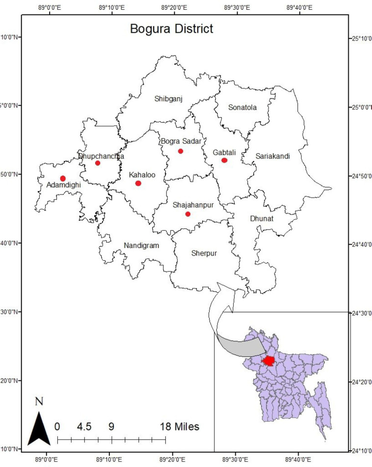
A total of 275 blood samples were collected randomly based on probability proportional to size techniques from 39 Sonali-type chickens (cross-bred between Rhode Island Red cocks and Fayoumi hens) farms in November 2019 in the Bogura district of Bangladesh. Bogura district is a Sonali chicken belt located in the northern part of Bangladesh (Fig. 1). During sample collection, farm location, flock size, and ages of chickens were considered. Samples were collected from six sub-districts (Bogura Sadar, Kahalu, Shajahanpur, Gabtoli, Adomdighi, and Duphachia) under the Bogura district of Bangladesh. With regard to flock size, farms were categorized into three groups, such as small (500–1,000 birds), medium (1,001–1,500 birds), and large (1,501–2,000 birds) flocks. Chickens were also categorized into three age groups (<30, 31–50, and >50 weeks of age) during sample collection. The chickens were reared in the open-floor system, appeared healthy and non-vaccinated against AEV but vaccinated for Newcastle diseases, infectious bursal diseases, and Marek’s disease virus with a standard schedule.
Figure 1. Map showing the sampling sites (red dots) in Bogura district of Bangladesh.
Collection of blood samples and separation of serum
Blood samples were collected from wing veins (1.5 ml blood/chicken) of apparently healthy chickens with 3-ml disposable plastic syringes without anticoagulant. After collection, blood samples were kept at room temperature in a standing position for an hour to allow clot formation. Then, the blood samples were kept in an ice-box and transferred to the laboratory, maintaining a cool chain. After collection, blood samples were subjected to centrifuge at 3,000 rpm for 5 min to remove the remaining clotted blood cells, clots, and other insoluble materials. Finally, clear supernatant serum was transferred in a sterilized Eppendorf tube and stored at −20°C until the ELISA was carried out.
Detection of AEV-specific antibody using indirect ELISA
The processed serum samples were tested for AEV-specific antibodies by indirect ELISA using a commercially available AEV antibody test kit (BioChek®, Netherlands) containing AEV antigen-coated plates. Briefly, for each sample, a two-step dilution procedure was used to make a final dilution of 1:500 in dilution buffer. Then, 100 μl of negative control was added into A1 and B1 wells; 100 μl of positive control was added into C1 and D1 wells; and 100 μl of reference control (BioChek® Reference Control 13) was added into E1 and F1 wells of AEV antibody-coated plate. The remaining 90 wells were filled separately with 100 μl of diluted serum samples. After that, the plate was covered with the lid and incubated at 22°C–27°C for 30 min. After incubation, the wells were aspirated and washed four times each with approximately 350 μl of the wash solution. During washing, the plate was inverted and tapped firmly on absorbent paper to remove moisture. Then, 100 μl of the conjugate was put into each well. Again, the plate was covered with a lid and incubated at 22°C–27°C for 30 min.
Meanwhile, substrate reagent was prepared by adding one tablet in 5.5 ml of substrate buffer. Following incubation, the plate was again emptied and washed four times with a wash solution. Then, 100 μl of substrate solutions were added into each well and the plate was incubated at 22°C–27°C for 15 min. The reaction was stopped by adding 100 μl stop solution after 15 min. Finally, each reaction’s optical density was measured at a 405 nm filter by an ELISA reader (Multiskan EX®, ThermoFisher, Waltham, MA). The results were analyzed by BioChek 15 software program (BioChek®, Reeuwijk, Netherlands) to calculate sample to positive (S/P) ratio and antibody titer where the S/P ratio and titer are 0.5 or more and 1,071 or more were considered positive, respectively. The test was duplicated for every sera sample for the accuracy of the result.
Statistical analysis
All questionnaire data and ELISA test results were incorporated in an MS Excel 2013 spreadsheet (Microsoft Corporation, Redmond, WA) and exported to STATA-IC-14.0 for further analysis [17]. Descriptive statistics were conducted to calculate the frequency and percentage of different variables such as farm location, flock size, and age with 95% confidence intervals (CI). Chi-square test (χ2 test) was carried out to evaluate the significant relationship of AEV seropositivity to different variables. In chi-square analysis, the probability value (p) less than 0.05 (p < 0.05) was considered statistically significant.
Results
The cross-sectional survey of AEV antibodies in Sonali parent chickens of the Bogura district of Bangladesh is presented in Table 1. Overall, 70.18% (193/275) of the chickens were positive for AEV antibodies. All 39 (100%) flocks were found positive for AEV-specific antibodies as at least one sample was positive from each flock.
Table 1. Serological detection of AEV antibodies in Sonali parent chickens based on the farm location, flock size, and age.
| Variable | Category | Number of flocks | Number of serum tested | Number of positive samples (%) | 95% CI | χ2 | p-value |
|---|---|---|---|---|---|---|---|
| Farm | Adomdighi | 5 | 37 | 17 (45.95) | 29.49–63.08 | 3.96 | 0.555 |
| Location | Dupchachia | 6 | 41 | 31 (75.61) | 59.70–87.64 | ||
| Gabtoli | 7 | 47 | 33 (70.21) | 55.11–82.66 | |||
| Kahalu | 7 | 49 | 35 (71.43) | 56.74–83.42 | |||
| Bogura Sadar | 6 | 46 | 42 (91.30) | 79.21–97.58 | |||
| Shajahanpur | 8 | 55 | 35 (63.64) | 49.56–76.19 | |||
| Flock size | Small (500–1,000) medium | 13 | 75 | 33 (44.00) | 32.55–55.94 | 6.67 | 0.036 |
| 11 | 110 | 86 (78.18) | 69.30–85.49 | ||||
| (1,001–1,500)large | 15 | 90 | 74 (82.22) | 72.74–89.48 | |||
| Age | (1,501–2,000) < 30 weeks | 9 | 88 | 42 (47.73) | 36.96–58.65 | 7.12 | 0.028 |
| 31–50 weeks> 50 weeks | 15 | 84 | 59 (70.24) | 59.27–79.73 | |||
| 15 | 103 | 92 (89.32) | 81.69–94.55 |
CI = Confidence interval; χ2 = chi-square.
*p-value less than 0.05 is considered significant.
The study was conducted in six sub-districts of the Bogura district based on farm location. Out of the six sub-districts, the highest prevalence was found in Bogura Sadar (91.30%), followed by 75.61% in Dupchachia, 71.43% in Kahalu, 70.2% in Gabtoli, 63.64% in Shajahanpur, and 45.95% in Adomdighi (Table 1). Farm locations did not differ statistically at p > 0.05. Regarding flock size, the detail seroprevalence of AEV in Sonali chickens was consequently 44% in small (500–1,000 chickens), 78.18% in medium (1,001–1,500 chickens), and 82.22% in large (1,501–2,000 chickens) flock. Table 1 illustrates that the prevalence of AEV in chickens was highest in the large flocks (82.22%) than in small flocks (44%). Flock size was significantly (p < 0.05) correlated to AEV infection in Sonali chickens.
The collected sera were grouped according to chickens’ age, such as less than 30 weeks, 31–50 weeks, and above 50 weeks. The estimated seroprevalence of AEV infection in Sonali chickens was 47.73% at below 30 weeks of age, 70.24% at within 31–50 weeks of age, and 89.32% above 50 weeks of ages, respectively (Table 1). The seroprevalence of AEV was significantly higher in adults than in the young chickens (p < 0.05).
Discussion
The AEV infection in poultry flocks caused severe economic losses in the poultry industry by increasing mortality and medication costs and decreasing egg production and hatchability [1,18]. The present cross-sectional study reported an overall 70.18% of Sonali parents’ chickens of Bogura district positive for AEV-specific antibodies during the laying period.
However, the tested chickens were healthy and non-vaccinated for AEV. A similar serological study was conducted in Tamil Nadu, India, by Sukumar and Sumitha [13] and they noted 79.35% seropositivie of AEV in a non-vaccinated commercial layer, which is closely related to the current result. Another study was carried out in Khartoum, Sudan, by Zahraa et al. [16] and reported 57.1% AEV seropositive layer chickens. Barros et al. [10] reported 100% AEV seropositive chickens in layer farms in Brazil during an outbreak.
The study indicated that larger flocks are more prone to AEV infection than smaller flocks. The results are also strongly supported by Schat and Skinner [19] that high-density flock during grow out contributes to the spread of the virus and clinical symptoms. In the present study, it was found that age was significantly correlated with the prevalence of AEV. Chickens during the laying period (>50 weeks) showed higher prevalence for AEV infection than those aged below 30 weeks. The findings are in agreement with several previous studies. Baksi [20], Hammami et al. [5], Sukumar and Sumitha [13], and Yu et al. [14] reported 25%, 75%, 89.43%, and 63.2% AEV seropositive chickens at adult age, respectively. Therefore, adult layer chickens are more sensitive to AEV infection than early laying period.
The current cross-sectional study revealed that the virus is widely circulated among the selected farms as the chickens are carrying anti-AEV antibodies. The chickens were non-symptomatic and non-vaccinated against AEV. Although non-symptomatic, they had a history of other vaccination against Newcastle diseases, infectious bursal diseases, and Marek’s disease virus. It is still uncommon to vaccinate the chickens against AEV, especially Sonali-type chickens, due to the absence of clinical signs prominently. All the Sonali parents’ flocks studied were reared in an open shed with a floor system for easy husbandry and mating [21,22]. A major predisposing factor of AEV transmission is its capacity to survive up to 4 weeks or more in the environment and transmit horizontally and vertically [23]. The virus can also easily be transmitted horizontally from contaminated feeds or litter by ingestion; direct chickens to chickens contact, fomites or poultry workers, and visitors are the potential risk factors [1]. Therefore, chicken rearing in-floor system may increase the chance of infection compared to rearing in the cage [16]. The AEV transmission in vertical routes also plays an essential role in causing increased early chicks mortality [24].
AEV diagnosis based on neurologic signs in adult chickens more than 3–5 weeks older is very hard as neurogenic signs are not very common in adult chickens. It seems that during this age, the humoral antibody can neutralize the AEV before reaching the central nervous system [24]. So, chickens can be non-symptomatic, although infected [8,25]. Westbury and Sinkovic [27] revealed that AEV might not neutralize chicks’ immune systems during the 1st week of age; thus, neurogenic signs develop. A maternally derived antibody from a vaccinated mother can neutralize AEV during the 1st week of age [24,26,27]. So, it is suggested that Sonali parents’ flock be vaccinated to save their chicks in early life from AEV infection as AEV is widely circulating in the environment. However, AEV can be controlled successfully by vaccination to commercial layer and breeders before their sexual maturity. The live embryo-propagated virus vaccine of AEV can be vaccinated in different routes like drinking water, spray, and wing-web [4,24]. It can also prevent AEV infection from day-old chicks by vertical transmission from infected parents [4,28–31].
To the best of our knowledge, this is the first study on Sonali parent chickens regarding AEV infection in Bangladesh using indirect ELISA. Therefore, further studies can be conducted focusing on isolation and molecular characterization of AEV to formulate prevention and control strategies.
Conclusion
The above findings conclude that AEV is widely circulated in Sonali parent chickens in the Bogura district of Bangladesh. Overcrowding and older age are identified as potential risk factors of AEV. Therefore, AE vaccination to Sonali parent flocks and further virological study of AEV are suggested.
List of abbreviations
95% CI: 95% confidence intervals; AEV: avian encephalomyelitis virus; ELISA: enzyme-linked immunosorbent assay.
Acknowledgments
We are thankful to the poultry farmers and laboratory persons for their essential help during sample collection and laboratory analysis. We are also grateful to Shariful Islam for making the map. The Central Poultry Laboratory, Nourish Poultry and Hatchery Ltd. were supported by their laboratory facilities during the research.
Conflict of interest
The authors declare that they have no competing interest in publishing the manuscript.
Authors’ contributions
M.Z.A.: designed the study, interpreted results, analyzes data, and wrote the manuscript. M.T.W.S., M.M.M., M.A.B., A.A.M.S., M.G., and Z.A.B.: Collected samples and carried out the experiments and laboratory analyses. S.A.K: supervised the overall research and laboratory management.
References
- [1].Tannock GA, Shafren DR. Avian encephalomyelitis: a review. Avian Pathol. 1994;23:603–20. doi: 10.1080/03079459408419031. https://doi.org/10.1080/03079459408419031. [DOI] [PubMed] [Google Scholar]
- [2].Marvil P, Knowles NJ, Mockett AP, Britton P, Brown TD, Cavanagh D. Avian encephalomyelitis virus is a picornavirus and is most closely related to hepatitis a virus. J Gen Virol. 1999;80(3):653–62. doi: 10.1099/0022-1317-80-3-653. https://doi.org/10.1099/0022-1317-80-3-653. [DOI] [PubMed] [Google Scholar]
- [3].Shafren DR, Tannock GA. Further evidence that the nucleic acid of Avian encephalomyelitis virus consists of RNA. Avian Dis. 1992;1:1031–3. https://doi.org/10.2307/1591568. [PubMed] [Google Scholar]
- [4].Calnek BW. Control of Avian encephalomyelitis: a historical account. Avian Dis. 1998;1:632–47. https://doi.org/10.2307/1592696. [PubMed] [Google Scholar]
- [5].Hammami N, Pacha MB, Boukais A, Rahal K. Serological study of Avian encephalomyelitis in laying hens and economic impact of vaccination in algeria. Agricultura. 2017;101(1–2):116–21. [Google Scholar]
- [6].Suarez DL. Avian encephalomyelitis. In: Swayne DE, Glisson JR, McDougald L, Nolan R, Suarez LK, Nair V, editors. Diseases of poultry. 13th. Ames, IA: Wiley-Blackwell; 2013. pp. 486–510. [Google Scholar]
- [7].Schaaf K. Immunization for the control of Avian encephalomyelitis. Avian Dis. 1959;2(3):279–89. https://doi.org/10.2307/1587528. [Google Scholar]
- [8].Taylor LW, Lowry DC, Raggi LG. Effects of an outbreak of Avian encephalomyelitis (epidemic tremor) in a breeding flock. Poult Sci. 1955;34:1036–45. https://doi.org/10.3382/ps.0341036. [Google Scholar]
- [9].Xie Z, Khan MI, Girshick T, Xie Z. Reverse transcriptase-polymerase chain reaction to detect Avian encephalomyelitis virus. Avian Dis. 2005;49:227–30. doi: 10.1637/7307-111804R. https://doi.org/10.1637/7307-111804R. [DOI] [PubMed] [Google Scholar]
- [10].Barros ME, Rocha BP, Souza FA, Mendonça FS, Evêncio-Neto J. Severe outbreak of Avian encephalomyelitis in laying hens in Northeastern Brazil. Braz J Poult Sci. 2019;21:1–4. https://doi.org/10.1590/1806-9061-2018-0749. [Google Scholar]
- [11].Ali MZ, Park JE, Shin HJ. Serological survey of avian metapneumovirus infection in chickens in Bangladesh. J Appl Poult Res. 2019;28:1330–4. https://doi.org/10.3382/japr/pfz050. [Google Scholar]
- [12].Jones EE. An encephalomyelitis in the chicken. Science. 1932;76:331–2. doi: 10.1126/science.76.1971.331. https://doi.org/10.1126/science.76.1971.331. [DOI] [PubMed] [Google Scholar]
- [13].Sukumar K, Sumitha P. Detection of Avian encephalomyelitis virus antibodies in commercial layer belt of Tamil Nadu. Indian Vet J. 2016;93:75–7. https://doi.org/10.1201/b19719-12. [Google Scholar]
- [14].Yu XH, Zhao J, Qin XH, Zhang GZ. Serological evidence of Avian encephalomyelitis virus infection associated with vertical transmission in chicks. Biologicals. 2015;43:512–4. doi: 10.1016/j.biologicals.2015.09.003. https://doi.org/10.1016/j.biologicals.2015.09.003. [DOI] [PubMed] [Google Scholar]
- [15].Bhuiyan ZA, Ali MZ, Moula MM, Giasuddin M, Khan ZU. Prevalence and molecular characterization of infectious bronchitis virus isolated from chicken in Bangladesh. Vet World. 2019;12(6):909–15. doi: 10.14202/vetworld.2019.909-915. https://doi.org/10.14202/vetworld.2019.909-915. [DOI] [PMC free article] [PubMed] [Google Scholar]
- [16].Zahraa F, Lutfi OH, Kheir SAM. Case report: anti-Avian encephalomyelitis virus antibodies in layer flocks in Khartoum State. Sudan J Vet Res. 2010;25:59–64. [Google Scholar]
- [17].StataCorp. College Station, TX: StataCorp LP; 2015. Stata statistical software: release 14. [Google Scholar]
- [18].Goto Y, Yaegashi G, Kumagai Y, Ogasawara F, Goto M, Mase M. Detection of Avian encephalomyelitis virus in chickens in Japan using RT-PCR. J Vet Med Sci. 2019;81:103–6. doi: 10.1292/jvms.18-0550. https://doi.org/10.1292/jvms.18-0550. [DOI] [PMC free article] [PubMed] [Google Scholar]
- [19].Schat KA, Skinner MA. Avian immunosuppressive diseases and immune evasion. In: Schat KA, Kaspers B, Kaiser P, editors. Avian immunology. 2nd. London, UK: Elsevier-Academic Press; 2013. pp. 275–97. https://doi.org/10.1016/B978-0-12-396965- 1.00016-9. [Google Scholar]
- [20].Baksi S. Estimation of seroprevalence of Avian encephalomyelitis in different parts of India. J Adv Res Biol. 2018;1:1–3. [Google Scholar]
- [21].Ali MZ, Moula MM, Bhuiyan ZA, Javed MT. A cross sectional survey of chicken astroviruses antibody in broiler and Sonali (cross-bred) chickens in selected areas in Bangladesh. Maced Vet Rev. 2020;43:75–80. https://doi.org/10.2478/macvetrev-2020-0016. [Google Scholar]
- [22].FAO. Animal Production and Health Working Paper. No. 14. Rome, Italy: 2015. [Mar 24;2020 ]. Comparative performance of Sonali chickens, commercial broilers, layers and local non-descript (deshi) chickens in selected areas of Bangladesh. [Google Scholar]
- [23].Martins PC, Silva PL. Encefalomielite aviária. In: Berchieri Júnior A, Silva EN, Di Fábio J, Sesti L, Zuanaze MAF, editors. Doenças das aves. 2nd. Campinas, Brazil: Fundação APINCO de Ciência e Tecnologia Avícolas; 2009. pp. 763–73. [Google Scholar]
- [24].Sentíes-Cué CG, Gallardo RA, Reimers N, Bickford AA, Charlton BR, Shivaprasad HL. Avian encephalomyelitis in layer pullets associated with vaccination. Avian Dis. 2016;60:511–5. doi: 10.1637/11306-102115-Case. https://doi.org/10.1637/11306-102115-Case. [DOI] [PubMed] [Google Scholar]
- [25].Roy P, Hemalatha S, Vairamuthu S, Purushothaman V, Chandramohan A, Karunamurthy G, et al. An outbreak of Avian encephalomyelitis in Tamil Nadu State of India. West Indian Vet J. 2009;9:8–10. [Google Scholar]
- [26].Ali MZ, Hasan B. Follow up of maternally derived antibodies titer against economically important viral diseases of chicken. Poult Sci J. 2018;6(2):149–54. https://doi.org/10.22069/PSJ.2018.15213.1340. [Google Scholar]
- [27].Westbury HA, Sinkovic B. The pathogenesis of infectious Avian encephalomyelitis: 4. The effect of maternal antibody on the development of the disease. Aust Vet J. 1978;54:81–5. doi: 10.1111/j.1751-0813.1978.tb00352.x. https://doi.org/10.1111/j.1751-0813.1978.tb00352.x. [DOI] [PubMed] [Google Scholar]
- [28].Calnek BW. Avian encephalomyelitis. In: Saif YM, Fadly AM, Glisson JR, McDouglas LR, Nolan LK, Swayne DE, editors. Diseases of poultry. 12th. Ames, IA: Wiley-Blackwell; 2008. pp. 430–41. [Google Scholar]
- [29].Back A. Back A, editor. Encefalomielite aviária. Manual de doenças de aves. (2nd) 2010:70–2. Editora Integração, Cafelândia, Brazil. [Google Scholar]
- [30].Ali MZ. Common respiratory diseases of poultry in Bangladesh: a review. SAARC J Agric. 2020;18(1):1–11. https://doi.org/10.3329/sja.v18i1.48377. [Google Scholar]
- [31].Ali MZ. The seroprevalence study of Reticuloendotheliosis virus infection in chicken in Bangladesh. Egypt J Vet Sci. 2018;49(2):179–86. https://doi.org/10.21608/ejvs.2018.5856.1051. [Google Scholar]