Figure 3.
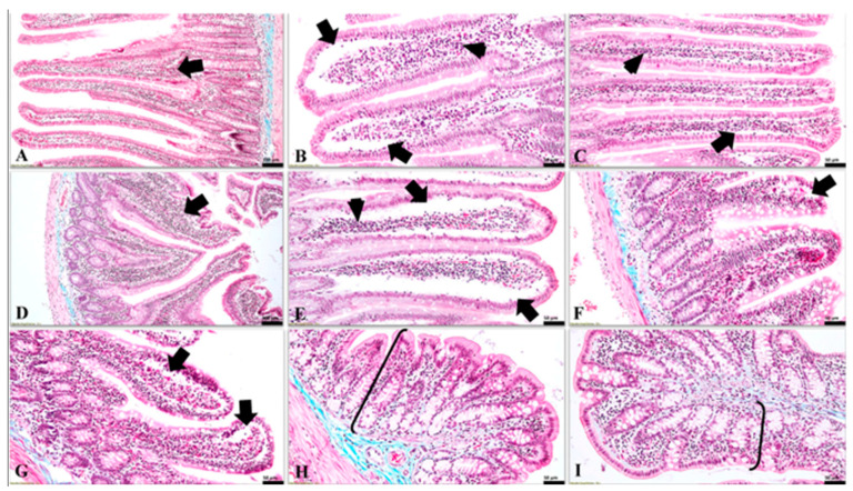
The microscopic features detected in the intestinal mucosa in all experimental groups (Goldner’s trichrome stain). (A) (G1, duodenum) normal aspect of the duodenal mucosa, and a moderate gut-associated lymphoid tissue (GALT) in lamina propria (arrow); (B) (G2 (ABX), duodenum) the presence of large subepithelial clefts (arrows), located mainly towards the apical pole of the duodenal villi along with abundant amount of GALT in the lamina propria (arrowhead); (C) (G3 (PRB), duodenum) noticeable subepithelial spaces (arrow) and moderate amounts of GALT in the lamina propria of the duodenum (arrowhead); (D) (G4 (ABX + PRB), duodenum) intestinal mucosa with a normal appearance and a prominent lymphoid tissue in the lamina propria of the duodenum (arrow); (E) (G5 (ABX/PRB), duodenum) large subepithelial clefts along the entire length of the duodenal villi (arrows) and an obvious mucosal-associated lymphoid tissue in the lamina propria (arrowhead); (F) (G2 (ABX), jejunum) intestinal villi in the jejunum with a discreet subepithelial space located mainly towards the apical poles and a moderate leukocytic infiltrate in the lamina propria; (G) (G3 (PRB), jejunum) jejunal villi with prominent subepithelial spaces towards their apical pole (arrow), in some cases these being visible up to the middle third of the villi; (H) (G2 (ABX), colon) and (I) (G3 (PRB), colon) absence of microscopic lesions in the mucosa (accolades) of the colon.

