Abstract
Pertussis toxin (Ptx) expression and secretion in Bordetella pertussis are regulated by a two-component signal transduction system encoded by the bvg regulatory locus. However, it is not known whether the metabolic pathways and growth state of the bacterium influence synthesis and secretion of Ptx and other virulence factors. We have observed a reduction in the concentration of Ptx per optical density unit midway in fermentation. Studies were conducted to identify possible factors causing this reduction and to develop culture conditions that optimize Ptx expression. Medium reconstitution experiments demonstrated that spent medium and a fraction of this medium containing components with a molecular weight of <3,000 inhibited the production of Ptx. A complete flux analysis of the intermediate metabolism of B. pertussis revealed that the sulfur-containing amino acids methionine and cysteine and the organic acid pyruvate accumulated in the media. In fermentation, a large amount of internal sulfate (SO42−) was observed in early stage growth, followed by a rapid decrease as the cells entered into logarithmic growth. This loss was later followed by the accumulation of large quantities of SO42− into the media in late-stage fermentation. Release of SO42− into the media by the cells signaled the decoupling of cell growth and Ptx production. Under conditions that limited cysteine, a fivefold increase in Ptx production was observed. Addition of barium chloride (BaCl2) to the culture further increased Ptx yield. Our results suggest that B. pertussis is capable of autoregulating the activity of the bvg regulon through its metabolism of cysteine. Reduction of the amount of cysteine in the media results in prolonged vir expression due to the absence of the negative inhibitor SO42−. Therefore, the combined presence and metabolism of cysteine may be an important mechanism in the pathogenesis of B. pertussis.
Bordetella pertussis is the causative agent of whooping cough. The development and utilization of vaccines against B. pertussis have greatly reduced the morbidity and mortality caused by this pathogen throughout the world. The toxicity associated with the whole-cell pertussis vaccines (39) has led to the development of a new generation of acellular vaccines containing one or more virulence factors (38, 41). Acellular vaccines that contain pertussis toxin (Ptx) require isolation and purification of active toxin from the media of batch fermentation (45). The number of doses per fermentation volume of acellular vaccine is estimated to be fivefold lower than that for a whole-cell vaccine (45). Therefore, there has been much interest in improving product yields through medium optimization, compound addition, and genetic manipulation of the B. pertussis strains.
B. pertussis is routinely cultured in Stainer-Scholte (SS) medium (46) or a modified derivative. These media contain glutamate as the primary source of carbon, because B. pertussis has an incomplete citric acid cycle and is not capable of using glucose. Growth in SS medium results in a final dry cell weight of 2 g/liter and a Ptx concentration of 5 to 10 mg/liter (51). During growth in these media, ammonium accumulates because of an imbalance in the nitrogen/carbon ratio of the components, which is one of the factors that limit cell density in fermentation and decrease the growth rate (1). Increasing the amount of glutamate in cultures has been shown to increase the final cell density, but not the amount of Ptx (51). However, adjusting the ionic composition of the media, maintaining pH at 7.0 (15), and growing cultures in pure oxygen have improved the Ptx yield in cultures containing a higher glutamate concentration. In addition, the presence of free fatty acids in the media has been reported to be either inhibitory or stimulatory for growth (13). Addition of heptakis-(2,6-O-dimethyl)-α-cyclodextrin to sequester free fatty acids has significantly increased Ptx production (15, 16, 58). Outer membrane vesicles produced in Stainer-Scholte cultures contain significant amounts of adenylate cyclase, lipopolysaccharide, and Ptx (18). Dissociation of Ptx from these vesicles may also improve yields.
Ptx and other vir-activated genes (vag genes), including the filamentous hemagglutinin (FHA) locus (fha) (5, 7), the pertactin gene (prn) (21), fimbrial genes (fim-1, fim-2, and fimX) (57), the adenylate cyclase gene (cyaA) (24), a gene encoding a porin-like protein (ompQ) (14), and a locus encoding serum resistance (brkAB) (53, 56) have been shown to be coordinately regulated at the level of transcription by a sensory transduction system encoded by the bvgAS (vir) locus (34) (2, 22, 50). Expression of the vir regulon in B. pertussis is affected by diverse environmental signals (31, 32, 35). The presence of sulfate anion (SO42−) or nicotinic acid (31, 32) or growth at low temperature (37) results in decreased expression of ptx and other virulence genes. Naturally occurring BvgS mutants (49), site-directed mutants (34), and serum-resistant mutants (12) have been shown to be less sensitive to these environmental signals.
In addition to the vag genes, there are a set of genes that are regulated in the opposite manner from the vag genes. These vir-repressed genes (vrg-6, vrg-8, vrg-24, vrg-53, and vrg-73) are expressed during the avirulent state (4, 22, 34). In several of these genes, the repression by bvg appears to be a result of binding of a 34-kDa vir-activated repressor to an intergenic sequence in the vrg coding DNA, which decreases transcription (3). Additional vir-repressed genes have been identified and characterized. Functional analysis of these two surface proteins may help to elucidate the physiologic role in modulation of B. pertussis (48).
Mutations in the genes responsible for the synthesis and secretion of an active Ptx molecule can also affect the virulence of the organism (53, 56). The genes encoding the Ptx subunits have been cloned and sequenced (27), and the operons for the Ptx secretory genes (ptl) have been characterized (54). Mutations in the S1 subunit of Ptx affect secretion because of destabilization of the molecule (9). Mutations in the ptl gene (55) also decrease Ptx secretion.
Transcription of the Ptx gene can be regulated by several mechanisms. Upon phosphorylation of BvgA, the transcription factor binds to specific promoter sequences that activate or repress transcription (40, 52). A kinetic study has indicated that fha and bvg are transcribed 10 min after signal transduction, but the ptx and cya genes are not transcribed until 2 to 4 h after induction (42, 43). The activation of Ptx and cya correlated with accumulation of high intracellular levels of BvgA (42). Transcription of ptx requires higher levels of phosphorylated BvgA than that of fha (47). Genetic and DNase I protection data support a model of Ptx activation in which phosphorylated BvgA binds to a high-affinity BvgA binding site (6, 28). Cooperative binding of BvgA dimers between the binding site and the promoter allows BvgA to interact with RNA polymerase (RNP) (5, 6). More recently, a Ptx accessory factor has been described that is required for transcription of the gene in Escherichia coli. In addition, B. pertussis mutants have been identified that overexpress the α subunit of RNA polymerase, which in turn down-regulates expression of the Ptx promoter (8, 28).
Isolation and construction of B. pertussis bvgs mutants (22, 34, 35) that are more resistant to antigenic modulation by exogenous SO42− and nicotinic acid have been helpful in studying ptx gene regulation. Still, investigators have yet to decipher the mechanism for the reduction in the concentration of Ptx per optical density unit ([Ptx]/OD) commonly observed in wild-type strains grown in shake flasks and fermentation containing standard and more complex media (15, 16). In this study, we show that B. pertussis autoregulates Ptx production through the metabolic conversion of cysteine to SO42− and pyruvic acid. Our data suggest that B. pertussis uses the bvgs sensory system in response to accumulation of both internal and external metabolic SO42−.
MATERIALS AND METHODS
Bacterial strains.
Wild-type B. pertussis strain CS-87 was used in these studies. This strain originated in China and was brought to the National Institute of Child Health and Human Development at the National Institutes of Health. Strain 9797, a Tohama I derivative, was procured from the American Type Culture Collection (Manassas, Va.). Organisms were stored at −70°C or maintained on Bordet-Gengou agar (BGA; Becton Dickinson, Sparks, Md.) in a humid incubator at 37°C.
Culture media.
The medium used to grow CS-87 was similar to the defined medium described by Stainer and Scholte (46). One liter of medium contained 10.7 g of monosodium glutamate, 0.24 g of l-proline, 2.5 g of NaCl, 0.5 g of KH2PO4, 0.2 g of KCl, 0.1 g of MgCl2 · 6H2O, 1.82 g of Tris base, 0.01 g of FeSO4 · H2O, 0.02 g of CaCl2 · 2H2O, 0.04 g of l-cysteine monohydrochloride, 0.1 g of glutathione (reduced), 0.02 g of l-ascorbic acid, and 0.004 g of niacin. The monosodium glutamate, proline, NaCl, KH2PO4, KCl, MgCl2 · 6H2O Tris base, and CaCl2 · 2H2O were prepared in a basal formulation (1×) and autoclaved. The remaining medium supplement was prepared in a concentrated form (100×) and filter sterilized. The final pH of the complete medim ranged from 7.2 to 7.5. For some experiments, a modified SS (MSS) medium was used. The MSS medium was supplemented with the following amounts of amino acids per liter: 0.40 g of l-arginine monohydrochloride, 0.10 g of l-asparagine, 0.04 g of l-aspartic acid, 0.1 g of l-cysteine monohydrochloride, 0.03 g of l-histidine, 0.10 g of l-isoleucine, 0.10 g of l-leucine, 0.08 g of l-lysine monohydrochloride, 0.03 g of l-methionine, 0.03 g of l-phenylalanine, 0.06 g of l-serine, 0.04 g of l-threonine, 0.01 g of l-tryptophan, and 0.04 g of l-valine. This amino acid supplement was prepared in a concentrated form (100×) and filter sterilized.
For MSS fermentation, the fermentor was filled with SS media, and the amino acid supplement was added to make a 1× concentration. Additional amino acid supplement was added to a 1× concentration when the OD of the fermentation reached >1.0. At this time, each liter of the SS and MSS cultures also received an additional 10.0 mg of FeSO4 · 6H20 and 5.0 g of monosodium glutamate. Fermentation experiments containing SS medium received 0.01 g of FeSO4 · 6H2O per liter and 5.0 g of monosodium glutamate per liter. Limiting cysteine experiments in batch mode (LCMSSB) contained 30% less cysteine, and in fed-batch mode experiments (LCMSSFB), cysteine was delivered at a rate of 20 mg/h. In some experiments, 1.0 M BaCl2 was added to the limiting cysteine MSS medium to a final concentration of 20 mM when the culture reached an OD of >1.0.
Growth conditions.
Shake flasks were inoculated with frozen seeds and grown 20 to 24 h before addition to the bioreactors. Shake flask cultures were transferred to the bioreactor when ODs were >1.0 at 600 nm. The bacteria were grown in triple-baffled Erlenmeyer flasks in a New Brunswick Innnova model 4300 shaking incubator maintained at 36.5°C or in a New Brunswick 20-liter BioFlo 4500 (New Brunswick Scientific, Edison, N.J.) run in batch mode with a working volume of 12 liters maintained at 36.5°C. The reactor was connected to an AFS Biocommand version 2.61 (New Brunswick Scientific), which collected data for pH, agitation, dissolved oxygen, temperature, and airflow rate and which had additional pumps for antifoam and pH control. The fermentation was maintained at a temperature of 36.5°C, with an aeration rate of 4.0 liters/min (pH 7.2) and a dissolved oxygen level of 40% by cascading the agitation rate from 200 rpm to 1,000 rpm. The pH was controlled by addition of 50% phosphoric acid, and foaming was controlled by addition of antifoam C (J. T. Baker, Phillipsburg, N.J.) as needed. The fermentation contained 11 liters of defined medium and was inoculated with 1 liter of an actively growing seed culture when the OD of the culture was >1.0. All chemicals were reagent grade and were obtained from J. T. Baker, Spectrum, New Brunswick, N.J., or Sigma, St. Louis, Mo., unless otherwise noted.
Fermentation sampling.
Samples were taken from a sterile port every 3 to 6 h. The OD of each sample was measured by using the Shimadzu UV 1601 spectrophotometer (Shimadzu, Columbia, Md.). A 30-ml sample was centrifuged at 9,000 rpm for 10 min at room temperature with an SA 600 rotor in a Sorvall centrifuge. The supernatants from the 30-ml samples were filter sterilized with a 0.2-μm-pore-diameter Millex-GV filter (Millipore, Bedford, Mass.). This supernatant was used for SO42−, Ptx, amino acid, and organic acid analyses. The cells were resuspended in 10 ml of distilled water, disrupted by nitrogen cavitation with a nitrogen bomb (Fike Metal Products, Blue Springs, Mo.), and centrifuged as before, and the supernatant was analyzed for internal SO42− concentration. A 5-ml sample was treated with 500 μl of 10% sodium dodecyl sulfate (SDS) and used for trichloroacetic acid precipitation of intracellular and extracellular proteins. The pellets were stored at −20°C, and the supernatant and the SDS-treated samples were stored at 2 to 8°C. Culture purity was verified by gram staining and plating on BGA and Trypticase soy agar (Becton-Dickinson).
Medium reconstitution experiments.
Culture supernatant from a B. pertussis culture grown to stationary phase in SS medium was lyophilized and was reconstituted with growth medium lacking NaCl, KH2PO4, KCl, Tris base, CalCl2 · 2H2O, and 0.1 g of MgCl2 · 6H2O. A fraction of this reconstituted spent medium was then added to a second culture of B. pertussis and was compared to the original medium used for Ptx production. Another fraction of the spent medium from the original culture was filtered through a membrane with a molecular weight cutoff of 3,000 (3,000 MW). Both the retentate and flowthrough were lyophilized and reconstituted as before. These mixtures were added to individual B. pertussis cultures at 18 h, and then samples for Ptx production were obtained at 23 and 28 h. The Ptx production and growth of these cultures containing reconstituted medium were then compared to Ptx production and growth in SS medium.
Nitrogen cavitation of cells.
Bacterial pellets from 30-ml culture supernatants were suspended in 10 ml of distilled water. The aqueous portion of the cells was isolated by first disrupting the cells with a nitrogen cavitation apparatus (Fike Metal Products) at a pressure of 1,500 lb/in2 and then removing cellular debris by centrifugation at 10,000 rpm in a Sorvall centrifuge with an SA 600 rotor.
Cross-streaking experiments.
Initial vertical streaks of strain CS-87 were made on individual BGA plates (Becton-Dickinson) and incubated at 36.5°C for 48 h. Secondary horizontal streaks were made when the sterile loop passed back and forth across the vertical streak. The cross-streaked plate was then incubated for 48 h at 36.5°C. Colonies were visualized with a Leica model MZ6 stereomicroscope (Leica, Buffalo, N.Y.) at a ×100 magnification, and the image was captured with a Digital Imaging System model IS-1000 (Alpha Innotech Co., San Leandro, Calif.).
Quantitative Ptx ELISA.
Microtiter plates (Nunc Maxi-sorp; Vangard International, Neptune, N.J.) were sensitized by adding 0.1 ml per well of fetuin (Sigma Chemical Co.) at 0.04 mg/ml in 0.1 M sodium carbonate (pH 9.6). The plates were incubated overnight at 4°C. The plates were blocked for 1 h at room temperature (RT) with 0.1% fetal bovine serum-phosphate-buffered saline (PBS; pH 7.5). The plates were washed, and samples diluted in PBS-Brij 35 plus 0.02% bovine serum albumin were added to the fetuin-coated plates and incubated for 1 h at RT. After incubation, plates were washed and reacted with a monoclonal antibody to Ptx (19, 20, 44) for 2 h at RT. The plates were washed and the secondary antibodies, alkaline phosphatase-conjugated goat anti-mouse immunoglobulin G (IgG) and IgM (Tago, Burlingame, Calif.) diluted 1:3,000 in PBS-Brij, were added to the plate and incubated for 2 h at RT. The plates were washed and developed with p-nitrophenyl phosphate (Sigma) in a solution containing 0.1 M diethanolamine, 1 mM MgCl2, 0.1 mM ZnCl2, and 0.02% azide (pH 9.8) and incubated for 1 h at RT before the A405 was read. Selective supernatants were assayed with a CHO cell assay (53) to confirm the toxin levels measured in the Ptx enzyme-linked immunosorbent assay (ELISA) (data not shown).
SO42− determination.
Extracellular and intracellular SO42− analyses were performed with a reproducible and quantitative assay (30). The barium chloride reagent was prepared by dissolving 40 mg of BaCl2 and 67.2 mg of NaHCO3 in 60 ml of distilled water. Fifty milliliters of 2N acetic acid was added, and the volume was brought to 500 ml with absolute ethanol. For the working BaCl2 reagent, 100 ml of this solution was diluted with 200 ml of absolute ethanol. The rhodizonate reagent was prepared by dissolving 100 mg of ascorbic acid in 20 ml of distilled water. The final volume was adjusted to 100 ml with absolute ethanol.
A microtiter plate format was designed by adding 20 μl of sample to each well followed by addition of 20 μl of barium chloride reagent and 40 μl of sodium rhodizonate reagent. The plates were agitated gently at RT for 30 s and read at 530 nm with an ELISA plate reader (Dynex Corp, Chantilly, Va.). The amount of SO42− per sample was determined by linear regression analysis of a standard curve generated by different concentrations of ammonium SO42− ranging from 2.5 μg/ml to 30.0 μg/ml.
Amino acid analysis.
The analysis and quantification of amino acids were made by reverse-phase high-pressure liquid chromatography (RP-HPLC) with an on-line pre-column derivitization, as provided for the AminoQuant column (Hewlett-Packard, Wilmington, Del.). Primary acids were derivatized by the ortho-phthalaldehyde (OPA) reagent, while secondary amino acids were derivatized by the FMOC (9-fluorenylmethoxy carbonyl) reagent. For the primary amino acids, the mobile phase consisted of sodium acetate-triethanolamine-tetrahydrofuran (pH 7.2) and was detected at 338 nm. Secondary amino acids were eluted with a sodium acetate-methanol-acetonitrile mobile phase (pH 7.2) and were detected at 262 nm. The quantification of each amino acid was performed by comparison of the amino acid standard curve at different concentrations.
Pyruvic acid analysis.
Pyruvic acid was detected with a model HP-1050 HPLC (Hewlett-Packard) in conjunction with the HP ChemStation version 2.0 software and containing a Bio-Rad Aminex HPX-87H column (Bio-Rad Laboratories, Burlingame, Calif.) with a mobile phase of 4 mM H2SO4. The column was equilibrated at 35°C, and the isocratic flow rate was 0.6 ml/min. The detection was done at 215 nm. Pyruvate was assessed by spiking the organic acid standard with pyruvate at a concentration of 2.5 g/liter. Pyruvic acid was quantified with a pyruvic acid analysis kit (Sigma Chemical Co.).
Statistical analysis.
The Ptx and sulfate concentrations of each sample were calculated by linear regression analysis of Ptx and ammonium sulfate standard curves with the Dynex Revelation 3. 2 statistical analysis package (Dynex, Chantilly, Va.). A Ptx standard curve was generated by using twofold dilutions of a Ptx internal standard from 1.5 μg/ml to 0.012 μg/ml. Culture supernatants were measured for Ptx by prediluting duplicate samples 1:10 and then adding twofold dilutions of the prediluted duplicates to the ELISA plate. The coefficient of variance of duplicate samples was <3% for each assay. The plate-to-plate variation standard was <10%, and the day-to-day variation of the standard was <10%.
The sulfate standard curve was generated by performing dilutions of ammonium sulfate (NH4)2SO4 in media from 35 μg/ml to 3 μg/ml. Undiluted culture supernatants and the concentrated intracellular component of the bacterium were measured in triplicate. The plate-to-plate coefficient of variance of the standard curve ranged from 3 to 12%, and the day-to-day coefficient of variance of the standard averaged <6%.
RESULTS
Determination of the presence of an inhibitor in culture supernatants.
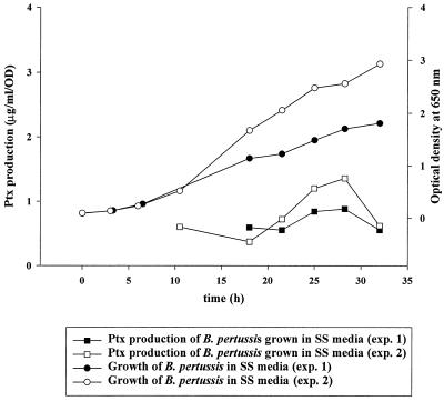
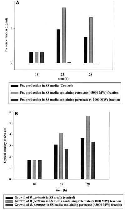
Studies of B. pertussis fermentation were initiated to assess the possibility of increasing Ptx productivity in batch fermentation. The ODs and [Ptx]/OD for two 20-liter fermentations with SS medium are shown in Fig. 1. ODs rise linearly to 32 h, while the [Ptx]/OD ratio begins to level off between 22 and 28 h, suggesting that Ptx production and/or secretion shuts down in the middle of fermentation. Experiments were then designed to determine the presence of an inhibitory component in the medium causing this down-regulation (Fig. 2). In the initial experiment, the spent medium from actively growing B. pertussis cultures at 24 h was lyophilized, reconstituted with the basic salts, and added to another actively growing culture at 18 h. This resulted in a decrease in Ptx production without any effect on growth of the culture (data not shown). The spent culture supernatant was then fractionated with a 3,000-MW-cutoff filter. The retentate fraction (fraction containing components with MW of >3,000) was added back to a B. pertussis culture in a shake flask at 18 h and then grown for an additional 10 h. This fraction demonstrated no inhibition of Ptx production (Fig 2A) or cell growth (Fig. 2B). However, when the <3,000-MW permeate (fraction containing components with MW of <3,000) was added to a culture at 18 h and then grown for an additional 10 h, Ptx production was inhibited (Fig. 2A), but cell growth was unaffected (Fig. 2B). As a control, cultures containing SS medium alone showed normal Ptx production and growth (Fig. 2A and B).
FIG. 1.
Decoupling of Ptx production with growth of B. pertussis strain CS-87 culture in SS medium. The bacteria were cultured in a New Brunswick 20-liter BioFlo 4500 fermentor running in batch mode with a working volume of 12 liters at 36.5°C. The OD of the cultures was measured at 650 nm. Culture purity was verified by Gram staining and plating on BGA and Trypticase soy agar. The OD of the cultures and the average [Ptx]/OD are compared. The average Ptx concentrations for two samples from each time point were used to calculate the [Ptx]/OD values. The coefficient of variance for each Ptx sample was <5%.
FIG. 2.
SS medium reconstitution experiment. The spent culture supernatant from actively growing B. pertussis at 24 h was fractionated with a 3,000-MW-cutoff filter. The retentate fraction (fraction containing components with MW >3,000) was added back to an actively growing B. pertussis cell culture at 18 h and grown to 28 h. Similarly, the flowthrough fraction (fraction containing components with MW <3,000) was added to an actively growing B. pertussis culture at 18 h and grown to 28 h. The Ptx concentration was determined on duplicate samples, and the error bars represent 1 standard deviation of the mean (A). The growth of the cultures is shown in panel B.
Identification of an inhibitor of adenylate cyclase-hemolysin toxin activity.
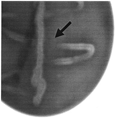
Since the adenylate cyclase-hemolysin (AC) toxin gene of B. pertussis is regulated in a similar manner to the ptx gene, a basic cross-streaking experiment was performed to determine if inhibitors are produced when the bacterium is grown on BGA plates. The bacterial colonies in areas closest to the initial streak showed less hemolytic activity than the ones away from the streak (Fig. 3). This suggested that a soluble component is secreted from colonies present in the initial streak and diffusing through the agar to inhibit adenylate cyclase-hemolysin (cyaA) toxin production in the fresh colonies generated by the cross-streaks.
FIG. 3.
B. pertussis cross-streaking experiments. Stereomicroscopic image of initial vertical streaks of B. pertussis strain CS-87 that were incubated for 48 h followed by secondary horizontal cross-streaks incubated for 48 h. Colonies were imaged with an Alpha imaging system. An arrow shows a zone devoid of adenylate cyclase-hemolysin toxin (CyaA) activity.
Identification of amino acid, pyruvic acid, and sulfur-containing metabolites in fermentation.
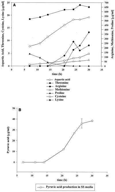
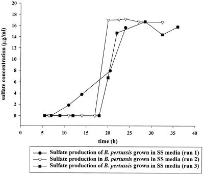
To identify the soluble factors amino acid, pyruvic acid, and sulfur-containing metabolites, amino acid and pyruvic acid analyses were performed with samples derived from a culture of B. pertussis grown in SS medium. Amino acid analysis of fermentation in SS medium shows that the concentrations of arginine, cysteine, and methionine increase over time, with the appearance of methionine and cysteine starting at 15 h. The amount of cysteine (2.5 μg/ml) was smaller than the amounts of methionine (150 μg/ml) and arginine (650 μg/ml) at 30 h (Fig. 4A). Amino acid and pyruvic acid analyses in fermentation showed that cysteine began to appear in the medium at 15 h (Fig. 4A) at the same time as the rapid accumulation of pyruvic acid (Fig. 4B) to a concentration of 40.0 μg/ml. Since SO42− is a known by-product of the cysteine metabolic pathway and an inhibitor of Ptx production, the medium was then analyzed for the presence of SO42−. The appearance of SO42− in the medium followed the pattern of the appearance of pyruvic acid at similar time points (Fig. 4B and 5). The concentration of SO42− in the medium was also similar to the concentration of pyruvic acid (Fig. 4B and 5).
FIG. 4.
Amino acid (A) and pyruvic acid (B) analyses of supernatants from B. pertussis strain CS-87 grown in SS medium. Culture supernatants were assayed at various time points in fermentation. These values represent the average amino acids and pyruvic acid concentrations in micrograms per milliliter determined by comparison to the calibration curves of the reference standards. The pyruvic acid analysis for the early and late time points represents the average of two independent experiments, and the error bars represent 1 standard deviation of the mean.
FIG. 5.
Sulfate analysis of permeates of cultures B. pertussis strain CS-87 grown in SS medium. Cells were centrifuged, and the resulting supernatant was filter sterilized with a 0.2-μm-pore-diameter filter before being assayed. These results represent the average of the samples tested from three independent cultures with a coefficient of variance of <12% for each sample tested.
Effects of adding reduced amounts of cysteine to the media on Ptx production.
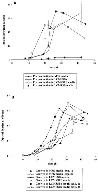
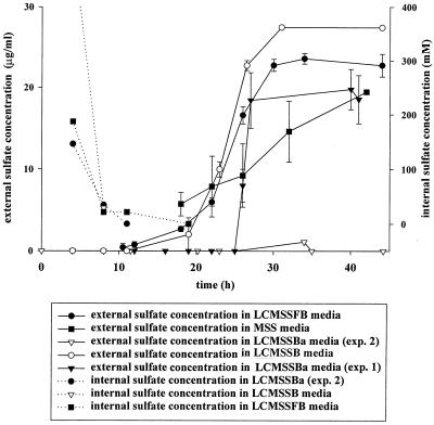
Fermentation of strain CS-87 was performed with MSS media to establish a baseline of Ptx and SO42− production. Ptx production ranged from 2 to 4 μg/ml (Fig. 6A), and external SO42− accumulation reached 15 μg/ml at 24 h (Fig. 7). Initial fermentation conditions were designed to determine if certain amino acids were required for growth of B. pertussis, particularly cysteine and methionine, two potential sources of sulfur. In these amino acid “rescue” experiments, cultures grown in shake flasks were added to the bioreactor containing MSS medium lacking arginine, methionine, and cysteine. When these cultures stopped dividing, arginine, methionine, and cysteine were sequentially added to determine which amino acid was required to restore growth. Only the addition of cysteine was capable of restoring growth. Addition of cysteine was followed by the release of SO42− in the medium (data not shown).
FIG. 6.
Ptx production (A) and cell growth (B) of B. pertussis strain CS-87 grown in various MSS media. The average and 1 standard deviation of the mean of two cultures grown in MSS and LCMSSBa media are represented (A). The averages from the LCMSSFB and LCMSSB cultures are also shown, for which the coefficient of variance of the assay is <25%.
FIG. 7.
Internal and external sulfate levels of B. pertussis CS-87 grown in MSS media. External sulfate levels were direct measurement of culture supernatants that were filter sterilized through a 0.2-μm-pore-diameter filter. Internal sulfate levels were a measurement of a threefold concentrate of the internal aqueous component of the cells. The concentration of internal sulfate was then calculated by taking into consideration the volume of water in all the cells at each time point and the threefold concentration factor. The averages for each sample are plotted for each time point. Error bars represent 1 standard deviation of the mean for the external sulfate analysis.
Since cysteine was required for growth and the addition of cysteine initiated production of sulfate, experiments were designed to reduce the concentration of cysteine in the medium. The first derivatives of MSS medium used in these experiments contained limiting amounts of cysteine in batch mode (LCMSSB). The vessel contained only 14% of the cysteine at the time of inoculation and only 50% of the cysteine at the time of supplement addition.
To determine if this formulation would lead to an increase in Ptx production without affecting the growth rate, a culture of B. pertussis strain CS-87 was used to inoculate the fermentor containing this media. Growth in LCMSSB medium was comparable to growth in MSS medium. The maximum ODs for MSS and LCMSSB media were 6.67 and 7.0 at 32 and 31 h, respectively (Fig. 6B). The maximum Ptx level was 70.5 μg/ml at 26.5 h in LCMSSB medium (Fig 6A), while the maximum Ptx level in MSS medium was 4.4 μg/ml at 32 h.
To determine if addition of limiting amounts of cysteine in fed-batch mode fermentation (LCMSSFB) would also increase Ptx yields, a fermentation experiment was designed to supply 660 mg of cysteine to the culture at a linear addition rate of 20 mg/h for 32 h. This method of adding cysteine to an actively growing culture resulted in a slower growth rate and reached a maximum OD of 4.88 at 34 h (Fig 6B). However, this addition strategy results in a similar increase in Ptx production, as observed in the batch mode yielding 54 μg/ml at 30 h (Fig 6A).
Effects of adding the Ba2+ ion to the medium on Ptx production.
Experiments were then designed that not only limited cysteine in the medium, but also precipitated excess internal and external SO42− with BaCl2 (LCMSSBa). In LCMSSBa fermentation, BaCl2 was added to the culture at 12 h to a final concentration of 20 mM. Addition of BaCl2 did not significantly affect the final OD of B. pertussis strain CS-87 compared to that with MSS medium (Fig. 6B). The ODs in the two LCMSSBa experiments were >4.0 after 36 h and >6.0 after 28 h (Fig. 6B), and the Ptx yields were greater than those observed in LCMSSB or LCMSSFB experiments (Fig. 6A). The amounts of Ptx produced in the two independent LCMSSBa experiments were 65 μg/ml after 32 h in experiment 1 and 80.8 μg/ml after 18 h in experiment 2.
Ptx production rates.
The highest specific production rates of the B. pertussis cells grown in medium that limited cysteine were 14.6 μg/ml/OD (LCMSSB) at 23 h and 14.1 μg/ml/OD (LCMSSFB) at 26 h, respectively. By combining smaller amounts of cysteine with precipitation of sulfate from the cultures by using BaCl2, Ptx production rates increased to 17.2 μg/ml/OD at 19.2 h in experiment 1 and 35.8 μg/ml/OD at 20.2 h in experiment 2. In comparison, the highest specific productivity in MSS medium was 1.0 μg/ml/OD at 22 h. Ptx production in LCMSSBa cultures reached the highest levels sooner in fermentation than those in cultures grown in medium that limited cysteine alone. Similar results in Ptx production were obtained with ATCC strain 9797 with these media (data not shown).
Effect of SO42− accumulation and release on Ptx production.
To determine if the decoupling of Ptx production with growth rate was signaled by the metabolism of cysteine, external SO42− levels were measured during fermentation. The accumulation of SO42− in LCMSSB and LCMSSFB cultures occurred later in fermentation than in MSS cultures and did not reach >10 μg/ml until after 24 h (Fig. 7). This correlated well with the time at which the Ptx production rate of the culture reached its maximum. In cultures containing BaCl2, SO42− accumulation was observed later in fermentation in experiment 1 and was largely undetectable in experiment 2 (Fig. 7). These results suggest that the concentration of BaCl2 in the medium was sufficient to sequester most of the internal and external SO42−.
The next experiments were designed to determine if intracellular SO42− levels could be used to predict Ptx production patterns. Intracellular SO42− levels of cells grown in medium that limited cysteine were rapidly diluted by cell division in early stages of fermentation (Fig. 7). The dissipation of internal SO42− patterned the growth rate of the culture (Fig. 6B and 7). In these cultures, the internal SO42− levels dropped below 20 mM between 10 and 14 h in fermentation (Fig. 7). Cells that eliminated internal SO42− more rapidly (Fig. 7) appear to produce Ptx sooner in fermentation (Fig. 6A), which resulted in an increase in the total Ptx yield.
DISCUSSION
Immunization with an acellular pertussis vaccine formulated with detoxified Ptx has been shown to be a simple, safe, and effective way to prevent whooping cough in infants (17). Formation of the active toxin requires the assembly of five different subunits in the periplasm (36) and secretion through a highly selective gated channel in the outer membrane composed of at least nine Ptl proteins (55). In the absence of adequate secretion machinery and/or the presence of inactive toxin, Ptx accumulates in the cell (11, 53, 55). Therefore, the bacteria must properly assemble the toxin as well as produce the appropriate number of functional secretion machinery. Virulence factors are synthesized in response to environmental signals under the control of the bvg regulatory locus by transactivation of its own autoregulated promoter and the promoter of FHA followed by transactivation of cyaA and Ptx (42). Isolation of spontaneous mutations in BvgS that are less sensitive to environmental signals results in the constitutive synthesis of multiple bvg-regulated loci. (34). Other than the effects of environmental stimuli on the BvgAS regulatory activity, B. pertussis has not been shown to self-modulate virulent factor expression in response to growth conditions by altering its metabolic pathways.
Studies have shown that virulence factor expression is influenced by temperature, pH, glutamate levels, free fatty acids, and environmental compounds like nicotinic acid and SO42− (15, 16, 23, 25, 26, 35, 37, 56). Addition of heptakis-(2,6-O-dimethyl)-α-cyclodextrin to the medium increases Ptx yields significantly (15, 16, 58). However, addition of this complex compound to the growth medium may pose adverse effects if it contaminates the formulated vaccine product. Our studies have shown that the simple reduction in the cysteine in growth medium enhanced Ptx production. Similar results were observed with ATCC strain 9797, a Tohama I derivative. This suggests that the bacterium responds to the cysteine concentrations in its environment as well as autoregulates virulence through recognition of exogenous and/or endogenous SO42−.
Addition of BaCl2 to the medium enhanced Ptx yields and appears to stabilize Ptx in the medium after cells enter the stationary growth phase (Fig. 6A). This suggests that the Ba2+ ion can compete for both intracellular and extracellular SO42− in actively growing cells to prevent recognition of SO42− by the bvg sensory system. This compound may also act to stabilize the toxin and/or prevent it from reassociating with membrane blebs in the medium. By modulating the concentration of cysteine and, in turn, SO42− in the medium, we have also observed an increase in the synthesis and secretion of other virulence factors like FHA, CyaA, and pertactin (data not shown). Additional experiments will focus on how various concentrations of cysteine affect each of these other components. Moreover, the rate of addition of cysteine may provide the cells with sufficient amounts of cysteine for growth yet minimize the conversion to SO42− and accumulation of SO42− within the cell and release into the medium.
Bacteria use a wide variety of metabolic enzymes to cause disease. B. pertussis cells have been able to coexist in the host environment due to their ability to adapt to the environment and evolve mechanisms to combat host defense mechanisms. These mechanisms allow B. pertussis to colonize and reproduce in the host and evade and disable the host defense system, as well as facilitate their transmission to a new host, propagating their infectious life cycle. B. pertussis has been shown to regulate its virulence genes by the ability to recognize diverse environmental signals. Based on our current understanding of the transmission and the etiology of whooping cough, there is very little knowledge of why this bacterium would turn its virulence factors on and off in the host. However, it is interesting to note that B. pertussis causes a severe infection in infants, whereas, most adult infections are less symptomatic. This might suggest the physiologic environment found in each might reflect differences in stimuli to the bacterium. Moreover, recent studies have shown that in vivo the Bvg states may not be the only important determinant for B. pertussis pathogenesis, suggesting an alternative mechanism for regulating virulence in the human host (29).
Investigators have postulated that the ionic composition of human blood plasma may serve as a signal stimulus for the initiation of virulence, since maximum expression of Ptx occurs at a sodium concentration at or above 140 mM, the human physiological sodium concentration (15). It has also been found that the concentration of cysteine in plasma is 70 to 108 μmol/liter in adults compared to 10 to 40 μmol/liter in the infant (10). Our studies on the growth of B. pertussis in fermentation suggest that the conversion of cysteine to SO42− may be an important mechanism used by the organism in regulating its virulence and expressing its virulence factors. Therefore, the amount and availability of cysteine in the physiological environment encountered by the organism may be one of the important factors in the differences found in the clinical manifestations of this disease in adult and infant populations.
The ability of B. pertussis to switch from bvg-activated to bvg-repressed genes suggests that the organism has evolved a quorum sensing mechanism(s) that controls its virulence. Recent studies suggest that this may not be the only mechanism, since the regulation of the bvg-repressed genes is not required for B. pertussis disease in the mouse aerosal challenge model, and regulation of the bvg-activated genes is not required for virulence (33). Examination and analysis of the metabolic utilization of the nutrients required for B. pertussis growth and virulence factor production suggest that quorum sensing of the external environment may not be the only mechanism. B. pertussis seems to have evolved a mechanism that may sense the internal metabolites in relationship to its growth phase as well. If there is a phase in which B. pertussis modulates expression of the bvg-regulated genes inside the host, it must occur late in infection at a time when the bacteria has established a disease state. Our results suggest that this may occur as the bacteria reach a certain cell density or growth state at which cysteine metabolism is highly active. The relationship between cysteine and the production of pyruvic acid and SO42− observed during different growth phases suggests that the metabolism of cysteine may play an important role in the virulence of the organism. Further examination of the feedback mechanism utilized by the bacterium will require the isolation and characterization of the enzymes involved in cysteine metabolism.
ACKNOWLEDGMENTS
We gratefully thank Susan Mindel, Kirk Bayones, John Salim, Wei Yuan, and Misha Donets of the Baxter Healthcare Corporation for technical assistance and Max Kristiansen for assistance with statistical analysis. We also gratefully thank Scott Stibitz (Center for Biologics Evaluation and Research, FDA, Bethesda, Md.), Mark S. Peppler and Trevor H. Stenson (University of Alberta, Edmonton, Canada), Jeffrey F. Miller (University of California, Los Angeles, Calif.), and Akshay Goel (Baxter Healthcare Corporation) for reagents, technical assistance, and scientific advice.
REFERENCES
- 1.Andorn N, Kaufman J B, Clem T R, Fass R, Shiloach J. Large-scale growth of Bordetella pertussis for production of extracellular toxin. Ann N Y Acad Sci. 1990;589:363–371. doi: 10.1111/j.1749-6632.1990.tb24258.x. [DOI] [PubMed] [Google Scholar]
- 2.Arico B, Miller J F, Roy C, Stibitz S, Monack D, Falkow S, Gross R, Rappuoli R. Sequences required for expression of Bordetella pertussis virulence factors share homology with prokaryotic signal transduction proteins. Proc Natl Acad Sci USA. 1989;86:6671–6675. doi: 10.1073/pnas.86.17.6671. [DOI] [PMC free article] [PubMed] [Google Scholar]
- 3.Beattie D T, Mahan M J, Mekalanos J J. Repressor binding to a regulatory site in the DNA coding sequence is sufficient to confer transcriptional regulation of the vir-repressed genes (vrg genes) in Bordetella pertussis. J Bacteriol. 1993;175:519–527. doi: 10.1128/jb.175.2.519-527.1993. [DOI] [PMC free article] [PubMed] [Google Scholar]
- 4.Beattie D T, Shahin R, Mekalanos J J. A vir-repressed gene of Bordetella pertussis is required for virulence. Infect Immun. 1992;60:571–577. doi: 10.1128/iai.60.2.571-577.1992. [DOI] [PMC free article] [PubMed] [Google Scholar]
- 5.Boucher P E, Murakami K, Ishihama A, Stibitz S. Nature of DNA binding and RNA polymerase interaction of the Bordetella pertussis BvgA transcriptional activator at the fha promoter. J Bacteriol. 1997;179:1755–1763. doi: 10.1128/jb.179.5.1755-1763.1997. [DOI] [PMC free article] [PubMed] [Google Scholar]
- 6.Boucher P E, Stibitz S. Synergistic binding of RNA polymerase and BvgA phosphate to the pertussis toxin promoter of Bordetella pertussis. J Bacteriol. 1995;177:6486–6491. doi: 10.1128/jb.177.22.6486-6491.1995. [DOI] [PMC free article] [PubMed] [Google Scholar]
- 7.Boucher P E, Yang M-S, Schmidt D M, Stibitz S. Genetic and biochemical analyses of BvgA interaction with the secondary binding region of the fha promoter of Bordetella pertussis. J Bacteriol. 2001;183:536–544. doi: 10.1128/JB.183.2.536-544.2001. [DOI] [PMC free article] [PubMed] [Google Scholar]
- 8.Carbonetti N H, Romashko A, Irish T J. Overexpression of the RNA polymerase alpha subunit reduces transcription of Bvg-activated virulence genes in Bordetella pertussis. J Bacteriol. 2000;182:529–531. doi: 10.1128/jb.182.2.529-531.2000. [DOI] [PMC free article] [PubMed] [Google Scholar]
- 9.Craig-Mylius K A, Stenson T H, Weiss A A. Mutations in the S1 subunit of pertussis toxin that affect secretion. Infect Immun. 2000;68:1276–1281. doi: 10.1128/iai.68.3.1276-1281.2000. [DOI] [PMC free article] [PubMed] [Google Scholar]
- 10.Diem K, Lentner C. Scientific tables. Documenta Geigy, 7th ed., no. 574. Basel, Switzerland: Ciba-Geigy, Ltd.; 1970. [Google Scholar]
- 11.Farizo K M, Huang T, Burns D L. Importance of holotoxin assembly in Ptl-mediated secretion of pertussis toxin from Bordetella pertussis. Infect Immun. 2000;68:4049–4054. doi: 10.1128/iai.68.7.4049-4054.2000. [DOI] [PMC free article] [PubMed] [Google Scholar]
- 12.Fernandez R C, Weiss A A. Serum resistance in bvg-regulated mutants of Bordetella pertussis. FEMS Microbiol Lett. 1998;163:57–63. doi: 10.1111/j.1574-6968.1998.tb13026.x. [DOI] [PubMed] [Google Scholar]
- 13.Field L H, Parker C D. Effects of fatty acids on growth of Bordetella pertussis in defined medium. J Clin Microbiol. 1979;9:651–653. doi: 10.1128/jcm.9.6.651-653.1979. [DOI] [PMC free article] [PubMed] [Google Scholar]
- 14.Finn T M, Amsbaugh D F. Vag8: a Bordetella pertussis bvg-regulated protein. Infect Immun. 1998;66:3985–3989. doi: 10.1128/iai.66.8.3985-3989.1998. [DOI] [PMC free article] [PubMed] [Google Scholar]
- 15.Frohlich B T, Bernardez Clark E R, Siber G R, Swartz R W. Improved pertussis toxin production by Bordetella pertussis through adjusting the growth medium's ionic composition. J Biotechnol. 1995;39:205–219. doi: 10.1016/0168-1656(95)00013-g. [DOI] [PubMed] [Google Scholar]
- 16.Frohlich B T, d'Alarcao M, Feldberg R S, Nicholson M L, Siber G R, Swartz R W. Formation and cell-medium partitioning of autoinhibitory free fatty acids and cyclodextrin's effect in the cultivation of Bordetella pertussis. J Biotechnol. 1996;45:137–148. doi: 10.1016/0168-1656(95)00155-7. [DOI] [PubMed] [Google Scholar]
- 17.Heron I, Chen F M, Fusco J. DTaP vaccines from North American vaccine (NAVA): composition and critical parameters. Biologicals. 1999;27:91–96. doi: 10.1006/biol.1999.0187. [DOI] [PubMed] [Google Scholar]
- 18.Hozbor D, Rodriguez M E, Fernandez J, Lagares A, Guiso N, Yantorno O. Release of outer membrane vesicles from Bordetella pertussis. Curr Microbiol. 1999;38:273–278. doi: 10.1007/pl00006801. [DOI] [PubMed] [Google Scholar]
- 19.Ibsen P, Heron I. Quantitation of pertussis toxin in an enzyme linked immunosorbent assay with improved specificity. Biologicals. 1990;18:123–126. doi: 10.1016/1045-1056(90)90022-r. [DOI] [PubMed] [Google Scholar]
- 20.Ibsen P H, Holm A, Petersen J W, Olsen C E, Heron I. Identification of B-cell epitopes on the S4 subunit of pertussis toxin. Infect Immun. 1993;61:2408–2418. doi: 10.1128/iai.61.6.2408-2418.1993. [DOI] [PMC free article] [PubMed] [Google Scholar]
- 21.Kinnear S M, Boucher P E, Stibitz S, Carbonetti N H. Analysis of BvgA activation of the pertactin gene promoter in Bordetella pertussis. J Bacteriol. 1999;181:5234–5241. doi: 10.1128/jb.181.17.5234-5241.1999. [DOI] [PMC free article] [PubMed] [Google Scholar]
- 22.Knapp S, Mekalanos J J. Two trans-acting regulatory genes (vir and mod) control antigenic modulation in Bordetella pertussis. J Bacteriol. 1988;170:5059–5066. doi: 10.1128/jb.170.11.5059-5066.1988. [DOI] [PMC free article] [PubMed] [Google Scholar]
- 23.Lacey B W. Antigenic modulation of Bordetella pertussis. J Hyg. 1960;58:57–93. doi: 10.1017/s0022172400038134. [DOI] [PMC free article] [PubMed] [Google Scholar]
- 24.Laoide B M, Ullmann A. Virulence dependent and independent regulation of the Bordetella pertussis cya operon. EMBO J. 1990;9:999–1005. doi: 10.1002/j.1460-2075.1990.tb08202.x. [DOI] [PMC free article] [PubMed] [Google Scholar]
- 25.Licari P, Siber G R, Swartz R. Production of cell mass and pertussis toxin by Bordetella pertussis. J Biotechnol. 1991;20:117–129. doi: 10.1016/0168-1656(91)90221-g. [DOI] [PubMed] [Google Scholar]
- 26.Licari P, Winberry L, Swartz R. The effect of pH on the production of pertussis toxin by Bordetella pertussis. J Biotechnol. 1991;17:189–193. doi: 10.1016/0168-1656(91)90009-k. [DOI] [PubMed] [Google Scholar]
- 27.Locht C. Molecular aspects of Bordetella pertussis pathogenesis. Int Microbiol. 1999;2:137–144. [PubMed] [Google Scholar]
- 28.Marques R R, Carbonetti N H. Genetic analysis of pertussis toxin promoter activation in Bordetella pertussis. Mol Microbiol. 1997;24:1215–1224. doi: 10.1046/j.1365-2958.1997.4371792.x. [DOI] [PubMed] [Google Scholar]
- 29.Martinez de Tejada G, Cotter P A, Heininger U, Camilli A, Akerley B J, Mekalanos J J, Miller J F. Neither the Bvg− phase nor the vrg6 locus of Bordetella pertussis is required for respiratory infection in mice. Infect Immun. 1998;66:2762–2768. doi: 10.1128/iai.66.6.2762-2768.1998. [DOI] [PMC free article] [PubMed] [Google Scholar]
- 30.Melnicoff M J, Godleski J J, Bercz J P. An automated method for the determination of sulfate. Res Commun Chem Pathol Pharmacol. 1976;14:377–386. [PubMed] [Google Scholar]
- 31.Melton A R, Weiss A A. Environmental regulation of expression of virulence determinants in Bordetella pertussis. J Bacteriol. 1989;171:6206–6212. doi: 10.1128/jb.171.11.6206-6212.1989. [DOI] [PMC free article] [PubMed] [Google Scholar]
- 32.Melton A R, Weiss A A. Characterization of environmental regulators of Bordetella pertussis. Infect Immun. 1993;61:807–815. doi: 10.1128/iai.61.3.807-815.1993. [DOI] [PMC free article] [PubMed] [Google Scholar]
- 33.Merkel T J, Stibitz S, Keith J M, Leef M, Shahin R. Contribution of regulation by the bvg locus to respiratory infection of mice by Bordetella pertussis. Infect Immun. 1998;66:4367–4373. doi: 10.1128/iai.66.9.4367-4373.1998. [DOI] [PMC free article] [PubMed] [Google Scholar]
- 34.Miller J F, Johnson S A, Black W J, Beattie D T, Mekalanos J J, Falkow S. Constitutive sensory transduction mutations in the Bordetella pertussis bvgS gene. J Bacteriol. 1992;174:970–979. doi: 10.1128/jb.174.3.970-979.1992. [DOI] [PMC free article] [PubMed] [Google Scholar]
- 35.Miller J F, Mekalanos J J, Falkow S. Coordinate regulation and sensory transduction in the control of bacterial virulence. Science. 1989;243:916–922. doi: 10.1126/science.2537530. [DOI] [PubMed] [Google Scholar]
- 36.Pizza M, Bartoloni A, Prugnola A, Silvestri S, Rappuoli R. Subunit S1 of pertussis toxin: mapping of the regions essential for ADP-ribosyltransferase activity. Proc Natl Acad Sci USA. 1988;85:7521–7525. doi: 10.1073/pnas.85.20.7521. [DOI] [PMC free article] [PubMed] [Google Scholar]
- 37.Prugnola A, Arico B, Manetti R, Rappuoli R, Scarlato V. Response of the bvg regulon of Bordetella pertussis to different temperatures and short-term temperature shifts. Microbiology. 1995;141:4–34. doi: 10.1099/13500872-141-10-2529. [DOI] [PubMed] [Google Scholar]
- 38.Robinson A, Irons L I, Ashworth L A. Pertussis vaccine: present status and future prospects. Vaccine. 1985;3:11–22. doi: 10.1016/0264-410x(85)90004-0. [DOI] [PubMed] [Google Scholar]
- 39.Ross E M. Immunisation against diphtheria, tetanus, pertussis and polio 5. Practitioner. 1985;229:795–799. [PubMed] [Google Scholar]
- 40.Roy C R, Miller J F, Falkow S. The bvgA gene of Bordetella pertussis encodes a transcriptional activator required for coordinate regulation of several virulence genes. J Bacteriol. 1989;171:6338–6344. doi: 10.1128/jb.171.11.6338-6344.1989. [DOI] [PMC free article] [PubMed] [Google Scholar]
- 41.Rutter D A, Ashworth L A, Day A, Funnell S, Lovell F, Robinson A. Trial of a new acellular pertussis vaccine in healthy adult volunteers. Vaccine. 1988;6:29–32. doi: 10.1016/0264-410x(88)90010-2. [DOI] [PubMed] [Google Scholar]
- 42.Scarlato V, Arico B, Prugnola A, Rappuoli R. Sequential activation and environmental regulation of virulence genes in Bordetella pertussis. EMBO J. 1991;10:3971–3975. doi: 10.1002/j.1460-2075.1991.tb04967.x. [DOI] [PMC free article] [PubMed] [Google Scholar]
- 43.Scarlato V, Prugnola A, Arico B, Rappuoli R. The bvg-dependent promoters show similar behaviour in different Bordetella species and share sequence homologies. Mol Microbiol. 1991;5:2493–2498. doi: 10.1111/j.1365-2958.1991.tb02094.x. [DOI] [PubMed] [Google Scholar]
- 44.Schou C, Au-Jensen M, Heron I. The interaction between pertussis toxin and 10 monoclonal antibodies. Acta Pathol Microbiol Immunol Scand C. 1987;95:177–187. doi: 10.1111/j.1699-0463.1987.tb00028.x. [DOI] [PubMed] [Google Scholar]
- 45.Sekura R D, Fish F, Manclark C R, Meade B, Zhang Y L. Pertussis toxin. Affinity purification of a new ADP-ribosyltransferase. J Biol Chem. 1983;258:14647–14651. [PubMed] [Google Scholar]
- 46.Stainer D W, Scholte M J. A simple chemically defined medium for the production of phase I Bordetella pertussis. J Gen Microbiol. 1970;63:211–220. doi: 10.1099/00221287-63-2-211. [DOI] [PubMed] [Google Scholar]
- 47.Steffen P, Goyard S, Ullmann A. Phosphorylated BvgA is sufficient for transcriptional activation of virulence-regulated genes in Bordetella pertussis. EMBO J. 1996;15:102–109. [PMC free article] [PubMed] [Google Scholar]
- 48.Stenson T H, Peppler M S. Identification of two bvg-repressed surface proteins of Bordetella pertussis. Infect Immun. 1995;63:3780–3789. doi: 10.1128/iai.63.10.3780-3789.1995. [DOI] [PMC free article] [PubMed] [Google Scholar]
- 49.Stibitz S, Aaronson W, Monack D, Falkow S. Phase variation in Bordetella pertussis by frameshift mutation in a gene for a novel two-component system. Nature. 1989;338:266–269. doi: 10.1038/338266a0. [DOI] [PubMed] [Google Scholar]
- 50.Stibitz S, Yang M S. Subcellular localization and immunological detection of proteins encoded by the vir locus of Bordetella pertussis. J Bacteriol. 1991;173:4288–4296. doi: 10.1128/jb.173.14.4288-4296.1991. [DOI] [PMC free article] [PubMed] [Google Scholar]
- 51.Thalen M, van den I J, Jiskoot W, Zomer B, Roholl P, de Gooijer C, Beuvery C, Tramper J. Rational medium design for Bordetella pertussis: basic metabolism. J Biotechnol. 1999;75:147–159. doi: 10.1016/s0168-1656(99)00155-8. [DOI] [PubMed] [Google Scholar]
- 52.Uhl M A, Miller J F. Autophosphorylation and phosphotransfer in the Bordetella pertussis BvgAS signal transduction cascade. Proc Natl Acad Sci USA. 1994;91:1163–1167. doi: 10.1073/pnas.91.3.1163. [DOI] [PMC free article] [PubMed] [Google Scholar]
- 53.Weiss A A, Goodwin M S M. Lethal infection by Bordetella pertussis mutants in the infant mouse model. Infect Immun. 1989;57:3757–3764. doi: 10.1128/iai.57.12.3757-3764.1989. [DOI] [PMC free article] [PubMed] [Google Scholar]
- 54.Weiss A A, Hewlett E L, Myers G A, Falkow S. Genetic studies of the molecular basis of whooping cough. Dev Biol Stand. 1985;61:11–19. [PubMed] [Google Scholar]
- 55.Weiss A A, Johnson F D, Burns D L. Molecular characterization of an operon required for pertussis toxin secretion. Proc Natl Acad Sci USA. 1993;90:2970–2974. doi: 10.1073/pnas.90.7.2970. [DOI] [PMC free article] [PubMed] [Google Scholar]
- 56.Weiss A A, Melton A R, Walker K E, Andraos-Selim C, Meidl J J. Use of the promoter fusion transposon Tn5 lac to identify mutations in Bordetella pertussis vir-regulated genes. Infect Immun. 1989;57:2674–2682. doi: 10.1128/iai.57.9.2674-2682.1989. [DOI] [PMC free article] [PubMed] [Google Scholar]
- 57.Willems R, Paul A H, van der Heide G, ter Avest A R, Mooi F R. Fimbrial phase variation in Bordetella pertussis: a novel mechanism for transcriptional regulation. EMBO J. 1990;9:2803–2809. doi: 10.1002/j.1460-2075.1990.tb07468.x. [DOI] [PMC free article] [PubMed] [Google Scholar]
- 58.Zealey G R, Loosmore S M, Yacoob R K, Cockle S A, Boux L K, Miller L D, Klein M H. Gene replacement in Bordetella pertussis by transformation with linear DNA. Bio/Technology. 1990;8:1025–1029. doi: 10.1038/nbt1190-1025. [DOI] [PubMed] [Google Scholar]


