Abstract
The last two decades have seen a shift in the way anatomy education is delivered. With the introduction of blended learning, cadaveric dissection is no longer the be all and end all and, in many cases, the continuing role of anatomical teaching artefacts has declined after decades of prominence. While some institutions have abandoned their archaic anatomical collections and medical museums completely, others have invested in their technological enhancement. We describe the integration of historical teaching artefacts into contemporary anatomy education through the development of an interactive online e‐platform and shed light on the enduring pedagogic value of past anatomical teaching specimen.
Keywords: anatomy education, blended learning, gross anatomy, medical museums, pedagogy
The last two decades have seen a shift in the way anatomy education is delivered. With the introduction of blended learning, cadaveric dissection is no longer the be all and end all and, in many cases, the continuing role of anatomical teaching artifacts has declined after decades of prominence. While some institutions have abandoned their archaic anatomical collections and medical museums completely, others have invested in their technological enhancement. We describe the integration of historical teaching artefacts into contemporary anatomy education through the development of an interactive online e‐platform and shed light on the enduring pedagogic value of past anatomical teaching specimen.

Anatomy is a cornerstone of medical education. Clinically applied anatomy carries professional relevance for students and cadaver‐led programmes are representative of students' first patient–clinician interaction. With the introduction of web‐based courses, computer simulations of the human body and the increased engagement of students with online resources, the last two decades have seen a paradigm shift in the way anatomy education is delivered (Barry et al., 2016). Pedagogical modalities of practical anatomy education have evolved to include radiology and digitised body simulations to complement cadaveric dissection and present‐day anatomical models. Moreover, the role of medical museums and the anatomical teaching specimen contained therein has completely eroded after centuries of prominence (Marreez et al., 2010). Prior to the advancement of tissue preservation techniques, anatomical collections of typical and pathological wax models, plaster casts, formalin‐filled specimen containers, osteology, illustrations and displays allowed for more engagement throughout a student's study than a post‐morbid and deteriorating dissection (Cooke, 2010). However, where possible, access to a medical museum and past teaching specimen collections has presented anatomists with an opportunity to develop strategies that rejuvenate historical teaching content for current generations of students (Mogali et al., 2019; Sugiura et al., 2019; Wakefield, 2007). As such, there are several ways in which student engagement with medical museums can be facilitated. Students can be invited into medical museums to be shown exceptional teaching resources as part of practical learning sessions, specimen may be brought to anatomy learning spaces such as dissection theatres or they may be digitised for “on demand” use by students through their virtual learning environments.
However, some institutions have abandoned their medical museums, and understandably so. Historic collections require ongoing maintenance, fiscal input and curatorial skill to continually preserve and store specimen. Others have decided it is worth the cost and invested in their dissemination through technological enhancement. For instance, the anatomical sciences have been supplemented by augmented reality at the University of Yamanashi, Japan (Sugiura et al., 2019) and a digital repository of historical specimen has been made available to students through the development of quick response (QR) codes at the Lee Kong Chain School of Medicine, Singapore (Mogali et al., 2019). Most of the students surveyed there found this system to be beneficial to their learning, from revision to accessing case histories associated with specimens. Notwithstanding the pedagogical positives, these infrastructural investments raise questions as to whether medical museums are themselves separate entities or perhaps represent opportunities for educational interfaces with anatomical facilities. Marreez et al. (2010) argued that the benefits of a museum cannot be achieved using multimedia simulations as museums improve observational and practical skills that are difficult to transfer to students virtually. But medical museums for students in years gone by were not museums. They were learning facilities where student collaborations were frequent and artefacts were highly valued as instruments for learning, due to their delicate nature and rarity. This was in part due to the limited number of students who could avail of a specimen, such as rare wax models, which ensured that small‐group engagement was a key component of learning activities (Cooke, 2010; Mitchell et al., 2011). The experience of a student entering a medical museum then, and now, is one that is immersive. The sights and smells invite visitors to observe. This represents a fundamentally different experience to the cadaver laboratory where learning is stimulated by tactile involvement. Students can reflect superficial layers to expose deeper structures and can orient musculature, viscera and neurovascular bundles in a way that imitates atlases or depictive diagrams. Alternatively, the medical museum is experiential and engages students in a different way. For example, larger artefacts may be rotated and displayed from multiple perspectives, but tactile engagement may be limited due to their encasement or presentation. This process decelerates the learner's pedagogical objectives encouraging them to take a step back and carefully inspect the object in front of them.
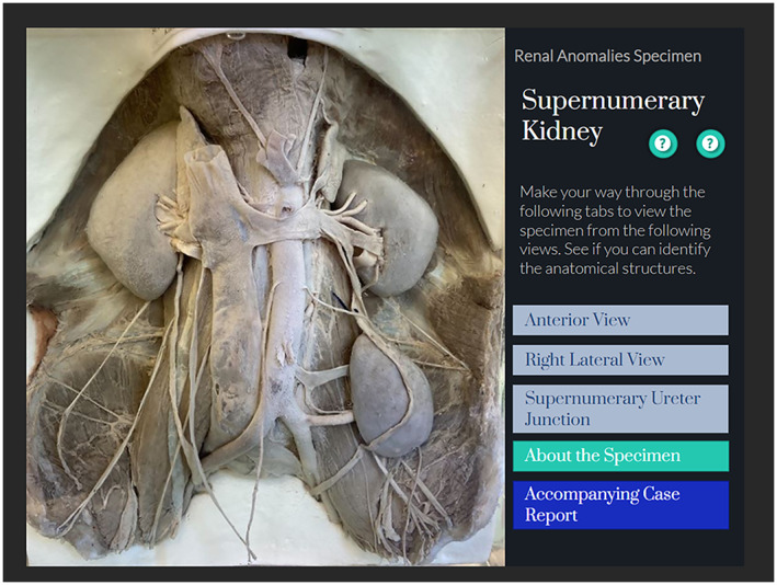
The Old Anatomy Museum at Trinity College Dublin houses an extensive collection of historical teaching artefacts, many of which retain extraordinary pedagogical value. However, due to their delicate nature, repairs and potentially sensitive content, these artefacts are rarely used for student learning. Artefacts include historic plaster casts, posters, cadaveric specimen and wax models that visually describe the human form from unique perspectives, revealing key insights into typical and pathological anatomy, which are very difficult to prosect. Learning from the past is also strengthened in the rarity of some specimen which gives students an appreciation of the dedication and skill required to prepare teaching tools of such exceptional quality. To exhibit these teaching artefacts for anatomy students, we have developed an interactive online platform which showcases digitised versions of these historical teaching tools (see Figure 1). This was achieved using the Articulate 360© online training tool, which is designed to enable content creators to share their content in an educational setting. The platform was then integrated into the University's virtual learning environment Blackboard©. Interactive labels, hover points and images from multiple angles simulate student attendance at a medical museum and mimic the experiential elements aforementioned, such as improvement of observational skills. Through the inclusion of insightful provenances and case histories, students are taken on a journey back in time to learn not only from the original donors and artifacts but also from the physicians and anatomists who treated and prepared the specimen, offering fascinating insights into the healthcare systems and the education values of the time. This platform is also available to view for the public, a condition of the funding for this project with a creative commons non‐commercial, no‐derivatives attribution. Previous studies have shown benefits from public engagement with similar applications (Jędrzejewski et al., 2020). Careful consideration was given to which specimens were to be showcased. Specifically, those over a 100 years old and without identifying features.
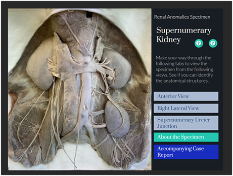
FIGURE 1.

Excerpt from historical anatomy tool at Trinity College Dublin. Students may view the historical artefact from a number of perspectives. Anatomical knowledge and relevant historical provenance is also provided. The dissection was prepared and discussed by Prof. Andrew Francis Dixon of Trinity College Dublin in 1911 (Dixon, 1911). Image of specimen courtesy of the Old Anatomy Museum, Trinity College Dublin
Advanced technical considerations such as the use of 2D images over 3D renderings of teaching specimen must be decided on the basis of practicality. For us, it was important to consider the cost, time and resources required for 3D renderings; however, there has been much success with 3D renderings of anatomical specimens (Moore et al., 2017; Venkatesh et al., 2013). A key goal was to enhance blended engagement within our Discipline as a response to an institutional learning strategy. Specific examples of relevant curricular content on the platform are signposted to students during lectures. Engagement has exceeded expectations and the response to the platform has been exceptionally positive.
What is pertinent to the usability of such digital resources is that they supplement existing curricular elements such as in‐person practical sessions and lectures and thus should not be seen as substitutes. Furthermore, in some cases, content that was originally intended to be delivered face‐to‐face has been transferred to virtual learning environments (Patra et al., 2022). This reallocation has been conducted with often little consideration as to whether the content is accessible, interactive or suitable for self‐directed learning. In this new era of blended learning, the concept of cognitive load is also particularly relevant. Cognitive load theory can loosely be defined as the amount of mental effort needed to learn or complete a task (Hadie et al., 2018). Specifically, extraneous cognitive load refers to factors in the environment that support or impede learning such as the way information is presented, and cognitive overload results when working memory becomes overwhelmed by too much information or information presented in incoherent ways (Van Nuland & Rogers, 2016). To reduce cognitive load, care was taken for our platform to be viewed as an optional resource for students and content does not directly form part of assessments. Consideration should therefore be made to ensure that the educational merit of historical artefacts outweighs the potential for cognitive overload, such that it does not overburden students in already congested medical courses maintaining the transactional distance between teaching staff and students (Stone & Barry, 2019).
In addition, the digitisation of both modern and historical specimens may present ethical challenges (Cornwall et al., 2016; Hennessy & Smith, 2020). For example, the provenance of some specimen and issues of consent may be problematic. The practice of illegally procuring cadavers for teaching purposes is the most widely known (Magee, 2001). The ethics of this time in history is beyond the scope of this article, however it is important to acknowledge the past. To allow students to gain an insight into the history behind each specimen in our platform their providence and other relevant documentation is provided for students to review at the click of a button.
Successful contemporary anatomy education effectively combines lecture‐based, practical and digital pedagogical resources to deliver diverse anatomical curricular in an era of increasing transactional distance and reductions in contact time (Barry et al., 2016; Stone & Barry, 2019). Recent pandemic‐related experiences in online learning have led to irreversible advances in learning modalities, and a general acceptance that blended learning enhances students' attitude, interest and motivation to learn both core knowledge and the professional relevance of the discipline (El Sadik & Al Abdulmonem, 2021; Nagaraj et al., 2021; Sarkar et al., 2021; Ullah et al., 2021). By digitising historical resources, students can learn from both past anatomists and the contemporary educators who identify the lasting teaching relevance of anatomical artefacts. In this way, anatomical sciences education may continually build upon its foundations while engaging with the ever‐growing catalogue of blended resources. But these blasts from the past and the success of their integration into modern curricula should always be evaluated with respect to student satisfaction and engagement, and to avoid the pitfalls of cognitive overload.
If you have the luxury of a medical museum at your institution, pop in and take a look. You never know what you might find and it's not as difficult as you might expect to generate some exceptionally insightful digital content. With digital natives now accounting for most undergraduate learners, breathing new life into the anatomic past may be one way to live up to their online expectations. In our experience, it became clear that the original purpose of the historical anatomy teaching specimen at TCD had not changed across generations of education, yet their distinctiveness and function provide an enduring pedagogical legacy for our students.
AUTHOR CONTRIBUTIONS
Dearbhla P. Cullinane, Conceptualization and writing of the original manuscript. Callum Franklin, Writing and editing of the revised manuscript. Denis S. Barry, Conceptualization, editing and writing of the original and revised manuscripts.
ACKNOWLEDGMENTS
This project was funded by the National Forum for the Enhancement of Teaching and Learning in Higher Education under the Teaching and Learning Enhancement Fund. Open access funding provided by IReL.
Cullinane, D.P. , Franklin, C. & Barry, D.S. (2023) Reviving the anatomic past: Breathing new life into historic anatomical teaching tools. Journal of Anatomy, 242, 701–704. Available from: 10.1111/joa.13802
DATA AVAILABILITY STATEMENT
Data sharing is not applicable to this article as no new data were created or analyzed in this study.
REFERENCES
- Barry, D.S. , Marzouk, F. , Chulak‐Oglu, K. , Bennett, D. , Tierney, P. & O'Keeffe, G.W. (2016) Anatomy education for the YouTube generation. Anatomical Sciences Education, 9(1), 90–96. [DOI] [PubMed] [Google Scholar]
- Cooke, R.A. (2010) A moulage museum is not just a museum. Virchows Archiv, 457(5), 513–520. [DOI] [PubMed] [Google Scholar]
- Cornwall, J. , Callahan, D. & Wee, R. (2016) Ethical issues surrounding the use of images from donated cadavers in the anatomical sciences. Clinical Anatomy, 29(1), 30–36. [DOI] [PubMed] [Google Scholar]
- Dixon, A.F. (1911) Supernumerary kidney: the occurrence of three kidneys in an adult male subject. Journal of Anatomy and Physiology, 45, 117–121. [PMC free article] [PubMed] [Google Scholar]
- Hadie, S.N.H. , Hassan, A. , Mohd Ismail, Z.I. , Ismail, H.N. , Talip, S.B. & Abdul Rahim, A.F. (2018) Empowering students' minds through a cognitive load theory‐based lecture model: a metacognitive approach. Innovations in Education and Teaching International, 55(4), 398–407. [Google Scholar]
- Hennessy, C.M. & Smith, C.F. (2020) Digital and social media in anatomy education. In: Biomedical Visualisation. Cham: Springer, pp. 109–122. [DOI] [PubMed] [Google Scholar]
- Jędrzejewski, Z. , Loranger, B. & Clancy, J.A. (2020) Virtual anatomy museum: facilitating public engagement through an interactive application. In: Biomedical visualisation. Cham: Springer, pp. 1–18. [DOI] [PubMed] [Google Scholar]
- Magee, R. (2001) Art macabre: resurrectionists and anatomists. ANZ Journal of Surgery, 71(6), 377–380. [PubMed] [Google Scholar]
- Marreez, Y.M.A.H. , Willems, L.N. & Wells, M.R. (2010) The role of medical museums in contemporary medical education. Anatomical Sciences Education, 3, 249–253. [DOI] [PubMed] [Google Scholar]
- Mitchell, P.D. , Boston, C. , Chamberlain, A.T. , Chaplin, S. , Chauhan, V. , Evans, J. et al. (2011) The study of anatomy in England from 1700 to the early 20th century. Journal of Anatomy, 219(2), 91–99. [DOI] [PMC free article] [PubMed] [Google Scholar]
- Mogali, S.R. , Vallabhajosyula, R. , Ng, C.H. , Lim, D. , Ang, E.T. & Abrahams, P. (2019) Scan and learn: quick response code enabled museum for mobile learning of anatomy and pathology. Anatomical Sciences Education, 12, 664–672. [DOI] [PubMed] [Google Scholar]
- Moore, C.W. , Wilson, T.D. & Rice, C.L. (2017) Digital preservation of anatomical variation: 3D‐modeling of embalmed and plastinated cadaveric specimens using uCT and MRI. Annals of Anatomy‐Anatomischer Anzeiger, 209, 69–75. [DOI] [PubMed] [Google Scholar]
- Nagaraj, C. , Yadurappa, S.B. , Anantharaman, L.T. , Ravindranath, Y. & Shankar, N. (2021) Effectiveness of blended learning in radiological anatomy for first year undergraduate medical students. Surgical and Radiologic Anatomy, 43(4), 489–496. [DOI] [PubMed] [Google Scholar]
- Patra, A. , Asghar, A. , Chaudhary, P. & Ravi, K.S. (2022) Integration of innovative educational technologies in anatomy teaching: new normal in anatomy education. Surgical and Radiologic Anatomy, 44(1), 25–32. [DOI] [PMC free article] [PubMed] [Google Scholar]
- El Sadik, A. & Al Abdulmonem, W. (2021) Improvement in student performance and perceptions through a flipped anatomy classroom: shifting from passive traditional to active blended learning. Anatomical Sciences Education, 14(4), 482–490. [DOI] [PubMed] [Google Scholar]
- Sarkar, S. , Sharma, S. & Raheja, S. (2021) Implementation of blended learning approach for improving anatomy lectures of phase I MBBS students–learner satisfaction survey. Advances in Medical Education and Practice, 12, 413–420. [DOI] [PMC free article] [PubMed] [Google Scholar]
- Stone, D.M. & Barry, D.S. (2019) Improving virtual learning interactions: reducing the transactional distance of online anatomy modules. Anatomical Sciences Education, 12(6), 686–687. [DOI] [PubMed] [Google Scholar]
- Sugiura, A. , Kitama, T. , Toyoura, M. & Mao, X. (2019) The use of augmented reality technology in medical specimen museum tours. Anatomical Sciences Education, 12, 561–571. [DOI] [PMC free article] [PubMed] [Google Scholar]
- Ullah, R. , Siddiqui, F. , Adnan, S. , Afzal, A.S. & Sohail Zafar, M. (2021) Assessment of blended learning for teaching dental anatomy to dentistry students. Journal of Dental Education, 85(7), 1301–1308. [DOI] [PubMed] [Google Scholar]
- Van Nuland, S.E. & Rogers, K.A. (2016) E‐learning, dual‐task, and cognitive load: the anatomy of a failed experiment. Anatomical Sciences Education, 9(2), 186–196. [DOI] [PubMed] [Google Scholar]
- Venkatesh, S.K. , Wang, G. , Seet, J.E. , Teo, L.L.S. & Chong, V.F.H. (2013) MRI for transformation of preserved organs and their pathologies into digital formats for medical education and creation of a virtual pathology museum. A pilot study. Clinical Radiology, 68(3), e114–e122. [DOI] [PubMed] [Google Scholar]
- Wakefield, D. (2007) The future of medical museums: threatened but not extinct. Medical Journal of Australia, 187, 380–381. [DOI] [PubMed] [Google Scholar]
Associated Data
This section collects any data citations, data availability statements, or supplementary materials included in this article.
Data Availability Statement
Data sharing is not applicable to this article as no new data were created or analyzed in this study.


