Abstract
Patient: Male, 60-year-old
Final Diagnosis: Renal calculi
Symptoms: Urinary frequency
Clinical Procedure: —
Specialty: Urology
Objective:
Unusual or unexpected effect of treatment
Background:
The removal of concurrent ureteral and renal stones within a single procedure has always been a challenge for urological surgeons. The incorporation of single-use digital flexible ureteroscopes into laparoscopic ureterolithotomy procedures has demonstrated effective removal of concurrent stones with a good clearance rate and decreased risk of bleeding and trauma. We report the successful removal of a unilateral upper ureteral stone and a smaller renal stone with this procedure.
Case Report:
A 60-year-old man visited the outpatient clinic with an ultrasonography report that revealed a large proximal ureteral stone with moderate hydronephrosis, accompanied by bilateral renal stones and prostatic hyperplasia. He had been experiencing urinary urgency for a year and was determined to undergo lithotomy. Due to his longstanding history of coronary artery disease and myocardial ischemia, the urologists decided that concurrent stone removal within an operation would be the best treatment. A preoperative computed tomography urogram measured the left ureteral and renal stones to be 2.0×0.8 cm and 0.6 cm, respectively. Both stones were successfully removed by laparoscopic ureterolithotomy using a single-use digital flexible ureteroscope. The patient had an uneventful recovery and remained well 1 month post-operation.
Conclusions:
The application of single-use digital flexible ureteroscopes for laparoscopic ureterolithotomy has demonstrated safety, efficiency, and cost-effectiveness. The authors believe that it is a safe alternative for the removal of concurrent ureteral and renal stones, especially in patients with multiple comorbidities.
Keywords: Case Reports, Laparoscopy, Ureteroscopes
Background
Ureteral calculi >2 cm are considered large and can result in ureteral obstruction if the calculus remains in the same location for ≥2 months [1]. In complicated cases, such as unilateral large ureteral stones with a concurrent small renal stone, urological surgeons find it difficult to extract the stones in a single session. Laparoscopic ureterolithomy (LU) has demonstrated a high stone-free rate (93.3–100%) after removing large upper ureteral stones within a procedure, yet can fail mainly due to ureteral stone migration [2]. Retrograde flexible ureteroscopy performed through a laparoscopic port and ureterostomy incision has been practiced in selected patients and can successfully remove the concurrent renal stone [3]. In this case, we detail a similar surgical experience, but with a disposable flexible ureteroscope, and further discuss its advantages.
Case Report
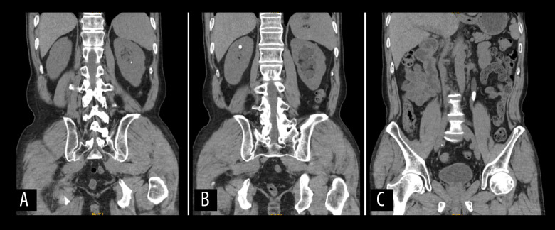
A 60-year-old man consulted our institute’s urologist with an ultrasonography report that revealed a large left proximal ureteral stone with moderate hydronephrosis, accompanied by bilateral renal stones and prostatic hyperplasia. The ultrasound was performed 10 days earlier at a local hospital, but the patient complained of having experienced urinary urgency for a year. The patient was afebrile and vital signs were stable. Physical examination was unremarkable for tenderness around the flank region and no biochemical abnormalities were detected. The patient was admitted due to his determination to undergo lithotomy. A pre-operative computerized tomography urogram revealed a large proximal ureteral stone (size 2.0×0.8 cm; maximum 1726 Hounsfield units [HU], mean 1045 HU) with a small renal stone (size 0.6 cm) in the left urinary tract (Figure 1). Given the patient’s 8-year history of coronary artery disease and myocardial ischemia with poor medical adherence, the urologists decided to perform elective laparoscopic ureterolithotomy, and achieve the removal of both stones within a single session with the aid of a single-use digital flexible ureteroscope.
Figure 1.
Preoperative computed tomography, revealing (A) a left renal calculus, (B) a right renal calculus, and (C) a left ureteral stone.
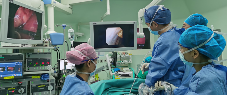
Under general anesthesia, the patient was placed in a right decubitus position with the left flank facing upwards. The procedure was performed through 3 ports. The camera port (10 mm trocar) was inserted 1 cm below the 12th intercostal space at the posterior axillary line. The first working port (10 mm trocar) was inserted 2 cm above the superior iliac crest at the mid-axillary line. The second working port was inserted at the junction between the anterior axillary line and 2 cm below the 12th intercostal space. The ureter was identified and isolated with a Harmonic ultrasonic scalpel (Johnson & Johnson, New Brunswick, NJ, USA). The large ureteral stone was extracted through the ureterostomy incision. A ureteral polyp was incidentally identified, dissected, and sent for biopsy. Afterwards, the first working port was replaced with the camera to guide the insertion of the single-use digital flexible ureteroscope (REDPINE, Guangzhou, China) through the second working port. The ureteroscope was delivered into the ureterostomy incision under the assistance of laparoscopic Kelly forceps and entered the renal pelvis via retrograde access (Figure 2). The renal stone was located and retrieved with a stone extraction basket (COOK Medical, Bloomington, IN, USA). The uretero-scope was withdrawn and an F5 double-J stent was inserted through the ureterostomy incision before closing with 4.0 Vicryl sutures. A retroperitoneal drain was placed before completing the operation. The procedure duration from skin to skin was 2 hours and 39 minutes. The estimated blood loss was 10 mL. The 2 stones (Figure 3) were sent for analysis and both were found to be composed of calcium oxalate monohydrate. Biopsy results of the ureteral polyp revealed that it was benign.
Figure 2.
External view of the operation shows the left laparoscopic monitor and right digital flexible ureteroscope monitor.
Figure 3.
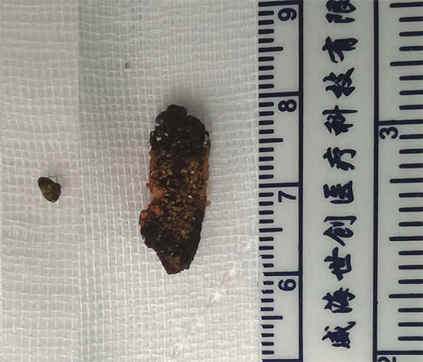
Specimen of the large ureteral calculus and smaller renal calculus.
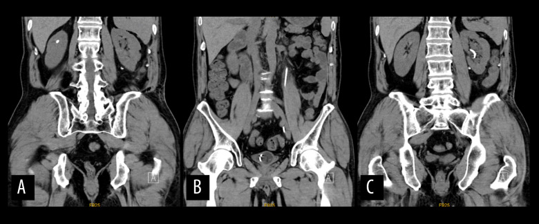
No postoperative complications were observed and the drain was removed 3 days later. The patient was discharged on the 6th postoperative day after removal of the Foley catheter. He was scheduled for follow-up a month later, and the 1-month postoperative kidney, ureter, and bladder X-ray (Figure 4) revealed no migration of the left stent. The stent was removed and the patient did not show discomfort or difficulty while passing urine.
Figure 4.
Postoperative computed tomography, revealing (A) a right renal calculus and (B, C) left double-J stent before removal.
Discussion
Percutaneous nephrolithotripsy (PCNL) and retrograde intrarenal surgery (RIRS) are alternative procedures for the removal of large upper ureteral stones and concurrent renal stones within the same session [4]. The American Urological Association and European Association of Urology strongly recommends PCNL as the first option for stones >20 mm for every location [5]. PCNL has the advantage of an early stone-free rate, but carrying out stone pulverization from within the ureter would increase the risk of bleeding. We reviewed several retrospective comparative studies. Güler et al compared LU, PCNL, and RIRS, and concluded with a preference towards PCNL [6]. However, PCNL resulted in a higher rate of need for blood transfusion compared with LU and RIRS. Topaloglu et al have also reported that PCNL resulted in a higher volume of bleeding than LU [7]. Kumar et al reported that retrieval of large stones by LU was much more effective than fragmentation by ureteroscopic lithotripsy and results in fewer complications [8]. Similarly, Tugcu et al found that post-LU complication rates were much lower than RIRS for the management of ureteral stones >15 mm [9]. Choi et al concluded that LU and RIRS both demonstrate similar effectiveness in the management of stones >15 mm [10].
In 2004, Ball et al documented the first combination of flexible scope with laparoscopy, also known as laparoscopic pyeloplasty with flexible nephroscopy, in patients with ureteropelvic junction obstruction and nephrolithiasis [11]. The flexible nephroscope was introduced to the renal pelvis or calyces via the trocar for stone retrieval. Stravodimos et al reported a case series on robot-assisted laparoscopic pyeloplasty with flexible nephroscopy to treat the same pathology and was able to achieve a 100% postoperative stone clearance rate [12]. Su et al reported favorable clinical outcomes for LU with flexible cystoscope in the management of unilateral ureteral stones and nephrolithiasis [13].
In 2009, Mongiat-Artus et al reported the first case of LU with flexible ureteroscopy for removal of renal and ureteral calculi, and similar cases followed suit [14–16]. All procedures were successful and yielded good clinical outcomes. This procedure allows complete stone extraction without pulverization from within the ureter. The flexible ureteroscope was later introduced through the ureteral incision to retrieve the renal stone. As the diameter of the ureteral segment above the obstruction had been chronically dilated, the application of a flexible ureteroscope sheath was unnecessary, even for the retrieval of larger nephroliths.
In the aforementioned cases, the flexible ureteroscopes were constantly reused. The equipment is costly, and repetitive use can easily damage the device. In 2013, Khan et al reported that a pressure leak test was effective in evaluating and extending the lifespan of repetitively used flexible ureteroscopes, and they have been promoting this technique since [17]. According to Legemate et al, epidural rupture-induced leakage was the main cause of damage in these flexible ureteroscopes [18]. During practice, the lens of the flexible ureteroscope is often clamped with a laparoscopic foreign body forceps to assist its delivery through the ureterostomy incision at a near-vertical angle. This can cumulatively damage the outer layer of the flexible ureteroscope and eventually lead to leakage. The refurbishment cost was approximately $590 per case [19]. However, this problem can be avoided by replacing traditional flexible ureteroscopes with disposable ones.
The LithoVue system single-use digital flexible ureteroscope (Boston Scientific, Marlborough, MA, USA) was launched in 2011 and has already demonstrated similar potential when compared with traditional ureteroscopes in several studies [20,21]. Mazzucchi et al stated that single-use flexible ureteroscopes were lighter and had superior quality of image when compared with fiberoptic ones [22]. Leveillee et al combined the single-use digital flexible ureteroscope and holmium laser fiber in the treatment of lower pole calculi [23]. A systematic review calculated that single-use scopes cost $1300–$3180 per procedure [24]. Although there was a partial overlap in ranges of costs with reusable scopes, other costs such as caseload, repair bills, added expenses when negotiating purchase prices, repair prices, and warranty conditions were not taken into consideration in the study.
China has also developed different single-use digital flexible ureteroscopes that have demonstrated favorable clinical outcomes [25–27]. The REDPINE Medical Instrument became commercially available in 2020. We used the same device (Figure 5) during the laparoscopic lithotomy procedure and were able to achieve a 273° rotation even with a lithotripsy basket attached, which has made nephrolithiasis extraction even more convenient. This case report documents our first experience in using the REDPINE single-use digital flexible ureteroscope to remove a unilateral large ureteral stone with nephrolithiasis in the same session with no obvious major or minor complications. However, the procedure lasted 159 minutes, which is longer than other experiences, which have a mean operating time of 70 minutes (range 35 to 129 minutes) [28].
Figure 5.
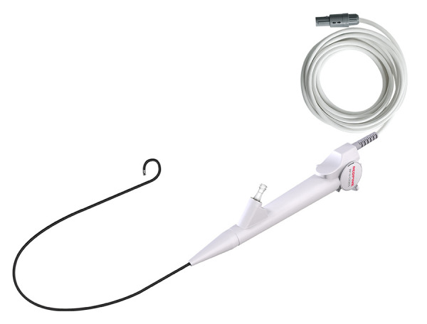
The REDPINE single-use digital flexible ureteroscope (use of this image has been permitted by the manufacturer).
It is undeniable that the development of single-use digital flexible ureteroscopes has eliminated the need for costly repairs and the occurrence of unpredictable performance that may delay the operation. To determine the efficacy and safety of the newer devices, more clinical trials are warranted. We believe that with more practice, the overall operation time can be reduced, which is beneficial for the surgeon and patient
Conclusions
Single-use digital flexible ureteroscopes provides an economical advantage over reusable digital ureteroscopes. By combining this type of ureteroscope with LU, we were able to achieve unilateral large ureteral stone and concurrent renal stone extraction within an operation. We believe that it is a clinically feasible and safe method that can be improved with practice.
Acknowledgments
The authors thank the patient, his family, and the physicians involved.
Footnotes
Publisher’s note: All claims expressed in this article are solely those of the authors and do not necessarily represent those of their affiliated organizations, or those of the publisher, the editors and the reviewers. Any product that may be evaluated in this article, or claim that may be made by its manufacturer, is not guaranteed or endorsed by the publisher
Research Location
Department of Urology, The University of Hong Kong – Shenzhen Hospital, Shenzhen, Guangdong, PR China.
Declaration of Figures’ Authenticity
All figures submitted have been created by the authors who confirm that the images are original with no duplication and have not been previously published in whole or in part.
References:
- 1.Bagley DH, Healy KA, Kleinmann N. Ureteroscopic treatment of larger renal calculi (>2 cm) Arab J Urol. 2012;10(3):296–300. doi: 10.1016/j.aju.2012.05.005. [DOI] [PMC free article] [PubMed] [Google Scholar]
- 2.Xu L, Zhang Y, Wang Z, Li G, Yu S. Comparison of combined laparoscopic ureterolithotomy and flexible ureteroscopy with percutaneous nephrolithotomy for removing large impacted upper ureteral stones with concurrent renal stones. Laparosc Endosc Robot Surg. 2018;1(2):37–41. [Google Scholar]
- 3.Lu GL, Wang XJ, Huang BX, et al. Comparison of mini-percutaneous nephrolithotomy and retroperitoneal laparoscopic ureterolithotomy for treatment of impacted proximal ureteral stones greater than 15 mm. Chin Med J (Engl) 2021;134(10):1209–14. doi: 10.1097/CM9.0000000000001417. [DOI] [PMC free article] [PubMed] [Google Scholar]
- 4.Karakoç O, Karakeçi A, Ozan T, et al. Comparison of retrograde intrarenal surgery and percutaneous nephrolithotomy for the treatment of renal stones greater than 2 cm. Turk J Urol. 2015;41(2):73–77. doi: 10.5152/tud.2015.97957. [DOI] [PMC free article] [PubMed] [Google Scholar]
- 5.Rodríguez-Monsalve Herrero M, Doizi S, et al. Retrograde intrarenal surgery: An expanding role in treatment of urolithiasis. Asian J Urol. 2018;5(4):264–73. doi: 10.1016/j.ajur.2018.06.005. [DOI] [PMC free article] [PubMed] [Google Scholar]
- 6.Güler Y, Erbin A. Comparative evaluation of retrograde intrarenal surgery, antegrade ureterorenoscopy and laparoscopic ureterolithotomy in the treatment of impacted proximal ureteral stones larger than 1.5 cm. Cent European J Urol. 2021;74(1):57–63. doi: 10.5173/ceju.2021.0174.R1. [DOI] [PMC free article] [PubMed] [Google Scholar]
- 7.Topaloglu H, Karakoyunlu N, Sari S, et al. A comparison of antegrade percutaneous and laparoscopic approaches in the treatment of proximal ureteral stones. Biomed Res Int. 2014;2014:691946. doi: 10.1155/2014/691946. [DOI] [PMC free article] [PubMed] [Google Scholar]
- 8.Kumar A, Vasudeva P, Nanda B, et al. A prospective randomized comparison between laparoscopic ureterolithotomy and semirigid ureteroscopy for upper ureteral stones >2 cm: A single-center experience. J Endourol. 2015;29(11):1248–52. doi: 10.1089/end.2013.0791. [DOI] [PubMed] [Google Scholar]
- 9.Tugcu V, Resorlu B, Sahin S, et al. Flexible ureteroscopy versus retroperito-neal laparoscopic ureterolithotomy for the treatment of proximal ureteral stones >15 mm: A single surgeon experience. Urol Int. 2016;96(1):77–82. doi: 10.1159/000430452. [DOI] [PubMed] [Google Scholar]
- 10.Choi JD, Seo SI, Kwon J, Kim BS. Laparoscopic ureterolithotomy vs ureteroscopic lithotripsy for large ureteral stones. JSLS. 2019;23(2):e2019.00008. doi: 10.4293/JSLS.2019.00008. [DOI] [PMC free article] [PubMed] [Google Scholar]
- 11.Ball AJ, Leveillee RJ, Patel VR, Wong C. Laparoscopic pyeloplasty and flexible nephroscopy: Simultaneous treatment of ureteropelvic junction obstruction and nephrolithiasis. JSLS. 2004;8(3):223–28. [PMC free article] [PubMed] [Google Scholar]
- 12.Stravodimos KG, Giannakopoulos S, Tyritzis SI, et al. Simultaneous laparoscopic management of ureteropelvic junction obstruction and renal lithiasis: The combined experience of two academic centers and review of the literature. Res Rep Urol. 2014;6:43–50. doi: 10.2147/RRU.S59444. [DOI] [PMC free article] [PubMed] [Google Scholar]
- 13.Su J, Zhu Q, Yuan L, et al. Combined laparoendoscopic single-site ureterolithotomy and flexible cystoscopy in the treatment of concurrent large upper ureteral and renal stones. Scand J Urol. 2017;51(4):314–18. doi: 10.1080/21681805.2017.1310129. [DOI] [PubMed] [Google Scholar]
- 14.Mongiat-Artus P, Almeida-Neto D, Meria P, et al. [Endoscopic removal of renal stones through laparoscopic access of the ureter and the pelvis] Prog Urol. 2009;19(1):21–26. doi: 10.1016/j.purol.2008.07.006. [in French] [DOI] [PubMed] [Google Scholar]
- 15.Chou SF, Hsieh PF, Lin WC, Huang CP. Laparoscopic ureterolithotomy and retrograde flexible ureteroscopy-assisted transperitoneal laparoscopic ureteroureterostomy for a huge ureteropelvic junction stone and multiple small renal stones: A CARE-compliant case report. Medicine (Baltimore) 2021;100(28):e26655. doi: 10.1097/MD.0000000000026655. [DOI] [PMC free article] [PubMed] [Google Scholar]
- 16.You JH, Kim YG, Kim MK. Flexible ureteroscopic renal stone extraction during laparoscopic ureterolithotomy in patients with large upper ureteral stone and small renal stones. Can Urol Assoc J. 2014;8(9–10):E591–94. doi: 10.5489/cuaj.1806. [DOI] [PMC free article] [PubMed] [Google Scholar]
- 17.Khan F, Mukhtar S, Marsh H, et al. Evaluation of the pressure leak test in increasing the lifespan of flexible ureteroscopes. Int J Clin Pract. 2013;67(10):1040–43. doi: 10.1111/ijcp.12149. [DOI] [PubMed] [Google Scholar]
- 18.Legemate JD, Kamphuis GM, Freund JE, et al. Durability of flexible uretero-scopes: A prospective evaluation of longevity, the factors that affect it, and damage mechanisms. Eur Urol Focus. 2019;5(6):1105–11. doi: 10.1016/j.euf.2018.03.001. [DOI] [PubMed] [Google Scholar]
- 19.Carey RI, Martin CJ, Knego JR. Prospective evaluation of refurbished flexible ureteroscope durability seen in a large public tertiary care center with multiple surgeons. Urology. 2014;84(1):42–45. doi: 10.1016/j.urology.2014.01.022. [DOI] [PubMed] [Google Scholar]
- 20.Kam J, Yuminaga Y, Beattie K, et al. Single use versus reusable digital flexible ureteroscopes: A prospective comparative study. Int J Urol. 2019;26(10):999–1005. doi: 10.1111/iju.14091. [DOI] [PubMed] [Google Scholar]
- 21.Yang E, Jing S, Niu Y, et al. Single-use digital flexible ureteroscopes as a safe and effective choice for the treatment of lower pole renal stones: Secondary analysis of a randomized-controlled trial. J Endourol. 2021;35(12):1773–78. doi: 10.1089/end.2021.0170. [DOI] [PubMed] [Google Scholar]
- 22.Mazzucchi E, Marchini GS, Berto FCG, et al. Single-use flexible uretero-scopes: Update and perspective in developing countries. A narrative review. Int Braz J Urol. 2022;48(3):456–67. doi: 10.1590/S1677-5538.IBJU.2021.0475. [DOI] [PMC free article] [PubMed] [Google Scholar]
- 23.Leveillee RJ, Kelly EF. Impressive performance: New disposable digital ureteroscope allows for extreme lower pole access and use of 365 μm holmium laser fiber. J Endourol Case Rep. 2016;2(1):114–16. doi: 10.1089/cren.2016.0051. [DOI] [PMC free article] [PubMed] [Google Scholar]
- 24.Ventimiglia E, Godínez AJ, Traxer O, Somani BK. Cost comparison of singleuse versus reusable flexible ureteroscope: A systematic review. Turk J Urol. 2020;46(Supp. 1):S40–S45. doi: 10.5152/tud.2020.20223. [DOI] [PMC free article] [PubMed] [Google Scholar]
- 25.Emiliani E, Mercadé A, Millan F, et al. First clinical evaluation of the new single-use flexible and semirigid Pusen ureteroscopes. Cent European J Urol. 2018;71(2):208–13. doi: 10.5173/ceju.2018.1620. [DOI] [PMC free article] [PubMed] [Google Scholar]
- 26.Patil A, Agrawal S, Singh A, et al. A Single-center prospective comparative study of two single-use flexible ureteroscopes: LithoVue (Boston Scientific, USA) and Uscope PU3022a (Zhuhai Pusen, China) J Endourol. 2021;35(3):274–78. doi: 10.1089/end.2020.0409. [DOI] [PubMed] [Google Scholar]
- 27.Qi S, Yang E, Bao J, et al. Single-use versus reusable digital flexible uretero-scopes for the treatment of renal calculi: A prospective multicenter randomized controlled trial. J Endourol. 2020;34(1):18–24. doi: 10.1089/end.2019.0473. [DOI] [PubMed] [Google Scholar]
- 28.Drake T, Ali A, Somani BK. Feasibility and safety of bilateral same-session flexible ureteroscopy (FURS) for renal and ureteral stone disease. Cent European J Urol. 2015;68(2):193–96. doi: 10.5173/ceju.2015.533. [DOI] [PMC free article] [PubMed] [Google Scholar]


