Abstract
Carotovoricin Er is a phage-tail-like bacteriocin produced by Erwinia carotovora subsp. carotovora strain Er, a causative agent for soft rot disease in plants. Here we studied binding and killing spectra of carotovoricin Er preparations for various strains of the bacterium (strains 645Ar, EC-2, N786, and P7) and found that the preparations contain two types of carotovoricin Er with different host specificities; carotovoricin Era possessing a tail fiber protein of 68 kDa killed strains 645Ar and EC-2, while carotovoricin Erb with a tail fiber protein of 76 kDa killed strains N786 and P7. The tail fiber proteins of 68 and 76 kDa had identical N-terminal amino acid sequences for at least 11 residues. A search of the carotovoricin Er region in the chromosome of strain Er indicated the occurrence of a DNA inversion system for the tail fiber protein consisting of (i) two 26-bp inverted repeats inside and downstream of the tail fiber gene that flank a 790-bp fragment and (ii) a putative DNA invertase gene with a 90-bp recombinational enhancer sequence. In fact, when a 1,400-bp region containing the 790-bp fragment was amplified by a PCR using the chromosomal DNA of strain Er as the template, both the forward and the reverse nucleotide sequences of the 790-bp fragment were detected. DNA inversion of the 790-bp fragment also occurred in Escherichia coli DH5α when two compatible plasmids carrying either the 790-bp fragment or the invertase gene were cotransformed into the bacterium. Furthermore, hybrid carotovoricin CGE possessing the tail fiber protein of 68 or 76 kDa exhibited a host range specificity corresponding to that of carotovoricin Era or Erb, respectively. Thus, a DNA inversion altered the C-terminal part of the tail fiber protein of carotovoricin Er, altering the host range specificity of the bacteriocin.
Hamon and Peron first described a substance(s) produced by Erwinia carotovora subsp. carotovora, the causative agent of soft rot disease in many plant species, that is bactericidal towards the same and related species of bacteria (7). Because of the proteinaceous nature of this bacteriocide and its narrow bactericidal spectrum, they assumed it to be a bacteriocin and designated it carotovoricin (7). Previous reports from this laboratory showed that E. carotovora subsp. carotovora Er produced carotovoricin Er as well as pectin lyase when it was exposed to nalidixic acid, mitomycin C, or bleomycin (27) and that a phage-tail-like particle was observed in the partially purified fraction of carotovoricin Er (16, 13). Recently, we developed a simple and efficient procedure for purification of intact carotovoricin Er and its major structural parts by use of sucrose density gradient centrifugation in the presence of 10 to 20% (vol/vol) ethanol and analyzed the highly purified carotovoricin Er for its morphology and components (21). Electron microscopy showed that carotovoricin Er consists of an antenna-like structure, a sheath-and-core part, a base plate, and several tail fibers (21). It was revealed that (i) carotovoricin Er has a length of 184 nm (from the distal end of the sheath-and-core part to the bottom of the base plate) and a diameter of 22 nm (at the sheath-and-core part); (ii) the antenna-like structure at the distal end of the sheath-and-core part is a rod structure with a length of 54 nm, which has never been reported for pyocin R (12) or xenorhabdicin (26); (iii) the sheath is a contractile cylindrical structure surrounding an inner core, which is visible in contracted carotovoricin Er; and (iv) the tail fibers have a length of 63 nm. Sodium dodecyl sulfate-polyacrylamide gel electrophoresis (SDS-PAGE) of the purified major parts of carotovoricin Er showed that the sheath, core, and tail fiber consist of single major proteins of 50, 19, and 68 kDa, respectively (21). In addition to these proteins, protein bands corresponding to 78, 76, 46, 44, 39, and 35 kDa were detected in the purified carotovoricin Er particle and were analyzed for their N-terminal amino acid sequences (21). Furthermore, we recently sequenced a 20-kbp chromosomal fragment of E. carotovora subsp. carotovora Er which contained the open reading frames coding for the major sheath protein (50 kDa), the major core protein (19 kDa), and the tail fiber protein (68 kDa) (DDBJ nucleotide sequence accession no. AB017338). The 20-kbp fragment also contained the structural genes for the proteins of 46, 44, and 35 kDa, which might be the proteins for the antenna-like and the base plate structures. Thus, carotovoricin Er is a phage-tail-like bacteriocin consisting of at least six proteins.
A previous paper from our laboratory has shown that carotovoricin Er binds to and kills several strains of E. carotovora subsp. carotovora, such as strains EC-2, P7, and 645Ar (14). However, our recent experiments showed that (i) a significant portion of the purified carotovoricin Er preparations failed to bind to EC-2 and P7 but that almost all carotovoricin Er particles bound to strain 645Ar under similar conditions and (ii) the unbound fraction of carotovoricin Er recovered after the incubation with EC-2 or P7 retained intact morphology and killing activity towards P7 or EC-2, respectively (unpublished data). These results prompted us to study whether or not the carotovoricin Er preparation contains multiple types of carotovoricin Er with different host specificities. In this paper, we show that E. carotovora subsp. carotovora Er produces two types of carotovoricin Er with different host specificities, which we named carotovoricin Era and Erb, and that the different host specificities of carotovoricins Era and Erb are due to alteration in the C-terminal part of the tail fiber protein by a DNA inversion.
MATERIALS AND METHODS
Bacterial strains, plasmids, and culture media.
Bacterial strains and plasmids used in this study are listed in Table 1. Bacteria were cultured in LB medium (10 g of polypeptone, 5 g of yeast extract, and 5 g of NaCl in 1 liter, pH 7.2) or in a modified M9 medium (14.7 g of Na2HPO412H2O, 3 g of KH2PO4, 5 g of NaCl, 1 g of NH4Cl, 0.0147 g of CaCl22H2O, 0.203 g of MgSO4, 0.0002 g of FeCl34H2O, and 2 g of glucose in 1 liter, pH 7.2) supplemented with casein acid hydrolysate (2 g/liter).
TABLE 1.
Bacterial strains and plasmids used in this study
| Strain or plasmid | Description | Source or reference |
|---|---|---|
| Strains | ||
| E. carotovora subsp. carotovora | ||
| Er | Producer of carotovoricins Era and Erb | 16 |
| 645 Ar | Sensitive to carotovoricin Era | 16 |
| EC-2 | Sensitive to carotovoricin Era | 14 |
| N786 | Sensitive to carotovoricin Erb | 14 |
| P7 | Sensitive to carotovoricin Erb | 14 |
| CGE234 M403 | Producer of carotovoricin CGE | 4 |
| T-29 | Sensitive to carotovoricin CGE | 4 |
| E. coli DH5α | supE44 ΔlacU169 (φ80 lacZΔM15) hsdR17 recA1 endA1 gyrA96 thi-1 relA1 | 8 |
| Plasmids | ||
| pTrc99A | Ampr pBR322 ori | 1 |
| pTrc99AfibA | pTrc99A containing fibA | This study |
| pTrc99AfibB | pTrc99A containing fibB | This study |
| pTrc99Aein | pTrc99A containing ein | This study |
| pACYC184 | Tetr Cmr p15A ori | 3 |
| pACYC184Ea | pACYC184 containing Ea | This study |
| pACYC184Eb | pACYC184 containing Eb | This study |
Assay for carotovoricin activity.
Test samples were serially diluted with 0.05 M sodium phosphate buffer, pH 7.2. A small portion (5 μl) of the dilutions were withdrawn and spotted on an LB soft agar plate (0.35% agar with a thickness of 2 mm) containing sensitive bacteria (2 × 10⁷ CFU/ml), and the plate was incubated at 30°C for 8 to 12 h. The reciprocal of the highest dilution that formed a clear zone in the LB soft agar was defined as the relative killing titer (units) of carotovoricin.
Production and purification of carotovoricin Er and carotovoricin CGE.
E. carotovora subsp. carotovora strain Er or CGE234 M403 was grown in LB medium at 30°C overnight. A portion (20 ml) of the culture was withdrawn, transferred into 600 ml of the modified M9 medium, and cultivated for several hours at 30°C. Mitomycin C (Wako Pure Chemical Industry, Osaka, Japan) was added at a final concentration of 0.4 μg/ml to an exponentially growing culture (with an optical density at 660 nm of 0.6), and the culture was incubated for a further 5 h at 30°C with shaking to induce carotovoricin production accompanied by cell lysis. Carotovoricin particles were purified from the cell lysates by a purification procedure including the use of 15 to 30% (wt/vol) sucrose linear gradient centrifugation in the presence of 20% (wt/vol) ethanol and 0.1 M NaCl, as described previously (21).
Fixation of bacterial cells with formaldehyde.
Exponentially growing cells of E. carotovora subsp. carotovora in LB medium were collected by centrifugation at 2,500 × g at room temperature for 5 min. The cells were suspended in LB medium and were treated with 2% formaldehyde at room temperature for 60 min. After fixation, the cells were washed twice with LB medium, and were suspended in LB medium at a concentration of 1010 cells/ml.
Isolation of two types of carotovoricin Er.
Carotovoricin Er particles were purified from the cell lysate of 2 liters of E. carotovora subsp. carotovora Er as described previously (21), and the carotovoricin Er preparation was divided into two parts. One-half of the carotovoricin Er preparation was mixed with 25 ml of formaldehyde-treated EC-2 cells, and the rest was mixed with 25 ml of formaldehyde-fixed N786 cells. The mixtures were rotated at room temperature for 30 min, followed by centrifugation at 2,500 × g at room temperature for 10 min. The supernatants obtained were centrifuged at 120,000 × g at 4°C for 2 h, and carotovoricin Er particles in the pellets were purified as described above (21).
Binding of carotovoricin Er to various strains of E. carotovora subsp. carotovora.
Carotovoricin Er was incubated with formaldehyde-fixed cells (500 μl) of E. carotovora subsp. carotovora at 0°C for 1 h. The mixtures were centrifuged at 12,000 × g at 0°C for 5 min to collect bacterial cells. The supernatants obtained were centrifuged at 300,000 × g at 4°C for 2 h to collect unbound carotovoricin particles.
SDS-PAGE and Western blotting.
SDS-PAGE was done as described by Laemmli (18) using 12.5% acrylamide gels. Proteins in the gel were electroblotted onto a polyvinylidene difluoride sheet, and the blotted protein bands were immunostained with specific antiserum raised against whole particles or the purified sheath of carotovoricin Er. The immunostained bands were visualized using alkaline phosphatase-conjugated anti-rabbit immunoglobulin G (Promega, Madison, Wis.), nitroblue tetrazolium (Wako Pure Chemicals), and 5-bromo-4-chloro-indolylphosphate (Wako Pure Chemicals). Antisera were raised in female New Zealand White rabbits, which were injected subcutaneously with the purified whole particle or sheath of carotovoricin Er (1 mg) emulsified in Freund's complete adjuvant (Difco Laboratories, Detroit, Mich.), followed by the injections of the purified whole particle or sheath of carotovoricin Er (0.5 mg) emulsified in Freund's incomplete adjuvant (Difco Laboratories) every 10 days for five times.
N-terminal amino acid sequences of proteins.
N-terminal amino acid sequences of proteins were analyzed for the blotted protein bands on a polyvinylidene difluoride sheet (19) by using a model 491 protein sequencer (Perkin-Elmer Applied Biosystems, Foster City, Calif.).
DNA manipulations.
All standard DNA manipulations were done as described by Sambrook et al. (23). PCR was done using Ex Taq polymerase (TaKaRa, Kyoto, Japan) for 30 cycles with the following temperature profile: 94°C for 30 s, 57°C for 30 s, and 72°C for 3 min. Six primers for PCR were synthesized according to the nucleotide sequence of the carotovoricin Er region of E. carotovora subsp. carotovora Er chromosome (DDBJ nucleotide sequence database accession no. AB017338).
Amplification of the DNA fragments containing invertible fragment E by PCR and analysis of the PCR products.
The fragment Er16098-17502, which contains invertible fragment E and a part of the putative DNA invertase gene (Fig. 2), was amplified by PCR using chromosomal DNA of strain Er as the template and the following primers: 5′-AGCCGTGGGCGCGTATTTATAGCGATCAAG-3′ (the forward primer), corresponding to the DNA fragment from nucleotides 16098 to 16127, and 5′-AGCAGCATCGAGTCCGGCCCGTGTCCGTTC-3′ (the reverse primer), corresponding to the DNA fragment from nucleotides 17502 to 17473. Because a single NcoI site is contained in fragment E, cleavage of the amplified products at the NcoI site was used for diagnosis of the DNA inversion of fragment E. The amplified DNA fragments were digested with NcoI restriction endonuclease and were then separated on a Tris-acetate-EDTA agarose gel (1%). DNA fragments were extracted from the gel using Quantum Prep Freeze 'N Squeeze DNA gel extraction spin columns (Bio-Rad Laboratories, Hercules, Calif.). DNA sequencing was done using an ABI Prism 373 DNA sequencer (Perkin-Elmer Applied Biosystems) and an ABI Prism Dye Terminator Cycle Sequencing Ready Reaction kit (Perkin-Elmer Applied Biosystems).
FIG. 2.
Tail fiber gene of carotovoricin Er and a possible DNA inversion system in the 20-kbp carotovoricin Er region of E. carotovora subsp. carotovora Er. Invertible segment E is illustrated in the same (A) or in the reverse (B) direction compared to that of the nucleotide sequence registered in the DDBJ nucleotide sequence database (accession no. AB017338), and the corresponding E fragment is designated Ea or Eb, respectively. Invertible segment E is flanked by the left inverted-repeat sequence (filled triangle) and by the right inverted-repeat sequence (open triangle). fibA and fibB are the tail fiber genes with a part of Ea and Eb, respectively. A putative DNA invertase gene, ein, is located directly to the right of E, and it contains a recombinational enhancer (hatched box) at the 5′ terminus. Nucleotides (nt) 16098 and 17502 are the nucleotide numbers of the 20-kbp carotovoricin Er region registered in the DDBJ nucleotide database (accession no. AB017338).
DNA inversion of segment E in Escherichia coli.
The invertible segment E and the invertase gene ein were separately cloned in compatible plasmids and transformed into E. coli DH5α to study DNA inversion of segment E in E. coli cells. The DNA fragment containing segment E (Er14307-17502) was amplified by PCR using the chromosomal DNA of strain Er as the template. The following forward and reverse primers were used: 5′-GATCTAGAATATTGGCCGCCTGAGCTACG-3′ (the forward primer, which corresponds to the DNA fragment from nucleotides 14307 to 14336 except in the italicized region, where 1 nucleotide was changed to create an XbaI site) and 5′-AGAAGCTTCGAGTCCGGCCCGTGTCCGTTC-3′ (the reverse primer, which corresponds to the DNA fragment from nucleotides 17502 to 17473, where 2 nucleotides were changed to create a HindIII site). The PCR product was digested with XbaI and HindIII and inserted into plasmid pACYC184 (3). The recombinant plasmid was transformed into E. coli DH5α (8). The presence of Ea and Eb was assayed for single colonies of the resultant transformants by a PCR amplification of the Er16098-17502 fragment, followed by NcoI digestion and agarose gel electrophoresis as described above. The DNA fragment containing the ein gene (Er16993-17739) was amplified by PCR using the chromosomal DNA of strain Er as the template. The following forward and reverse primers were used: 5′-CGGAGTTGCTTGCGGCCATGGTAA-3′ (the forward primer, which corresponds to the DNA fragment from nucleotides 16993 to 17016; the italicized region indicates the NcoI site) and 5′-ACCAAGCTTCTGGTCACTCTGGCGCAGAGG-3′ (the reverse primer, which corresponds to the DNA fragment from nucleotides 17739 to 17710, where 2 nucleotides were changed to create a HindIII site). The PCR products containing the ein gene were digested with NcoI and HindIII and cloned into the pTrc99A plasmid to construct plasmid pTrc99Aein. pTrc99Aein was transformed into E. coli DH5α harboring either pACYC184Ea or pACYC184Eb. The presence of Ea and Eb in the resultant transformants was assayed as described as above.
Expression of the tail fiber genes of carotovoricins Era and Erb in E. carotovora subsp. carotovora strain CGE234 M403.
The DNA fragment containing the tail fiber gene of carotovoricin Era or Erb was amplified by PCR using the chromosomal DNA of strain Er as the template. To amplify the DNA fragment containing the tail fiber gene of carotovoricin Era (fibA), the following primers were used: 5′-GATCTAGAATATTGGCCGCCTGAGCTACG-3′ (the forward primer, which is the same oligonucleotide as that described above for amplification of segment E) and 5′-ATAAGCTTACCATGGCCGCAAGCAACTCCG-3′ (the reverse primer, which corresponds to the DNA fragment from nucleotides 17022 to 16993, where 2 nucleotides were changed to create a HindIII site). To amplify the DNA fragment containing the tail fiber gene of carotovoricin Erb (fibB), we used a reverse primer (5′-GGTAAGCTTTTGGGATTTAATCACTGTGCC-3′, which corresponds to the DNA fragment from nucleotides 16586 to 16557 except in the italicized region, where 2 nucleotides were changed to create a HindIII site) in a combination with the same forward primer used for amplification of the tail fiber gene of carotovoricin Era (fibA). The PCR products were digested with XbaI and HindIII and were inserted into pTrc99A plasmids to construct plasmids pTrc99AfibA and pTrc99AfibB, respectively. pTrc99AfibA and pTrc99AfibB were transformed into strain CGE234 M403. Production of carotovoricin CGE by the transformants was induced with mitomycin C, and normal and hybrid carotovoricin CGE particles were purified as described previously (21).
RESULTS AND DISCUSSION
E. carotovora subsp. carotovora Er produces two types of carotovoricin Er.
Carotovoricin Er was purified from the lysate of mitomycin C-treated E. carotovora subsp. carotovora Er as described previously (21) and was assayed for its bactericidal activity towards various strains of E. carotovora subsp. carotovora (strains Er, 645Ar, EC-2, N786, and P7). The carotovoricin Er preparation exhibited killing activity towards all of the tested strains except towards the producing strain Er, although the bactericidal titer of the carotovoricin Er preparation varied with different strains (Table 2). To study whether or not there exist multiple types of carotovoricin Er, the killing spectrum of the carotovoricin Er preparation was assayed after incubation with formaldehyde-treated cells of strain Er, 645Ar, EC-2, N786, or P7 at 30°C for 30 min. When the carotovoricin Er preparation was preincubated with the formaldehyde-fixed cells of strain EC-2, the preparation lost the ability to kill 645Ar and EC-2 but it retained the ability to kill N786 and P7 (Table 2). In contrast, the carotovoricin Er preparation which was preincubated with fixed cells of N786 or P7 killed 645Ar and EC-2 but failed to kill N786 and P7 (Table 2). The residual bactericidal activity of the carotovoricin Er preparation which was recovered after incubation with fixed cells of EC-2 or N786 was abolished by further incubation with N786 or EC-2, respectively (data not shown). These results suggested that the carotovoricin Er preparations contained two types of carotovoricin Er with different host range specificities.
TABLE 2.
Bactericidal spectra of carotovoricin Er preparations after incubation with various strains of E. carotovora subsp. carotovoraa
| Preincubation strain | Residual killing activity in units
|
|||
|---|---|---|---|---|
| 645Ar | EC-2 | N786 | P7 | |
| None | 300 | 50 | 60 | 40 |
| Er | 300 | 50 | 60 | 40 |
| N786 | 300 | 50 | 0 | 1 |
| P7 | 300 | 50 | 0 | 1 |
| EC-2 | 1 | 0 | 60 | 40 |
| 645Ar | 0 | 0 | 0 | 0 |
A carotovoricin Er preparation (1 μl of 1 mg of protein/ml) was incubated with formaldehyde-fixed bacteria (100 μl) at 30°C for 30 min, followed by centrifugation at 12,000 × g at 0°C for 5 min. The supernatants obtained were assayed for killing activity to various strains as described in Materials and Methods. The experiments were repeated five times, and a representative set of data is shown.
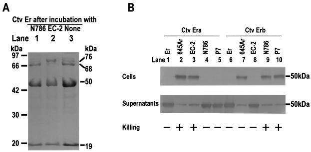
To isolate different types of carotovoricin Er, a partially purified preparation of carotovoricin Er was incubated with a large number of formaldehyde-treated cells of strain EC-2, N786, or P7 at 30°C for 30 min, followed by centrifugation at 12,000 × g for 5 min. Carotovoricin Er particles recovered from the supernatants were further purified by a procedure including the use of sucrose density gradient centrifugation in the presence of 20% (vol/vol) ethanol as described in Materials and Methods. Purified carotovoricin Er was analyzed for its protein components by using SDS-PAGE. When the partially purified preparation of carotovoricin Er was further purified after incubation with strain N786, it exhibited three major protein bands corresponding to 68, 50, and 19 kDa on SDS-PAGE (Fig. 1A, lane 1). These results were consistent with our previous results showing that the tail fiber, sheath, and inner core of carotovoricin Er consisted of major proteins of 68, 50, and 19 kDa, respectively (21). Similar results were obtained when the carotovoricin Er preparation was incubated with strain P7 instead of strain N786 (data not shown). In contrast, when the carotovoricin Er preparation was purified after incubation with strain EC-2, purified carotovoricin Er gave three major protein bands corresponding to 76, 50, and 19 kDa but no intense band corresponding to 68 kDa (Fig. 1A, lane 2). The protein band corresponding to 76 kDa was also detected for the carotovoricin Er preparation before the incubation with strain N786, P7, or EC-2, although it was 1/5 to 1/10 less intense than the band corresponding to 68 kDa (Fig. 1A, lane 3). Protein bands of purified carotovoricin Er (Fig. 1A, lanes 1 and 2) were blotted onto a polyvinylidene difluoride sheet and were analyzed for their N-terminal amino acid sequences. We found that (i) the 11 N-terminal residues of the 76-kDa protein (i.e., Ala-Asn-Leu-Ser-Glu-Asn-Pro-Gln-Trp-Val-Asp-) were identical to those of the 68-kDa tail fiber protein and (ii) the other major proteins of 68, 50, and 19 kDa had N-terminal amino acid sequences identical to those of the purified tail fiber, sheath, and inner core, respectively. These results showed that E. carotovora subsp. carotovora Er produces two types of carotovoricin Er with identical sheath and core parts but different tail fiber proteins with identical N-terminal regions. Two types of carotovoricin Er exhibiting the protein bands corresponding to 68 and 76 kDa on SDS-PAGE were designated carotovoricin Era and Erb, respectively. Carotovoricin Era and Erb were tested for their abilities to bind to and kill strains 645Ar, EC-2, N786, and P7, as described in Materials and Methods. As shown in Fig. 1B, carotovoricin Era bound to and killed strains 645Ar and EC-2 (lanes 2 and 3) but it did not bind to Er, N786, or P7 (lanes 1, 4, and 5). In contrast, carotovoricin Erb exhibited the ability to bind to and kill N786 and P7 (Fig. 1B, lanes 9 and 10) but not Er and EC-2 (Fig. 1B, lanes 6 and 8). The results indicated that carotovoricin Era and Erb have different host specificities, probably due to their different tail fiber proteins.
FIG. 1.
Major components of two types of carotovoricin Er (A) and binding of carotovoricin Era and Erb to various strains of E. carotovora subsp. carotovora (B). (A) A portion of a carotovoricin Er preparation was incubated with a large number of formaldehyde-fixed cells of strain N786 (lane 1) or EC-2 (lane 2), followed by centrifugation at 2,500 × g at room temperature for 10 min. Carotovoricin particles were purified from the supernatants as described in Materials and Methods, and 10 μg of protein from the purified carotovoricin preparation was loaded onto an SDS–12.5% polyacrylamide gel. For comparison, the carotovoricin Er preparation before the incubation with strain N786 or EC-2 was also loaded onto the same gel (lane 3). (B) Purified carotovoricin Era (Fig. 1A, lane 1) or Erb (Fig. 1A, lane 2) was incubated with formaldehyde-fixed cells of various strains at 0°C for 60 min. After centrifugation at 2,500 × g at 0°C for 10 min, the precipitates (the bacterial cells) and the supernatants obtained were subjected to Western blotting using antisheath antiserum, as described in Materials and Methods. The results of killing of various strains by carotovoricin Era or Erb were from Table 2. Ctv, carotovoricin. +, killed. −, not killed.
Carotovoricin Era and Erb failed to bind to the strain that produced them, Er (Fig. 1B), implying that the resistance of strain Er to the bacteriocin is not due to the presence of an immunity system but is due to the lack of a receptor(s) for carotovoricin Er. It was also noted that carotovoricin Era and Erb bound to strain 645Ar but that only carotovoricin Era killed the strain (Fig. 1B). It will be interesting to study whether strain 645Ar has two different receptors for two types of carotovoricin Er. It also remains to be studied whether carotovoricin Er utilizes lipopolysaccharide of E. carotovora subsp. carotovora as the receptor. Pyocin R was shown to bind to lipopolysaccharide of the host bacterium (11).
A DNA inversion in the tail fiber gene is responsible for the production of two types of carotovoricin Er.
It has been demonstrated that bacteriophages Mu and P1 alter the C-terminal regions of tail fiber proteins by DNA inversion, leading to alteration of their host range (5, 6, 10, 17, 24, 28). Since two properly oriented recombination sites (inverted repeats), a DNA invertase, and a recombinational enhancer are essential for a DNA inversion system (15), we searched the corresponding sequences on the 20-kbp fragment from the E. carotovora subsp. carotovora Er chromosome (DDBJ nucleotide sequence database accession no. AB017338), which includes the genes encoding the major proteins of the sheath, core, and tail fiber of carotovoricin Er. As illustrated in Fig. 2, (i) the 20-kbp fragment contains a pair of 26-bp inverted-repeat sequences which are located inside and downstream of the gene encoding the tail fiber protein of 68 kDa; (ii) the inverted repeats flank a 790-bp segment, or the putative invertible segment, which covers the region corresponding to the C-terminal part of the tail fiber protein; (iii) a segment corresponding to a DNA invertase gene is located immediately downstream of the 790-bp segment; and (iv) the putative DNA invertase gene contains a recombinational-enhancer-like segment in its 5′ region (Fig. 2).
The invertible segments in the previously reported DNA inversion systems were designated H for the phase variation in Salmonella enterica serovar Typhimurium (29), G in bacteriophage Mu (5, 17, 28), C in bacteriophage P1 (10), and P in the E. coli chromosomal element e4 (22), and the corresponding DNA invertases were Hin, Gin, Cin, and Pin, respectively. The invertible 790-bp segment was designated E (for carotovoricin Er), and the E segments with forward and reverse orientations registered in the DDBJ nucleotide sequence database were referred to as Ea and Eb, respectively (Fig. 2). The putative DNA invertase for E was designated Ein. Based on the alignment of the 26-bp inverted repeats for Ein with those for Gin (22), Cin (9), Hin (30), and Pin (22) (data not shown), we defined a consensus sequence of the inverted repeats for the DNA invertases as follows: 5′-TT-TC-AAACCT-GGTTT-GAGAA-3′, where at least 7 of 10 inverted repeats have the same nucleotide in each position. The left inverted repeat for Ein (i.e., 5′-CTCCCGCAAACCTCGGTTTTGGGGAC-3′) displayed identity in 16 of the 20 nucleotides of the consensus sequence, and the right inverted repeat for Ein (i.e., 5′-TTCTCGCAAACCTCGGTTTTGGAGAA-3′) was identical with the consensus sequence. Alignment of the deduced amino acid sequence for Ein with those for Gin, Cin, Hin, and Pin (9, 22, 30) indicated that there was a considerable degree of identity between the amino acid residues of Ein and the other DNA invertases, ranging from 61 to 67% (data not shown). Interestingly, many differences in the DNA sequence were found in the third nucleotide of codons, leaving the amino acid sequences unaltered. This implies a selective pressure to keep the protein functional. The 90-bp segments of the recombinational enhancer within ein showed more than 60% identity to those of the other DNA invertase genes (data not shown). Thus, the existence of a DNA inversion system for production of two types of carotovoricin Er was strongly suggested, and inversion of segment E would lead to the production of two different tail fiber proteins with identical N-terminal regions.
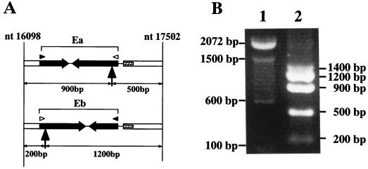
To demonstrate the occurrence of DNA inversion in E. carotovora subsp. carotovora Er, we attempted to detect the Ea and Eb segments in the Er16098-17502 region of the bactrial chromosome (Fig. 2). The Ea-containing fragment (Era16098-17502) included a single NcoI site (DDBJ nucleotide sequence database accession no. AB017338), which, when cleaved, would divide the 1,403-bp fragment into two segments of approximately 900 and 500 bp (Fig. 3A). In contrast, the Eb-containing fragment (Erb16098-17502) would be cleaved by NcoI into two fragments of approximately 1,200 and 200 bp (Fig. 3A). The fragment corresponding to Er16098-17502 was amplified by a PCR using chromosomal DNA of strain Er as the template, as described in Materials and Methods. The amplified products were digested by NcoI and were loaded onto an agarose gel. The NcoI digestion of the fragment produced five DNA bands corresponding to approximately 1,400, 1,200, 900, 500, and 200 bp (Fig. 3B). Sequencing of the fragments showed that the fragments of 900 and 500 bp were derived from NcoI-digested Era16098-17502, while the segments of 1,200 and 200 bp were from NcoI-digested Erb16098-17502. Similar results were obtained when fresh single colonies (n = 20) of strain Er were used as the source of the DNA template for the amplification of the Er16098-17502 fragment by PCR (data not shown). These results indicated that the E fragment was invertible in vivo. The tail fiber genes containing Ea and Eb were designated fibA and fibB, respectively. The open reading frames for FibA and FibB comprised 2,001 bp and 2,142 bp, respectively, coding for 666 and 713 amino acid residues. Calculated molecular masses were 70,792 and 75,910 Da for FibA and FibB, respectively, and they shared identical 568-residue polypeptides from the N termini. Thus, the DNA inversion changed the C-terminal one-fifth portion of the tail fiber protein of carotovoricin Er.
FIG. 3.
Existence of the invertible segments Ea and Eb in the chromosome of E. carotovora subsp. carotovora Er. (A) The 1,403-bp DNA fragments containing the putative invertible segments Ea and Eb (from nucleotides [nt] 16098 to 17502; see also Fig. 2) are illustrated. Filled arrows in the upward direction indicate the NcoI sites, which divide the 1,403-bp fragments containing Ea or Eb into 900- and 500-bp fragments or 200- and 1,200-bp fragments, respectively. Nucleotides 16098 and 17502 are the nucleotide numbers of the 20-kbp carotovoricin Er region registered in the DDBJ nucleotide database (accession no. AB017338). (B) The 1,403-bp DNA fragments were amplified by PCR using the chromosomal DNA of E. carotovora subsp. carotovora Er as the template. The PCR products obtained were digested with NcoI restriction endonuclease and were separated on a 1% agarose gel. Lane 1, size markers; lane 2, NcoI digests of the amplified DNA fragments from nucleotides 16098 to 17502.
Furthermore, the function of ein was tested using an E. coli system where invertible segment E and the ein gene were separately carried by compatible plasmids; the DNA fragment containing segment E (Er14307-17502) and the DNA fragment containing the ein gene (Er16993-17739) were amplified by PCR using the chromosomal DNA of strain Er as the template and were cloned into pACYC184 and pTrc99A, respectively. After transformation of E. coli DH5α with the recombinant plasmid(s), the Ea and the Eb fragments in the transformants were detected by an amplification of the Er16098-17502 fragments by PCR, followed by NcoI digestion and agarose gel electrophoresis as described above. Single colonies (n = 20) of the transformants harboring only recombinant pACYC184 gave either a combination of 900- and 500-bp fragments or a combination of 1,200- and 200-bp fragments (data not shown). However, when E. coli DH5α carrying pACYC184Ea or pACYC184Eb (i.e., pACYC184 containing Ea or Eb) was further transformed with pTrc99Aein (i.e., pTrc99A containing ein), the resultant transformants gave the bands corresponding to 1,200, 900, 500, and 200 bp (data not shown). These results indicated that the function of ein was necessary for DNA inversion of segment E and that the DNA inversion took place in the forward and backward directions.
Tail fiber proteins determine the host range specificity of carotovoricin.
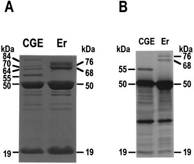
To study if the tail fiber proteins determine the host specificity of carotovoricin Er, we expressed the tail fiber gene fibA or fibB in E. carotovora subsp. carotovora CGE234 M403 to produce hybrid carotovoricin CGE possessing FibA or FibB and tested the host specificity for the hybrid carotovoricins. E. carotovora subsp. carotovora CGE234 M403 is nonpathogenic to plants but retains bactericidal activity towards the same bacterial species, probably because of its ability to produce two types of bacteriocin, high-molecular-weight and low-molecular-weight bacteriocins (4, 25). In this study, we purified the high-molecular-weight bacteriocin from the mitomycin C-treated cells of strain CGE234 M403 according to the procedure for purification of carotovoricin Er (21). Electron microscopy of the purified high-molecular-weight bacteriocin showed the same phage-tail-like morphology as that of carotovoricin Er (data not shown). SDS-PAGE for the purified bacteriocin indicated major protein bands corresponding to 84, 70, 64, 55, 50, and 19 kDa (Fig. 4A). The 20 N-terminal amino acid residues of the 50- and the 19-kDa proteins were 100% identical to those of the sheath and the core proteins of carotovoricin Er, respectively. Furthermore, the 15 N-terminal amino acid residues of the 64-kDa protein (i.e., Ala-Asn-Leu-Ser-Glu-Asn-Pro-Gln-Trp-Val-Asp-Ser-Ile-Tyr-Gln-) were identical with those of the tail fiber protein of carotovoricin Er, except for Ser in the 12th position (i.e., the tail fiber proteins of carotovoricin Er, FibA and FibB, have Gly for Ser in the same position). However, the 64-kDa protein was not immunostained with the antiserum raised against carotovoricin Er (Fig. 4B), implying that the C-terminal region of the 64-kDa protein is different from those of carotovoricin Era and Erb. Based on these results, the high-molecular-weight bacteriocin from strain CGE234 M403 was designated carotovoricin CGE and we determined that carotovoricin CGE has a structure similar to that of carotovoricin Er except for the tail fiber protein, implying that the tail fiber protein of carotovoricin CGE might be interchangeable with those of carotovoricin Er. Since Western blotting of purified carotovoricin CGE using antiserum against carotovoricin Er revealed a prominent band corresponding to 55 kDa (Fig. 4B), we determined the amino acid sequence for the 11 N-terminal residues of the 55-kDa protein: Ala-Asn-Leu-Ser-Glu-Gln-Glu-Ser-Trp-Ile-Asp-. The 5 N-terminal amino acid residues of the 55-kDa protein are identical to those of the tail fiber protein of carotovoricin CGE, but the rest of the amino acid residues are different from those of the tail fiber protein. Furthermore, an open reading frame coding for the 64-kDa protein is present in the 20-kbp carotovoricin CGE region of strain CGE234 M403, while no open reading frame for the 55-kDa protein is present in the same region (M. Hirota et al., unpublished results). Therefore, the 64-kDa protein may be the major tail fiber protein of carotovoricin CGE, but it remains unclear if the 55-kDa protein is a structural component of carotovoricin CGE.
FIG. 4.
SDS-PAGE (A) and Western blotting (B) for the purified carotovoricin CGE. (A) Purified carotovoricin CGE (15 μg of protein) was electrophoresed in an SDS-polyacrylamide gel. The protein bands were stained with Coomassie brilliant blue R-250. For comparison, purified carotovoricin Er (20 μg of protein) was also electrophoresed in the same gel. (B) Purified carotovoricin CGE (3 μg of protein) and carotovoricin Er (4 μg of protein) were subjected to SDS-PAGE, followed by Western blotting using specific antiserum raised against purified carotovoricin Er as described in Materials and Methods. Note that the antiserum did not stain the 64-kDa tail fiber protein of carotovoricin CGE. CGE, carotovoricin CGE; Er, carotovoricin Er.
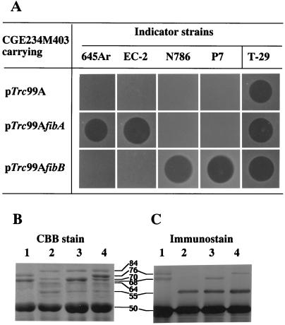
Carotovoricin CGE exhibited no ability to kill strains Er, 645Ar, EC-2, N786, and P7 (Fig. 5A), indicating that carotovoricin CGE has a host specificity different from that of carotovoricin Er, possibly due to a different C-terminal region of the tail fiber protein. To produce hybrid carotovoricin CGE with the corresponding tail fiber protein, strain CGE234 M403 was transformed with the multicopy vector pTrc99A carrying fibA or fibB. Carotovoricin particles were purified from the lysate of the mitomycin C-treated transformants and were analyzed for bactericidal activity towards various indicator strains. As shown in Fig. 5A, the purified carotovoricin preparation from the transformant harboring pTrc99AfibA killed strains 645Ar, EC-2, and T-29 (the indicator strain for carotovoricin CGE) but did not kill strains N786 and P7. In contrast, carotovoricin preparation from the transformant harboring pTrc99AfibB exhibited activity bactericidal to strains N786, P7, and T-29 but no ability to kill strains 645Ar and EC-2 (Fig. 5A). Carotovoricin preparations from all of the transformants killed strain T-29 (Fig. 5A), indicating that normal carotovoricin CGE was also produced by the transformants. SDS-PAGE indicated that the transformants carrying plasmid pTrc99AfibA or pTrc99AfibB produced carotovoricins possessing a 68- or 76-kDa protein, respectively (Fig. 5B, lanes 3 and 4). Furthermore, Western blotting using antiserum specific to purified carotovoricin Er showed that FibA (68 kDa) or FibB (76 kDa) protein was present in the carotovoricin preparation obtained from the transformant carrying pTrc99AfibA or pTrc99AfibB, respectively (Fig. 5C, lanes 3 and 4). These results showed that the transformants carrying pTrc99AfibA or pTrc99AfibB produced hybrid carotovoricin CGE possessing FibA or FibB, respectively, and that the tail fiber proteins, more specifically their C-terminal parts, determined host range specificity. It has been demonstrated that in many of the well-known tailed bacteriophages, such as λ, P1, Mu, and T4, the N-terminal part of tail fiber proteins is bound to the base plate or to the tail shaft and the C-terminal part is the distal part that binds to a specific receptor (2, 20). In Mu and P1 phages, DNA inversion of segments G and C results in the formation of tail fiber proteins with a constant N-terminal part and two alternative C-terminal parts, alternating the host range specificities of the phages. With these results taken together, it is quite conceivable that the host range specificity of carotovoricin Er is determined by the C-terminal part of the tail fiber protein. The production of carotovoricins aid E. carotovora subsp. carotovora in surviving competition among the same and closely related species of bacteria in nature, and alteration in the C-terminal part of tail fiber proteins by DNA inversion is an effective way to furnish carotovoricin with a wider bactericidal spectrum.
FIG. 5.
Killing spectra of the transformants of E. carotovora subsp. carotovora CGE234 M403 carrying fibA or fibB (A) and production of hybrid carotovoricin CGE possessing FibA or FibB (B and C). The tail fiber genes of carotovoricin Era and Erb (fibA and fibB, respectively) were expressed in E. carotovora subsp. carotovora strain CGE234 M403, and carotovoricin particles were purified and analyzed as described in Materials and Methods. (A) Purified carotovoricins from the transformants carrying pTrc99A, pTrc99AfibA, or pTrc99AfibB were spotted on an LB soft agar plate containing E. carotovora subsp. carotovora strain 645Ar, EC-2, N786, P7, or T-29, and a clear zone was observed after incubation at 30°C for 8 to 12 h. (B) Purified carotovoricin particles (15 μg of protein) from the transformants carrying pTrc99A, pTrc99AfibA, or pTrc99AfibB were electrophoresed in an SDS-polyacrylamide gel (lane 2, 3, or 4, respectively). The protein bands were stained with Coomassie brilliant blue R-250 (CBB). Purified carotovoricin Er (12 μg of protein) was also electrophoresed in the same gel (lane 1). (C) Purified carotovoricin particles (3 μg of protein) from the transformants carrying pTrc99A, pTrc99AfibA, or pTrc99AfibB were subjected to SDS-PAGE, followed by Western blotting using a specific antiserum against carotovoricin Er (lane 2, 3, or 4, respectively). Lane 1, purified carotovoricin Er (3 μg of protein).
ACKNOWLEDGMENTS
We thank Elisabeth Hofstad for her help in the preparation of the manuscript.
This work was supported in part by a grant-in-aid for scientific research from the Ministry of Education, Science, Sports and Culture of Japan. H. A. Nguyen was the recipient of a Japanese government scholarship from the Ministry of Education, Science, Sports, and Culture of Japan.
REFERENCES
- 1.Amann E, Ochs B, Abel K J. Tightly regulated tac promoter vectors useful for the expression of unfused and fused proteins in Escherichia coli. Gene. 1988;69:301–315. doi: 10.1016/0378-1119(88)90440-4. [DOI] [PubMed] [Google Scholar]
- 2.Casjens S, Hendrix R. Control mechanisms in dsDNA bacteriophage assembly. In: Calendar R, editor. The bacteriophages. New York, N.Y: Plenum Press; 1988. pp. 15–91. [Google Scholar]
- 3.Chang A C, Cohen S N. Construction and characterization of amplifiable multicopy DNA cloning vehicles derived from the P15A cryptic miniplasmid. J Bacteriol. 1978;134:1141–1156. doi: 10.1128/jb.134.3.1141-1156.1978. [DOI] [PMC free article] [PubMed] [Google Scholar]
- 4.Chuang D Y, Kyeremeh A G, Gunji I, Takahara Y, Ehara Y, Kikumoto T. Identification and cloning of an Erwinia carotovora subsp. carotovora bacteriocin regulator gene by insertional mutagenesis. J Bacteriol. 1999;181:1953–1957. doi: 10.1128/jb.181.6.1953-1957.1999. [DOI] [PMC free article] [PubMed] [Google Scholar]
- 5.Giphart-Gassler M, Plasterk R H A, van de Putte P. G inversion in bacteriophage Mu: a novel way of gene splicing. Nature. 1982;297:339–342. doi: 10.1038/297339a0. [DOI] [PubMed] [Google Scholar]
- 6.Grundy F J, Howe M M. Involvement of the invertible G segment in bacteriophage Mu tail fiber biosynthesis. Viology. 1984;134:296–317. doi: 10.1016/0042-6822(84)90299-x. [DOI] [PubMed] [Google Scholar]
- 7.Hamon Y, Peron Y. Les propriete antagonistes reciproques parmi les Erwinia. Discussion de la position taxonomique de ce genre. C R Acad Sci. 1961;253:913–915. [PubMed] [Google Scholar]
- 8.Hanahan D. Studies on transformation of Escherichia coli with plasmids. J Mol Biol. 1983;166:557. doi: 10.1016/s0022-2836(83)80284-8. [DOI] [PubMed] [Google Scholar]
- 9.Hiestand-Nauer R, Iida S. Sequence of the site-specific recombinase gene Cin and of its substrates serving in the inversion of the C segment of bacteriophage P1. EMBO J. 1983;2:1733–1740. doi: 10.1002/j.1460-2075.1983.tb01650.x. [DOI] [PMC free article] [PubMed] [Google Scholar]
- 10.Iida S. Bacteriophage P1 carries two related sets of genes determining its host range in the invertible C segment of its genome. Virology. 1984;134:421–434. doi: 10.1016/0042-6822(84)90309-x. [DOI] [PubMed] [Google Scholar]
- 11.Ikeda K, Egami F. Receptor substance for pyocin R. I. Partial purification and chemical properties. J Biochem. 1969;65:603–609. doi: 10.1093/oxfordjournals.jbchem.a129053. [DOI] [PubMed] [Google Scholar]
- 12.Ishii S, Nishi Y, Egami F. The fine structure of a pyocin. J Mol Biol. 1965;13:428–431. doi: 10.1016/s0022-2836(65)80107-3. [DOI] [PubMed] [Google Scholar]
- 13.Itoh Y, Izaki K, Takahashi H. Purification and characterization of a bacteriocin from Erwinia carotovora. J Gen Appl Microbiol. 1978;24:27–39. [Google Scholar]
- 14.Itoh Y, Izaki K, Takahashi H. Simultaneous synthesis of pectin lyase and carotovoricin induced by mitomycin C, nalidixic acid or ultraviolet light irradiation in Erwinia carotovora. Agric Biol Chem. 1980;44:1135–1140. [Google Scholar]
- 15.Johnson R C, Simon M I. Hin-mediated site-specific recombination requires two 26 bp recombination sites and a 60 bp recombinational enhancer. Cell. 1985;41:781–789. doi: 10.1016/s0092-8674(85)80059-3. [DOI] [PubMed] [Google Scholar]
- 16.Kamimiya S, Izaki K, Takahashi H. Bacteriocins in Erwinia aroideae with tail like structure of bacteriophages. Agric Biol Chem. 1977;41:911–912. [Google Scholar]
- 17.Kamp D, Kahmann R, Zipser D, Broker T R, Chow L T. Inversion of the G DNA segment of phage Mu controls phage infectivity. Nature. 1978;271:577–580. doi: 10.1038/271577a0. [DOI] [PubMed] [Google Scholar]
- 18.Laemmli U K. Cleavage of structural proteins during the assembly of the head of bacteriophage T4. Nature. 1970;277:680–685. doi: 10.1038/227680a0. [DOI] [PubMed] [Google Scholar]
- 19.Matsudaira P. Sequence from picomole quantities of proteins electroblotted onto polyvinylidene difluoride membranes. J Biol Chem. 1987;262:10035–10038. [PubMed] [Google Scholar]
- 20.Montag D, Schwarz H, Henning U. A component of the side tail fiber of Escherichia coli bacteriophage λ can functionally replace the receptor-recognizing part of a long tail fiber protein of the unrelated bacteriophage T4. J Bacteriol. 1989;171:4378–4384. doi: 10.1128/jb.171.8.4378-4384.1989. [DOI] [PMC free article] [PubMed] [Google Scholar]
- 21.Nguyen A H, Tomita T, Hirota M, Sato T, Kamio Y. A simple purification method and morphology and component analyses for carotovoricin Er, a phage-tail-like bacteriocin from the plant pathogen Erwinia carotovora Er. Biosci Biotechnol Biochem. 1999;63:1360–1369. doi: 10.1271/bbb.63.1360. [DOI] [PubMed] [Google Scholar]
- 22.Plasterk R H A, Brinkman A, van de Putte P. DNA inversions in the chromosome of Escherichia coli and in bacteriophage Mu: relationship to other site-specific recombination systems. Proc Natl Acad Sci USA. 1983;80:5355–5358. doi: 10.1073/pnas.80.17.5355. [DOI] [PMC free article] [PubMed] [Google Scholar]
- 23.Sambrook J, Fritsch E F, Maniatis T. Molecular cloning: a laboratory manual. 2nd ed. Cold Spring Harbor, N.Y: Cold Spring Harbor Laboratory Press; 1989. [Google Scholar]
- 24.Sandmeier H. Acquisition and rearrangement of sequence motifs in the evolution of bacteriophage tail fibres. Mol Microbiol. 1994;12:343–350. doi: 10.1111/j.1365-2958.1994.tb01023.x. . (Review.) [DOI] [PubMed] [Google Scholar]
- 25.Takahara Y. Development of the microbial pesticide for soft-rot disease. PSJ Biocontrol Rep. 1994;4:1–7. . (In Japanese.) [Google Scholar]
- 26.Thaler J O, Baghdiguian S, Boemare N E. Purification and characterization of xenorhabdicin, a phage-tail-like bacteriocin, from the lysogenic strain F1 of Xenorhabdus nematophilus. Appl Environ Microbiol. 1995;61:2049–2052. doi: 10.1128/aem.61.5.2049-2052.1995. [DOI] [PMC free article] [PubMed] [Google Scholar]
- 27.Tomizawa H, Takahashi H. Stimulation of pectolytic enzyme formation of Erwinia aroideae by nalidixic acid, mitomycin C and bleomycin. Agric Biol Chem. 1971;35:191–200. [Google Scholar]
- 28.van de Putte P, Cramer S, Giphart-Gassler M. Invertible DNA determines host specificity of bacteriophage Mu. Nature. 1980;286:218–222. doi: 10.1038/286218a0. [DOI] [PubMed] [Google Scholar]
- 29.Zieg J, Silverman M, Hilman M, Simon M. Recombinational switch for gene expression. Science. 1977;196:170–172. doi: 10.1126/science.322276. [DOI] [PubMed] [Google Scholar]
- 30.Zieg J, Simon M. Analysis of the nucleotide sequence of an invertible controlling element. Proc Natl Acad Sci USA. 1980;77:4196–4201. doi: 10.1073/pnas.77.7.4196. [DOI] [PMC free article] [PubMed] [Google Scholar]


