Abstract
The affinities of extinct organisms are often difficult to resolve using morphological data alone. Chemical analysis of carbonaceous specimens can complement traditional approaches, but the search for taxon-specific signals in ancient, thermally altered organic matter is challenging and controversial, partly because suitable positive controls are lacking. Here, we show that non-destructive Fourier Transform Infrared Spectroscopy (FTIR) resolves in-situ molecular fingerprints in the famous 407 Ma Rhynie chert fossil assemblage of Aberdeenshire, Scotland, an important early terrestrial Lagerstätte. Remarkably, unsupervised clustering methods (principal components analysis and K-mean) separate the fossil spectra naturally into eukaryotes and prokaryotes (cyanobacteria). Additional multivariate statistics and machine-learning approaches also differentiate prokaryotes from eukaryotes, and discriminate eukaryotic tissue types, despite the overwhelming influence of silica. We find that these methods can clarify the affinities of morphologically ambiguous taxa; in the Rhynie chert for example, we show that the problematic “nematophytes” have a plant-like composition. Overall, we demonstrate that the famously exquisite preservation of cells, tissues and organisms in the Rhynie chert accompanies similarly impressive preservation of molecular information. These results provide a compelling positive control that validates the use of infrared spectroscopy to investigate the affinity of organic fossils in chert.
Subject terms: Palaeontology, Biogeochemistry, Palaeontology, Eukaryote, Prokaryote
It can be challenging to identify extinct organisms with morphology alone. Here, the authors use non-destructive Fourier Transform Infrared Spectroscopy to determine the molecular fingerprints of eukaryotes and prokaryotes from the 407 Ma Rhynie chert fossil assemblage of Aberdeenshire, Scotland.
Introduction
Vibrational methods such as Raman and Fourier Transform Infrared (FTIR) microspectroscopy are increasingly popular tools for the investigation of fossil organic matter1. These methods are widely available, rapid, and essentially non-destructive, and they can be applied to fossils in situ without removing the sedimentary matrix. Raman spectroscopy uses a monochromatic laser and relies on the Stokes Raman effect whereby incident photons are shifted to a lower frequency by an amount corresponding to the vibration of a specific chemical bond. FTIR spectroscopy uses a polychromatic infrared light source and a Michelsen interferometer; a Fourier transform of the resulting interferogram generates an intelligible transmission or reflectance spectrum with bands at specific wavenumbers, again corresponding to specific chemical bonds. These methods can detect methyl, methylene, carbonyl, carboxyl, aromatic and other moieties in ancient organic fossilization products and thereby shed light both on the original composition of the precursor biomolecules and on the pathways of chemical alteration during and after fossilization2,3.
Previously, Raman spectroscopy has been used to survey phylogenetic and physiological diversity of organically preserved Phanerozoic metazoans4, to differentiate vertebrate from invertebrate tissues in the Carboniferous Mazon Creek Lagerstatte5, to identify colour pigments in fossilized dinosaur eggs6, to investigate the biogenicity of Precambrian microfossils7 and to determine kerogen maturity (e.g., refs. 8–10). Similarly, FTIR spectroscopy and chemometrics have been applied in chemotaxonomy11,12, the determination of kerogen maturity (see review in ref. 13), the characterization of individual microfossils (e.g., refs. 2,14–17) and the identification of bacterial, eukaryotic and archaeal lipid fossilization products in various Precambrian successions3,18–20.
These studies suggest that vibrational microspectroscopy can reveal physiological and phylogenetic information about organic fossils independently of other lines of evidence; this would be especially useful in the study of fossil microbes, the morphology of which, even when well preserved, is often not diagnostic of their phylogenetic affinities. In principle, such evidence could elucidate the diversity of important organic Lagerstätten in a way that traditional palaeontological methods have been unable to achieve. An essential first step, however, is to confirm the relationships between the phylogenetic affinity and microspectroscopic fingerprints of ancient, thermally altered microfossils within a single assemblage.
To achieve this, the Lower Devonian (~407 Ma) Rhynie chert assemblage of Aberdeenshire, Scotland, famous for its exceptionally preserved fossils since they were discovered more than a century ago, provides ideal material. These fossils open a unique window on an early terrestrial biotic community and include some of the oldest known terrestrial arthropods, land plants, fungi, heterocystous cyanobacteria, and various other microbial eukaryotes and prokaryotes (see review in ref. 21). They are composed of brown-black carbonaceous matter permineralized and encased by transparent microcrystalline quartz, which derives from silica precipitated by terrestrial hot springs22. As such, they can be examined in transmitted light under an optical microscope, revealing exquisite, micron-scale, morphological detail. To date, almost all studies of the Rhynie chert biota have focused on the identification and systematic description of new taxa using this approach21,23. Chemical investigations have been limited but promising24–26. FTIR spectroscopy has confirmed the presence of aliphatic CH bands and subordinate amide, C=O and COOH bands, but taxon-specific differences within the assemblage have not yet been reported25,26.
In this work, we analyze 49 individual fossils from the Rhynie chert assemblage using attenuated total reflectance (ATR) FTIR microspectroscopy. The acquired infrared spectra are then compared using multivariate and machine-learning approaches. We show that differences between prokaryotes (cyanobacteria) and eukaryotes and between eukaryotic tissue types can be identified based on the fossilization products of lipids, sugar, and protein.
Results
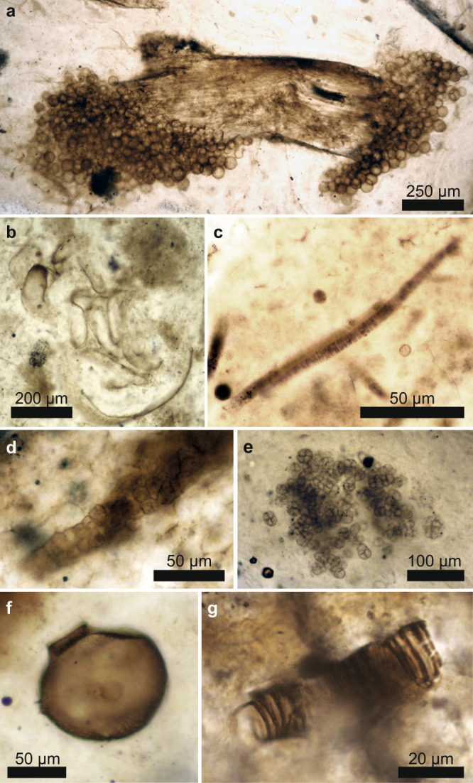
The fossils (Fig. 1 and Supplementary Fig. 1) are located at the surface of polished thin sections and represent nine individual fossil cyanobacteria (Rhyniosarcina devonica27, Palaeolyngbya kerpii28, and a multiseriate Stigonema-like taxon), six unclassified arthropod cuticles, four unclassified plant spores, eleven plant cortices (Aglaophyton majus, Rhynia gwynne-vaughanii) and one set of rhizoids, four peronosporomycetes (Frankbaronia polyspora), two specimens of the amoebozoan Palaeoleptochlamys hassii23 and ten fungi (six Palaeomyces sp.; four glomeromycotan vesicles preserved within plants). We also analyzed two specimens of the enigmatic tubular nematophyte, Nematoplexus rhyniensis29, which has been interpreted as either a (lichenized) fungus or a plant29,30. These individuals were chosen for their phylogenetic diversity, excellent preservation and lack of visible carbonization, and presence at the thin-section surface.
Fig. 1. Rhynie chert fossils.

Photomicrographs of representative Rhynie chert eukaryotic (a, b, f, g) and prokaryotic (c–e) groups of organisms. Palaeomyces sp. (fungi) on plant stalk (a); Arthropod (b); Palaeolyngbya kerpii (cyanobacterium, c); Stigonema-like cyanobacteria (d); Rhyniosarcina devonica (cyanobacterium, e); Palaeoleptochlamys hassii (putative arcellinid amoeba, f); Nematoplexus rhyniensis (nematophyte, g). Photomicrographs were taken by ER, SM, and CCL at the University of Edinburgh.
ATR-FTIR spectra exhibit typical characteristics of organic fossils preserved in chert (Supplementary Fig. 3). Absorption below 1400 cm−1 is masked by intense absorption bands of silica (Si–O stretch). Seven absorption bands at ~1995, 1870, 1793, 1684, 1610, 1525, and 1492 cm−1 are present in every specimen and are associated with Si–O overtones in quartz19. Broad absorption between 3800 and 3000 cm−1 is attributed to molecular H2O in the chert matrix, various OH groups, and NH stretching vibrations31. In addition, absorption bands of organic matter are recognized in the interval 3000–2800 cm−1 and 1800–1400 cm−1 and are interpreted as characteristic absorptions for different CH, C=O, COOH, and nitrogen-moieties (see Supplementary Table 1 and Supplementary Note). These organic groups represent the fossilization products of biomass that was originally dominated by lipids, proteins, and sugars. Long-chained aliphatic lipids are known to be very resistant to degradation and prone to fossilization32,33. Although less resistant to diagenetic degradation, protein, and sugar are modified via oxidative crosslinking forming carbonyl and carboxyl rich peroxidation products as well as N- and S-heterocyclic polymers5,33.
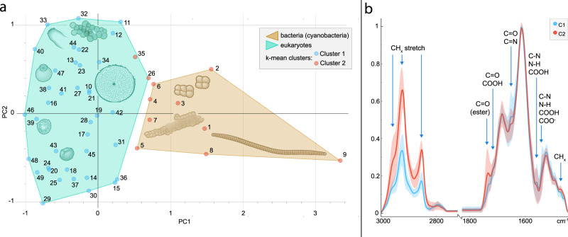
After preprocessing (see Methods), spectra were submitted to two unsupervised methods of clustering: K-mean clustering and principal component analysis (PCA). Both methods naturally sorted the data between eukaryotes and prokaryotes, i.e., cyanobacteria (Fig. 2). In the PCA, these domains are separated on PC1 (49% variance); the loading (Supplementary Fig. 5) shows that the separation is due chiefly to variation in the CH region corresponding to differences in lipid composition. PC2 (25% variance) loading corresponds to fossilization products of sugar and protein (C=O, COOH, N-moities) but is less informative to sort the data, probably due to heterogeneities prior to permineralization (e.g., microbial degradation of plant pieces). K-mean clustering suggested two clusters corresponding to a similar sorting to PC1 with 96% accuracy (only 2 eukaryotes (numbers 26 and 35) were sorted as prokaryotes; Fig. 2a, b).
Fig. 2. Unsupervised analysis of Rhynie chert fossils.
a K-means clustering (blue/red dots) and score plot for Principal Component Analysis (PCA) separating eukaryotes from prokaryotes on PC1 (convex hulls). Numbers correspond to the specimen numbers in Supplementary Data 1. The cartoons represent an arthropod fragment, a plant spore, a plant axis, Palaeoleptochlamys hassii (putative arcellinid amoeba), Frankbaronia polyspora (putative peronosporomycete), and three cyanobacteria: Palaeolyngbya kerpii, Rhyniosarcina devonica, and an unidentified Stigonema-like taxon. Cartoons were drawn from Rhynie chert organism photographs taken by ER, SM, and CCL. Source data are provided in the Source Data file. b Average spectra from k-mean clustering showing the differentiation between the groups. Organic bands correspond to absorption of lipids (bands numbers 1–3, 9 in Supplementary Table 1) and fossilization products of sugar and protein (bands 4–8); see Supplementary Note. Red and blue area represent the full variability of spectra between the specimens of each group (C1 and C2). Thick lines in each area represent average spectra for each group (mean values of each intensity at each wavenumber).
Subsequently, spectra were divided into two classes, eukaryotes and prokaryotes (based on their reported affinity). Nematophytes were excluded from the model. They were then submitted to supervised machine learning algorithms (K-nearest neighbour, logistic regression, and Random Forest). The K-nearest neighbour algorithm correctly classified all spectra from the test set, validating the approach to discriminate between domains using infrared data. Inclusion of the nematophyte data in the model predicted a eukaryotic affinity for these fossils.
The chemometric ratio R3/2, i.e., the ratio of the peak intensities corresponding to CH3 and CH2, has been proposed as a domain indicator of bacteria, archaea and possibly eukaryotes3 because membrane lipids in archaea are characterized by shorter and more highly branched aliphatic chains than those of bacteria and eukaryotes3,18,19. However, discriminating the three domains in natural assemblages based only on this ratio may be difficult34. Our specimens yield R3/2 values between 0.25 and 0.63, in accordance with values expected for non-fossil bacterial and eukaryotic lipids. However, the measured ratios do not discriminate between prokaryotes (average 0.43; σx̅ = 0.03) and eukaryotes (0.42; σx̅ = 0.01), unlike in living cells3. Mann–Whitney U tests did not reveal significant differences between the ratios obtained for prokaryotes and eukaryotes (p-value = 0.76652). As suggested by Igisu et al. (2012)34, R3/2 cannot be applied unequivocally as a domain discriminant (eukaryote vs prokaryote) to all organic fossils. Possibly the Rhynie chert is unusual in this respect; other, more mature, fossil assemblages may be more dominated by the fossilization products of lipids, whereas eukaryotes in the Rhynie chert contain a range of fossilization products that can influence the R3/2 ratio3. Alternatively, another Rhynie chert study suggests that prokaryotes and eukaryotes may be discriminated when Raman I-1350/1600 ratio (degree of structural order) is plotted against R3/2 ratios26.
The intensity ratio of carbonyl ester and methylene (ester/CH2) has also been proposed to discriminate different living bacteria and archaea based on the main differences in their lipids35. This method has been successful in distinguishing bacteria with ester-linked lipids (E. coli, B. subtilis, M. lysodeikticus) from bacteria with ester and ether lipids (T. commune)35. Archaeal cell membranes are constituted of glycerol-ether lipids, whereas bacterial and eukaryotic membranes consist mainly of glycerol-ester lipids36. A variable contribution of carbonyl ester groups is observed in fossils; thus this ratio might constitute a chemometric proxy for domain discrimination. Mann–Whitney U tests showed significant differences in ester/CH2 ratio between prokaryotes and eukaryotes from the Rhynie material (p-value = 0.02). The eukaryotes as a whole show higher values than prokaryotes, consistent with the former being richer in ester-linked lipids36. However, the large variation between eukaryote groups (Supplementary Fig. 2) suggests that the ester/CH2 ratio alone may not reliably discriminate between fossil domains.
We tested alternative ratios, C=O/CH2 (carbonyl/CH2) and N/CH2 (Nitrogen-moieties/CH2), corresponding to the variation in contribution of products from the fossilization of sugars and proteins. These ratios differentiate the two domains in Rhynie material (Mann–Whitney U test, p-value = 0.00002 (C=O/CH2) and 0.003 (N/CH2)). Higher values for eukaryotes are consistent with the result of multivariate analyses showing a lower contribution of lipids to their general composition (therefore a lower CH2 contribution). Products from the fossilization of saccharide-rich tissues would contain various amounts of carboxyl (COOH), carbonyl (C=O), and N-moieties5,37 and, in the cell wall, preservation of carbohydrate and protein material can be favoured by the reconformation of long-chained aliphatic moieties38. Sugars and proteins in bacterial extracellular polymeric substances (EPS) and cell walls may be more prone to degradation, while the esterification of carboxyl-rich EPS at the silica-kerogen interface, together with existing EPS esters39,40, may explain the high absorption from ester C=O in the bacterial spectra (Fig. 2B). Taken together, ester/CH2, C=O/CH2, and N/CH2 chemometric ratios constitute useful tools to narrow down the origins of fossil material in the Rhynie chert.
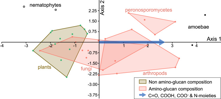
To understand how variations in sugar-protein fossilization products might discriminate between the eukaryotes, we adapted for ATR-FTIR data the methods used for Raman data by refs. 4,6,41 and 5. We transformed the intensity of informative organic bands (as revealed by the multivariate analyses) for each specimen (excluding the plant spores) into a matrix and conducted a discriminant analysis (Fig. 3). Living opisthokont biomass (fungi and animals) is rich in amino-sugar (chitin, chitosan) but fungi are also rich in β-glucan42. Modern Peronosporomycota, although containing cellulose, have a cell wall with up to 10% of N-acetylglucosamine-based carbohydrates43. Plant cells are richer in non-amino-sugars44. On the other hand, plant spores are composed of resistant aromatic polymers termed sporopollenin45. Discriminant factors along the first axis correspond to fossilization products of sugar and protein (C=O, COOH and N-moities). The rightward distribution of chitin-bearing organisms on the first axis may be explained by a higher contribution of amino-compounds from the degradation of chitinous tissues. The position of Palaeoleptochlamys hassii suggests a composition enriched in amine-containing fossilization products. Strullu-Derrien et al.23 pointed out an organic (non-agglutinated) composition for the test of this taxon and interpreted it as an arcellinid amoebozoan (testate amoeba). The ATR-FTIR results are consistent with an organic arcellinid affinity46. For the enigmatic tubular nematophyte, Nematoplexus, our FTIR data support a eukaryotic affinity, according to both unsupervised and machine-learning methods, suggesting a cyanobacterial-dominated lichen is the less likely option. Its position on the discriminant analysis (Fig. 3) indicates the composition is most similar to plant fossilization products (non-amino-sugars), excluding the possibility its composition was chitinous.
Fig. 3. Canonical Correspondence Analysis of Rhynie chert fossils.
Specimens are distributed according to the organic bands relative to fossilization products of sugars and protein (carbonyl, carboxyl, and N-moieties), showing rightward an enrichment in N-moieties (axis 1). See Supplementary Figure 9 for detail on the specific bands responsible. Source data are provided in the Source Data file.
The remarkable quality of preservation in the Rhynie chert is well attested and extends to very fine cellular features, soft-tissues, and mutual associations between taxa in life positions (e.g., refs. 47,48; see review in ref. 21) all of which were facilitated by rapid silicification23. The excellent morphological preservation, together with the excellent chemical preservation reported here, suggest a low thermal maturity profile. A previous study26 using Raman geothermometry has estimated the thermal maturity of Rhynie chert carbonaceous matter as between 190–310 and 245–345 °C, depending on the geothermometer used. However, the Raman spectra presented in that study are strikingly similar to the low thermal maturity spectrum presented by Lahfid et al.8, suggesting a similar temperature (ca. 160 °C), more consistent with the quality of fossil preservation and our FTIR results; in particular, the Raman spectra in Qu et al.26 show strong bands for disordered carbon (D3, D4, D58–10).
Although the fossils analyzed here evidently followed similar taphonomic and diagenetic pathways and are all extremely well preserved, some minor variation between them is expected, for example, in respect of the microenvironmental conditions in which they fossilized or the extent of microbial degradation (although only well-preserved tissues were included in the dataset). In addition, the thin sections were not all prepared at the same laboratory; the eukaryotes are in nine thin sections obtained from two different sample collections, and the cyanobacteria are in four thin sections obtained from three different sample collections. Nevertheless, the overall spectra are very similar, and taxon-specific signatures appear not to be confounded by any of these variables.
Organic matter in some ancient chert-hosted microfossils may have been replaced at a late stage by mobile hydrocarbons49,50. There is no doubt that the organic matter comprising Rhynie chert fossils is original; its colour and texture varies between tissue types and there is no evidence for late-stage replacement. We interpret the differences observed to be variations in the original precursors of the fossil organic matter, modified through diagenesis, rather than resulting from external contamination or preparation. Indeed, our results imply that taxon-specific microspectrosopic data could be used in future to determine the syngenicity, biogenicity and likely affinities of organically preserved structures in other cherts, as hydrocarbons of different composition are unlikely to replace different taxa selectively.
This study shows that the remarkable quality of morphological preservation characteristic of the ~407 Ma-old Rhynie chert fossil assemblage is accompanied by similarly impressive preservation of chemical information. Contrary to many older (i.e., Precambrian) cherts to which vibrational spectroscopic methods have been applied, in the Rhynie chert one can distinguish visually between prokaryotes and eukaryotes, often with well documented taxa21, and thus confirm that the spectroscopic data map exceptionally well to phylogenetic affinity. These results therefore provide a positive control for future studies of more ambiguous material and have three additional implications. Firstly, the chemometric ratio R3/2 is not an infallible guide to domain-level affinity (as already pointed out in a previous study34); here it does not significantly separate prokaryotes and eukaryotes even within a single, thermally immature fossil assemblage (although other ratios based on the occurrence of sugar and protein fossilization products do achieve this). Secondly, unsupervised methods can sort FTIR spectra into eukaryotes and prokaryotes (without preselecting preferred absorption bands), while machine learning (supervised) methods offer the potential for predictive chemotaxonomy and the resolution of ambiguous fossil affinities (as in the case of Nematoplexus). Finally, we show that FTIR spectra obtained from chert-hosted fossils retain compositional specificity despite the overwhelming silica signal. Overall, these results open a unique window on the diversity of early terrestrial life while validating the use of ATR-FTIR spectroscopy, a rapid, widely available, non-destructive in situ method, for the resolution of biomolecular differences between ancient organically preserved fossils in chert.
Methods
Sample preparation
Samples were borrowed from the School of Geosciences, University of Aberdeen (hand samples), the Oxford University Museum of Natural History (polished thin sections), and the National Museum of Scotland (NMS, hand samples). Polished thin sections from the Aberdeen samples were prepared at University College Dublin. Polished thin sections from the NMS samples were prepared by Ivan Febbrari at the University of Edinburgh. Thin-section numbers corresponding to individual fossils are provided in Supplementary Data 1.
Optical microscopy
Optical microscopy was conducted on a Leica DM2700P at the UK Centre for Astrobiology (University of Edinburgh). Photographs were acquired in transmitted light using ×10 and ×20 objectives and the Leica Application Suite (4.0) software.
ATR-FTIR
Attenuated Total Reflectance-FTIR was conducted at room temperature on a Smiths IlluminateIR microscope equipped with liquid nitrogen cooled MCT detector providing spectral resolution of 4 cm−1, using a diamond coated ATR objective (magn x36) at the School of Chemistry (University of Edinburgh). Backgrounds were taken in the air before analysis. Spectra were acquired in reflection mode for each specimen by combining 128 accumulations in the range 4000–650 cm−1 with an aperture of 100 µm with the software Qual ID 2.51 (Smiths).
Data processing
FTIR spectra were preprocessed before multivariate and machine learning analyses in the software Quasar 1.6.0 (Orange51,52). For analyses, spectra were truncated to 3000 and 1400 cm−1 in order to remove the uninformative intervals due to OH absorptions (4000–3000 cm−1) and intense vibration of silica (1400–650 cm−1). Similarly, the interval between 2700 and 1760 cm−1 was cut to remove the influence of atmospheric CO2, ATR diamond absorption and high wavenumber silica overtones. Rubberband baseline correction and Min-Max normalization (see below) were applied to the spectra before analyses. For inspection and band assignment, average second derivative spectra for each domain (Eukaryote and Prokaryote) were obtained by applying a Savitzky–Golay filter of polynomial 2 with a window of 9 (Supplementary Fig. 4).
Normalization
To minimize the influence of thickness and differences in organic matter concentration, normalization of the data (spectra) is necessary53. We performed a min-max normalization by artificially setting the minimal intensity at 0 and the maximal intensity at 1 (corresponding to the 1615 cm−1 silica band). The normalization was conducted in the preprocessing widget of the software Quasar. Such normalization results in the expression of all other absorption bands relative to the most intense band (in our case the ratio X/Silica).
Sanity checks were performed on the PCA results to ensure that the detected clustering was a result of compositional differences and not simply the changes in the abundance of organic matter in regard to silica (presence of a larger fossil for example). In the latter case, the ratio organic/silica would follow broadly the same trend for each organic band. To test this, we calculated the ratios CH2/silica and C=O/silica by dividing the intensity of the 2920 cm−1 (CH2) and 1650 cm−1 (C=O) absorption bands by the 1615 cm−1 Si–O band. Results were implemented on PCA score plots. We can see by comparing Supplementary Figures 6 and 7 that the increase of CH2/silica ratio rightward (PC1) is not followed by the increase of the C=O/silica ratio; note also that the cyanobacteria, which might intuitively be suspected to preserve less organic material than plants, arthropods, etc., are actually characterized by higher CH2/silica ratios, not lower. A similar observation can be made for PC2, along which C=O/silica increases but not CH2/silica. In addition, the other bands of silica does not contribute clearly to the loadings (Supplementary Fig. 5). Loadings for PC1 and PC2 display features characteristic of lipid moieties and soft tissue fossilization products, respectively. The clustering of the different specimens is therefore compositional. Close inspection of Fig. 2B shows that the same is true of the k-means clustering: C1 is characterized by smaller CH stretching peaks but stronger peaks for several other organic moieties, so the primary difference between the two clusters is not simply the amount of organic matter.
Data analyses
Semi-quantitative ratios
Calculations of methyl/methylene ratio (R3/2) were conducted using the method introduced by3. After baselining of the spectra between ca. 3000 and 2800 cm−1, intensity of CH3 asymmetric stretch (2960 cm−1) is divided by intensity of CH2 asymmetric stretch (2925 cm−1). Other parameters were obtained by dividing the intensity at 1650 cm−1 (C=O); 1735 cm−1 (C=O in ester group); 1560 cm−1 (NH bending, CN stretching, COOH) by the intensity at 2925 cm−1 (CH2 asymmetric stretch). Statistical tests to test the difference between prokaryotes (n = 9) and eukaryotes (n = 40) for each of the ratio results were performed in PAST 4.09. A Shapiro–Wilk test shows that the data do not consistently follow a normal distribution. Therefore, non-parametric Mann–Whitney U tests were applied to compare the two groups (eukaryotes and prokaryotes).
Multivariate analyses
Principal component analysis (PCA) and K-means clustering (KMC) were performed in Quasar. KMC was performed with random initialization and 300 iterations. PCA was performed on 6 components, explaining 96% of the variance. Only PC1 (49%) and PC2 (25%) were considered informative. Loadings for PC1 and PC2 are available in Supplementary Figure 5. Additional PCA include all fossils and 4 matrix samples is shown of Supplementary Fig. 8. Discriminant analysis was performed in PAST 4.09 using absorption band intensities at ca. 1460, 1540, 1560, 1650, 1715, 1735, 2851, 2920, 2960 cm−1.
Supervised analyses (machine learning)
Classification and predictions were done using Quasar. Preprocessed spectra were divided into a training set containing 60% of the spectra (29) and a test set containing 40% of the spectra (18). In order to build a consistent model, the training set was submitted to three different learners: (1) logistic regression with L2 ridge regularization (C=1); (2) k-nearest neighbour (kNN) using Euclidean distances and 5 neighbours; and (3) Random Forest (RF) with 50 trees. The three methods were applied with a leave-one-out cross validation. The classification accuracy on the training set was 97% for Random Forest with a ROC-AUC (Receiving Operator Characteristic Area Under the Curve) of 0.98. Logistic regression and kNN both provided 90% classification accuracy and a ROC-AUC of 0.98 and 0.97, respectively. We then used the three models to predict the classes within the test set. The classification accuracies were 94% for Random Forest (ROC-AUC 0.9) and Logistic Regression (ROC-AUC 1) and 100% for kNN (ROC-AUC 1).
Reporting summary
Further information on research design is available in the Nature Portfolio Reporting Summary linked to this article.
Supplementary information
Description of Additional Supplementary Files
Acknowledgements
This research was supported by The Royal Society (UK) and the Belgium Wallonia Brussels programme WBI.WORLD (C.C.L.) and the CONACYT (National Council of Science and Technology of Mexico) Postgraduate Scholarship for Studying Abroad (E.R). We thank J. Parnell at the University of Aberdeen, the National Museums Scotland and the University of Oxford Museum of Natural History for facilitating access to samples. I. Febbrari is thanked for the preparation of thin sections. S. Hetherington and M. Krings are thanked for their help identifying cyanobacterial and plant taxa. D. Marosi-McMahon is thanked for the help with statistical comparison tests.
Source data
Author contributions
C.C.L., S.M., and P.J.O. conceived the study. S.M., N.C.F., and P.J.O. acquired and prepared samples. S.M., E.R., and C.C.L. acquired photomicrographs. C.C.L. and A.V.G. acquired the ATR data. C.C.L. processed the data and conducted multivariate and supervised analyses with input from S.M. and E.R. C.C.L., and S.M. drafted the manuscript and figures. All authors reviewed and edited the manuscript.
Peer review
Peer review information
Nature Communications thanks Jasmina Wiemann and the other, anonymous, reviewer(s) for their contribution to the peer review of this work. Peer reviewer reports are available.
Data availability
Thin-sections are available from their respective collections: Specimen codes: 2829p: Oxford University Museum of Natural History. Rfa, Rfe, Rff, Rfg, ALG2, AGLyon 2019, AGL127, Agl75 2019-1: University of Aberdeen. NMSRC9: National Museums of Scotland. Raw spectra are available on Edinburgh DataShare at 10.7488/ds/3806. Source data are provided as a Source Data file. Source data are provided with this paper.
Competing interests
The authors declare no competing interests.
Footnotes
Publisher’s note Springer Nature remains neutral with regard to jurisdictional claims in published maps and institutional affiliations.
Contributor Information
C. C. Loron, Email: v1cloron@ed.ac.uk
S. McMahon, Email: sean.mcmahon@ed.ac.uk
Supplementary information
The online version contains supplementary material available at 10.1038/s41467-023-37047-1.
References
- 1.Marshall A, Marshall CP. Vibrational spectroscopy of fossils. Palaeontology. 2015;58:201–211. doi: 10.1111/pala.12144. [DOI] [Google Scholar]
- 2.Marshall CP, Javaux EJ, Knoll AH, Walter MR. Combined micro-Fourier transform infrared (FTIR) spectroscopy and micro-Raman spectroscopy of Proterozoic acritarchs: a new approach to palaeobiology. Precambrian Res. 2005;138:208–224. doi: 10.1016/j.precamres.2005.05.006. [DOI] [Google Scholar]
- 3.Igisu M, et al. Micro-FTIR spectroscopic signatures of bacterial lipids in Proterozoic microfossils. Precambrian Res. 2009;173:19–26. doi: 10.1016/j.precamres.2009.03.006. [DOI] [Google Scholar]
- 4.Wiemann J, Crawford JM, Briggs DE. Phylogenetic and physiological signals in metazoan fossil biomolecules. Sci. Adv. 2020;6:eaba6883. doi: 10.1126/sciadv.aba6883. [DOI] [PMC free article] [PubMed] [Google Scholar]
- 5.McCoy VE, et al. Chemical signatures of soft tissues distinguish between vertebrates and invertebrates from the Carboniferous Mazon Creek Lagerstätte of Illinois. Geobiology. 2020;18:560–565. doi: 10.1111/gbi.12397. [DOI] [PubMed] [Google Scholar]
- 6.Wiemann J, et al. Fossilization transforms vertebrate hard tissue proteins into N-heterocyclic polymers. Nat. Commun. 2018;9:1–9. doi: 10.1038/s41467-018-07013-3. [DOI] [PMC free article] [PubMed] [Google Scholar]
- 7.Foucher F, Ammar MR, Westall F. Revealing the biotic origin of silicified Precambrian carbonaceous microstructures using Raman spectroscopic mapping, a potential method for the detection of microfossils on Mars. J. Raman Spectrosc. 2015;46:873–879. doi: 10.1002/jrs.4687. [DOI] [Google Scholar]
- 8.Lahfid A, et al. Evolution of the Raman spectrum of carbonaceous material in low‐grade metasediments of the Glarus Alps (Switzerland) Terra Nova. 2010;22:354–360. doi: 10.1111/j.1365-3121.2010.00956.x. [DOI] [Google Scholar]
- 9.Sforna MC, Van Zuilen MA, Philippot P. Structural characterization by Raman hyperspectral mapping of organic carbon in the 3.46 billion-year-old Apex chert, Western Australia. Geochim. Cosmochim. Acta. 2014;124:18–33. doi: 10.1016/j.gca.2013.09.031. [DOI] [Google Scholar]
- 10.Baludikay BK, et al. Raman microspectroscopy, bitumen reflectance and illite crystallinity scale: comparison of different geothermometry methods on fossiliferous Proterozoic sedimentary basins (DR Congo, Mauritania, and Australia) Int. J. Coal Geol. 2018;191:80–94. doi: 10.1016/j.coal.2018.03.007. [DOI] [Google Scholar]
- 11.D’Angelo JA, Escudero LB, Volkheimer W, Zodrow EL. Chemometric analysis of functional groups in fossil remains of the Dicroidium flora (Cacheuta, Mendoza, Argentina): implications for kerogen formation. Int. J. Coal Geol. 2011;87:97–111. doi: 10.1016/j.coal.2011.05.005. [DOI] [Google Scholar]
- 12.Vajda V, et al. Molecular signatures of fossil leaves provide unexpected new evidence for extinct plant relationships. Nat. Ecol. Evol. 2017;1:1093–1099. doi: 10.1038/s41559-017-0224-5. [DOI] [PubMed] [Google Scholar]
- 13.Lis GP, Mastalerz M, Schimmelmann A, Lewan MD, Stankiewicz BA. FTIR absorption indices for thermal maturity in comparison with vitrinite reflectance R0 in type-II kerogens from Devonian black shales. Org. Geochem. 2005;36:1533–1552. doi: 10.1016/j.orggeochem.2005.07.001. [DOI] [Google Scholar]
- 14.Steemans P, Lepot K, Marshall CP, Le Hérissé A, Javaux EJ. FTIR characterisation of the chemical composition of Silurian miospores (cryptospores and trilete spores) from Gotland, Sweden. Rev. Palaeobot. Palynol. 2010;162:577–590. doi: 10.1016/j.revpalbo.2010.07.006. [DOI] [Google Scholar]
- 15.Dutta S, Hartkopf-Fröder C, Witte K, Brocke R, Mann U. Molecular characterization of fossil palynomorphs by transmission micro-FTIR spectroscopy: Implications for hydrocarbon source evaluation. Int. J. Coal Geol. 2013;115:13–23. doi: 10.1016/j.coal.2013.04.003. [DOI] [Google Scholar]
- 16.Loron CC, et al. Early fungi from the Proterozoic era in Arctic Canada. Nature. 2019;570:232–235. doi: 10.1038/s41586-019-1217-0. [DOI] [PubMed] [Google Scholar]
- 17.Bonneville S, et al. Molecular identification of fungi microfossils in a Neoproterozoic shale rock. Sci. Adv. 2020;6:eaax7599. doi: 10.1126/sciadv.aax7599. [DOI] [PMC free article] [PubMed] [Google Scholar]
- 18.Igisu M, Komiya T, Ikemoto Y, Geng Y, Uehara H. Studying microscale distributions of aliphatic CH bonds in Neoproterozoic prokaryotic fossils using SR micro-FTIR. Geochem. J. 2017;51:589–594. doi: 10.2343/geochemj.2.0486. [DOI] [Google Scholar]
- 19.Igisu M, et al. Spatial distribution of organic functional groups in Ediacaran acritarchs from the Doushantuo Formation in South China as revealed by micro-FTIR spectroscopy. Precambrian Res. 2022;373:106628. doi: 10.1016/j.precamres.2022.106628. [DOI] [Google Scholar]
- 20.Hickman‐Lewis K, Westall F, Cavalazzi B. Diverse communities of Bacteria and Archaea flourished in Palaeoarchaean (3.5–3.3 Ga) microbial mats. Palaeontology. 2020;63:1007–1033. doi: 10.1111/pala.12504. [DOI] [Google Scholar]
- 21.Garwood RJ, Oliver H, Spencer AR. An introduction to the Rhynie chert. Geol. Mag. 2020;157:47–64. doi: 10.1017/S0016756819000670. [DOI] [Google Scholar]
- 22.Trewin NH, Fayers SR, Kelman R. Subaqueous silicification of the contents of small ponds in an Early Devonian hot-spring complex, Rhynie, Scotland. Can. J. Earth Sci. 2003;40:1697–1712. doi: 10.1139/e03-065. [DOI] [Google Scholar]
- 23.Strullu-Derrien C, Kenrick P, Goral T, Knoll AH. Testate amoebae in the 407-million-year-old Rhynie chert. Curr. Biol. 2019;29:461–467. doi: 10.1016/j.cub.2018.12.009. [DOI] [PubMed] [Google Scholar]
- 24.Boyce CK, Hazen RM, Knoll AH. Nondestructive, in situ, cellular-scale mapping of elemental abundances including organic carbon in permineralized fossils. Proc. Natl Acad. Sci. USA. 2001;98:5970–5974. doi: 10.1073/pnas.101130598. [DOI] [PMC free article] [PubMed] [Google Scholar]
- 25.Preston LJ, Genge MJ. The Rhynie chert, Scotland, and the search for life on Mars. Astrobiology. 2010;10:549–560. doi: 10.1089/ast.2008.0321. [DOI] [PubMed] [Google Scholar]
- 26.Qu Y, Engdahl A, Zhu S, Vajda V, McLoughlin N. Ultrastructural heterogeneity of carbonaceous material in ancient cherts: Investigating biosignature origin and preservation. Astrobiology. 2015;15:825–842. doi: 10.1089/ast.2015.1298. [DOI] [PubMed] [Google Scholar]
- 27.Taylor TN, Krings M. A colony-forming microorganism with probable affinities to the Chroococcales (Cyanobacteria) from the Lower Devonian Rhynie chert. Rev. Palaeobot. Palynol. 2015;219:147–156. doi: 10.1016/j.revpalbo.2015.04.003. [DOI] [Google Scholar]
- 28.Krings M. Palaeolyngbya kerpii sp. nov., a large filamentous cyanobacterium with affinities to Oscillatoriaceae from the Lower Devonian Rhynie chert. PalZ. 2019;93:377–386. doi: 10.1007/s12542-019-00475-w. [DOI] [Google Scholar]
- 29.Lyon AG. IV.—On the fragmentary remains of an organism referable to the nematophytales, from the Rhynie chert, Nematoplexus rhyniensis gen. et sp. nov. Earth Environ. Sci. Trans. R. Soc. Edinb. 1962;65:79–87. doi: 10.1017/S0080456800012382. [DOI] [Google Scholar]
- 30.Kondas M. Nematophytes. Geol. Today. 2018;34:73–78. doi: 10.1111/gto.12225. [DOI] [Google Scholar]
- 31.Coates, J. Encyclopedia of Analytical Chemistry (ed. Meyers, R. A.) 10881–10882 (John Wiley & Sons Ltd., 2000).
- 32.Briggs, D. E. Experimental taphonomy. Palaios10, 539–550 (1995).
- 33.Killops, V. J., & Killops, S. D. Introduction to Organic Geochemistry (John Wiley & Sons., 2013).
- 34.Igisu M, et al. Domain‐level identification and quantification of relative prokaryotic cell abundance in microbial communities by micro‐FTIR spectroscopy. Environ. Microbiol. Rep. 2012;4:42–49. doi: 10.1111/j.1758-2229.2011.00277.x. [DOI] [PubMed] [Google Scholar]
- 35.Hedrick DB, Nivens DE, Stafford C, White DC. Rapid differentiation of archaebacteria from eubacteria by diffuse reflectance Fourier-transform IR spectroscopic analysis of lipid preparations. J. Microbiol. Methods. 1991;13:67–73. doi: 10.1016/0167-7012(91)90033-M. [DOI] [Google Scholar]
- 36.Řezanka T, Kolouchová I, Gharwalová L, Palyzová A, Sigler K. Lipidomic analysis: from archaea to mammals. Lipids. 2018;53:5–25. doi: 10.1002/lipd.12001. [DOI] [PubMed] [Google Scholar]
- 37.Mohsin GF, et al. Structural characterization of melanoidin formed from D-glucose and L-alanine at different temperatures applying FTIR, NMR, EPR, and MALDI-ToF-MS. Food Chem. 2018;245:761–767. doi: 10.1016/j.foodchem.2017.11.115. [DOI] [PubMed] [Google Scholar]
- 38.Vandenbroucke M, Largeau C. Kerogen origin, evolution and structure. Org. Geochem. 2007;38:719–833. doi: 10.1016/j.orggeochem.2007.01.001. [DOI] [Google Scholar]
- 39.Kaplan Can H, Gurbuz F, Odabaşı M. Partial characterization of cyanobacterial extracellular polymeric substances for aquatic ecosystems. Aquat. Ecol. 2019;53:431–440. doi: 10.1007/s10452-019-09699-z. [DOI] [Google Scholar]
- 40.Hantal G, Brochard L, Dias Soeiro Cordeiro MN, Ulm FJ, Pellenq RJM. Surface chemistry and atomic-scale reconstruction of kerogen–silica composites. J. Phys. Chem. C. 2014;118:2429–2438. doi: 10.1021/jp406329n. [DOI] [Google Scholar]
- 41.Wiemann, J. et al. Fossil biomolecules reveal an avian metabolism in the ancestral dinosaur. Nature606, 1–5 (2022). [DOI] [PubMed]
- 42.Watkinson, S. C., Boddy, L. & Money, N. The Fungi (Academic Press, 2015).
- 43.Mélida H, Sandoval-Sierra JV, Diéguez-Uribeondo J, Bulone V. Analyses of extracellular carbohydrates in oomycetes unveil the existence of three different cell wall types. Eukaryot. Cell. 2013;12:194–203. doi: 10.1128/EC.00288-12. [DOI] [PMC free article] [PubMed] [Google Scholar]
- 44.Heredia A, Jiménez A, Guillén R. Composition of plant cell walls. Z. f.ür. Lebensm.-Unters. und Forsch. 1995;200:24–31. doi: 10.1007/BF01192903. [DOI] [PubMed] [Google Scholar]
- 45.de Leeuw JW, Versteegh GJ, van Bergen PF. Biomacromolecules of algae and plants and their fossil analogues. Plant Ecol. 2006;182:209–233. doi: 10.1007/s11258-005-9027-x. [DOI] [Google Scholar]
- 46.Kosakyan A, Gomaa F, Lara E, Lahr DJ. Current and future perspectives on the systematics, taxonomy, and nomenclature of testate amoebae. Eur. J. Protistol. 2016;55:105–117. doi: 10.1016/j.ejop.2016.02.001. [DOI] [PubMed] [Google Scholar]
- 47.Claridge MF, Lyon AG. Lung-books in the Devonian Palaeocharinidae (Arachnida) Nature. 1961;191:1190–1191. doi: 10.1038/1911190b0. [DOI] [Google Scholar]
- 48.Hetherington AJ, Dolan L. Rhynie chert fossils demonstrate the independent origin and gradual evolution of lycophyte roots. Curr. Opin. Plant Biol. 2019;47:119–126. doi: 10.1016/j.pbi.2018.12.001. [DOI] [PubMed] [Google Scholar]
- 49.Rasmussen B, Muhling JR. Evidence for widespread oil migration in the 1.88 Ga Gunflint Formation, Ontario, Canada. Geology. 2019;47:899–903. doi: 10.1130/G46469.1. [DOI] [Google Scholar]
- 50.Rasmussen B, Muhling JR, Fischer WW. Ancient oil as a source of carbonaceous matter in 1.88-billion-year-old Gunflint stromatolites and microfossils. Astrobiology. 2021;21:655–672. doi: 10.1089/ast.2020.2376. [DOI] [PubMed] [Google Scholar]
- 51.Toplak M, et al. Infrared orange: connecting hyperspectral data with machine learning. Synchrotron Rad. N. 2017;30:40–45. doi: 10.1080/08940886.2017.1338424. [DOI] [Google Scholar]
- 52.Toplak M, et al. Quasar: easy machine learning for biospectroscopy. Cells. 2021;10.9:2300. doi: 10.3390/cells10092300. [DOI] [PMC free article] [PubMed] [Google Scholar]
- 53.Trevisan J, Angelov PP, Carmichael PL, Scott AD, Martin FL. Extracting biological information with computational analysis of Fourier-transform infrared (FTIR) biospectroscopy datasets: current practices to future perspectives. Analyst. 2012;137:3202–3215. doi: 10.1039/c2an16300d. [DOI] [PubMed] [Google Scholar]
Associated Data
This section collects any data citations, data availability statements, or supplementary materials included in this article.
Supplementary Materials
Description of Additional Supplementary Files
Data Availability Statement
Thin-sections are available from their respective collections: Specimen codes: 2829p: Oxford University Museum of Natural History. Rfa, Rfe, Rff, Rfg, ALG2, AGLyon 2019, AGL127, Agl75 2019-1: University of Aberdeen. NMSRC9: National Museums of Scotland. Raw spectra are available on Edinburgh DataShare at 10.7488/ds/3806. Source data are provided as a Source Data file. Source data are provided with this paper.


