1. PATIENT PRESENTATION
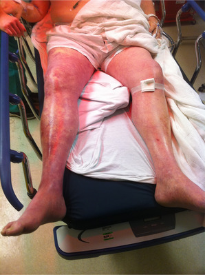
A 66‐year‐old‐male with a history of type 2 diabetes and hypertension presented as a transfer for rapid progression of lower extremity pain, swelling, and blue‐purple discoloration of the entire limb (Figure 1) with concern for a possible necrotizing infection. His symptoms began earlier in the day and progressed over just a few hours. He had no known thromboembolic risk factors. Initial vital signs were notable only for a heart rate of 115.
FIGURE 1.

Right lower limb with swelling, blotchy blue and purple discoloration, and pain consistent with phlegmasia cerulea dolens.
Point‐of‐care ultrasound revealed extensive deep venous thrombosis burden confirmed by formal duplex sonography. The patient was admitted on a heparin infusion and underwent EKOSTM catheter directed tissue plasminogen therapy with stent placement. He was admitted for 5 days and successfully discharged on oral anticoagulation back to his baseline level of function.
2. DIAGNOSIS
Phlegmasia cerulea dolens
Phlegmasia cerulea dolens (PCD) is a rare ischemic complication of massive venous thromboembolism with high rates of morbidity and mortality. Patients present with limb edema, pain, and cyanosis that can quickly develop into compartment syndrome, limb ischemia, and venous gangrene with amputation and death rates as high as 50% and 40%, respectively. 1 PCD tends to affect the iliofemoral segment of the lower extremities. 2 Non‐traumatic cases of PCD are most commonly associated with malignancy. 3 The preferred imaging modality is Doppler ultrasound and use of point‐of‐care ultrasound by physicians can expedite treatment of PCD. 4 Management includes limb elevation, intravenous fluids, and either systemic anticoagulation, catheter‐directed thrombolysis, thrombectomy, or a combination of these definitive therapies. 5
Byars D, Landon J, Spurrell M. Man with leg pain. JACEP Open. 2023;4:e12924. 10.1002/emp2.12924
REFERENCES
- 1. Chaochankit W, Akaraborworn O. Phlegmasia cerulea dolens with compartment syndrome. Ann Vasc Dis. 2018;11(3):355‐357. doi: 10.3400/avd.cr.18-00030 [DOI] [PMC free article] [PubMed] [Google Scholar]
- 2. Gardella L, Faulk JB. Phlegmasia alba and cerulea dolens. StatPearls [Internet]. StatPearls Publishing; 2022. Updated 2022 Oct 3. Jan‐. Available from. https://www.ncbi.nlm.nih.gov/books/NBK563137/ [PubMed] [Google Scholar]
- 3. Bazan HA, Reiner E, Sumpio B. Management of bilateral phlegmasia cerulea dolens in a patient with subacute splenic laceration. Ann Vasc Dis. 2008;1(1):45‐48. doi: 10.3400/avd.AVDcr07002 [DOI] [PMC free article] [PubMed] [Google Scholar]
- 4. Schroeder M, Shorette A, Singh S, Budhram G. Phelgmasia cerulea dolens diagnosed by point‐of‐care ultrasound. Clin Pract Cases Emerg Med. 2017;1(2):104‐107. doi: 10.5811/cpcem.2016.12.32716 [DOI] [PMC free article] [PubMed] [Google Scholar]
- 5. Said A, Sahlieh A, Sayed L. A comparative analysis of the efficacy and safety of therapeutic interventions in phlegmasia cerulea dolens. Phlebology. 2021;36(5):392‐400. doi: 10.1177/0268355520975581 [DOI] [PubMed] [Google Scholar]


