Abstract
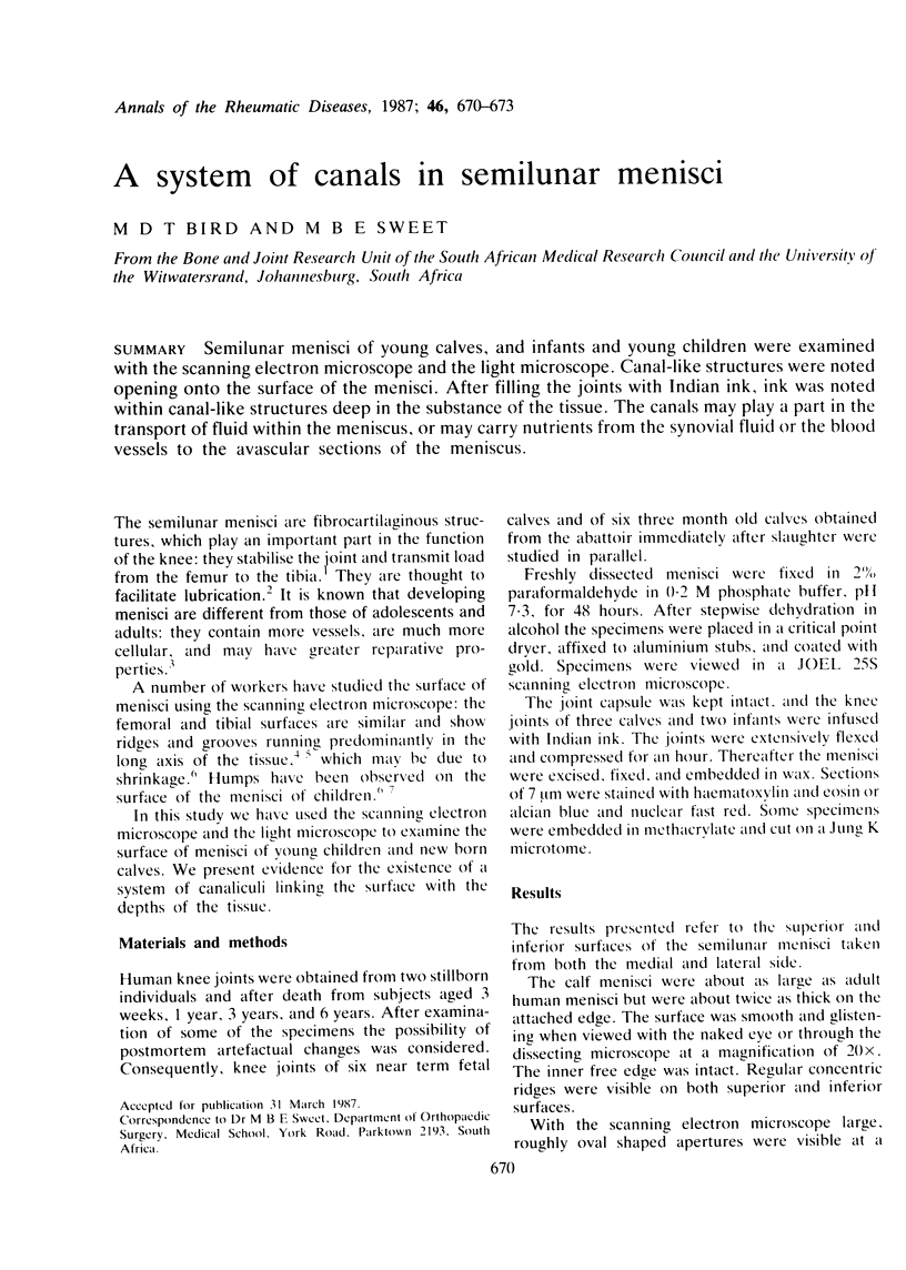
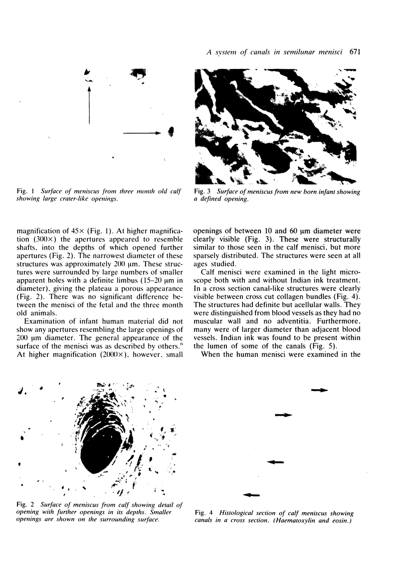
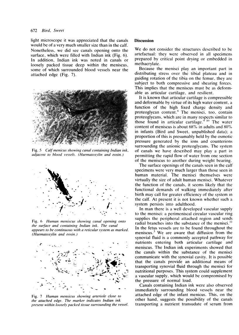
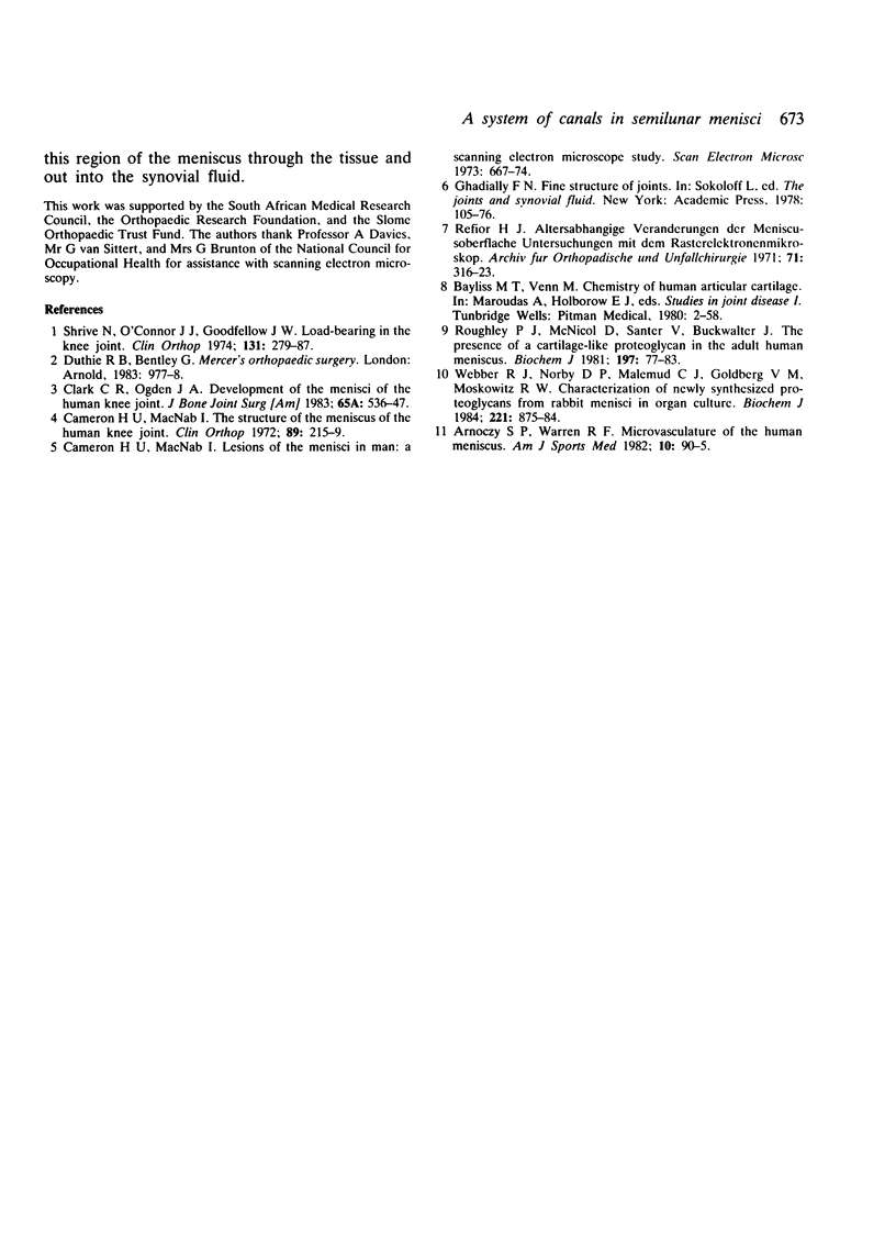
Semilunar menisci of young calves, and infants and young children were examined with the scanning electron microscope and the light microscope. Canal-like structures were noted opening onto the surface of the menisci. After filling the joints with Indian ink, ink was noted within canal-like structures deep in the substance of the tissue. The canals may play a part in the transport of fluid within the meniscus, or may carry nutrients from the synovial fluid or the blood vessels to the avascular sections of the meniscus.
Full text
PDF


Images in this article
Selected References
These references are in PubMed. This may not be the complete list of references from this article.
- Arnoczky S. P., Warren R. F. Microvasculature of the human meniscus. Am J Sports Med. 1982 Mar-Apr;10(2):90–95. doi: 10.1177/036354658201000205. [DOI] [PubMed] [Google Scholar]
- Cameron H. U., Macnab I. The structure of the meniscus of the human knee joint. Clin Orthop Relat Res. 1972;89:215–219. [PubMed] [Google Scholar]
- Refior H. J. Altersabhängige Veränderungen der Meniscusoberfläche--Untersuchungen mit dem Rasterelektronemikroskop. Arch Orthop Unfallchir. 1971;71(4):316–323. [PubMed] [Google Scholar]
- Roughley P. J., McNicol D., Santer V., Buckwalter J. The presence of a cartilage-like proteoglycan in the adult human meniscus. Biochem J. 1981 Jul 1;197(1):77–83. doi: 10.1042/bj1970077. [DOI] [PMC free article] [PubMed] [Google Scholar]
- Shrive N. G., O'Connor J. J., Goodfellow J. W. Load-bearing in the knee joint. Clin Orthop Relat Res. 1978 Mar-Apr;(131):279–287. [PubMed] [Google Scholar]


