



Abstract
Objectives:
We sought to develop a rigorous, systematic protocol for the dissection and preservation of human hearts for biobanking that expands previous success in postmortem transcriptomics to multiomics from paired tissue.
Background:
Existing cardiac biobanks consist largely of biopsy tissue or explanted hearts in select diseases and are insufficient for correlating whole organ phenotype with clinical data.
Methods:
We demonstrate optimal conditions for multiomics interrogation (ribonucleic acid (RNA) sequencing, untargeted metabolomics) in hearts by evaluating the effect of technical variables (storage solution, temperature) and simulated postmortem interval (PMI) on RNA and metabolite stability. We used bovine (n=3) and human (n=2) hearts fixed in PAXgene or snap-frozen with liquid nitrogen.
Results:
Using a paired Wald test, only two of the genes assessed were differentially expressed between left ventricular samples from bovine hearts stored in PAXgene at 0 and 12 hours PMI (FDR q<0.05). We obtained similar findings in human left ventricular samples, suggesting stability of RNA transcripts at PMIs up to 12 hours. Different library preparation methods (mRNA poly-A capture vs. rRNA depletion) resulted in similar quality metrics with both library preparations achieving >95% of reads properly aligning to the reference genomes across all PMIs for bovine and human hearts. PMI had no effect on RNA Integrity Number or quantity of RNA recovered at the time points evaluated. Of the metabolites identified (855 total) using untargeted metabolomics of human left ventricular tissue, 503 metabolites remained stable across PMIs (0, 4, 8, 12 hours). Most metabolic pathways retained several stable metabolites.
Conclusions:
Our data demonstrate a technically rigorous, reproducible protocol that will enhance cardiac biobanking practices and facilitate novel insights into human CVD.
Condensed abstract:
Cardiovascular disease (CVD) is the leading cause of mortality worldwide. Current biobanking practices insufficiently capture both the diverse array of phenotypes present in CVDs and the spatial heterogeneity across cardiac tissue sites. We have developed a rigorous and systematic protocol for the dissection and preservation of human cardiac biospecimens to enhance the availability of whole organ tissue for multiple applications. When combined with longitudinal clinical phenotyping, our protocol will enable multiomics in hearts to deepen our understanding of CVDs.
Keywords: Biobank, Multiomics, RNA-Seq, Metabolomics, Postmortem interval, Cardiovascular
1. Introduction
Cardiovascular diseases (CVDs) remain the predominant cause of death in the United States and confer significant economic repercussions with ∼$214 billion dollars spent annually [1]. The Framingham Heart Study and other large cohort studies have been pivotal in delineating the association of risk factors with the development of CVD [2,3]. Recently, several epidemiologic studies have highlighted the association of greater time of CVD risk factor exposure with increased cumulative lifetime risk of CVD and reduced longevity [4–7]. However, the relation of CVD risk factors across the lifespan on metabolism and gene expression in cardiac tissue is not well understood, as no longitudinal cohorts, including the National Heart, Lung, and Blood Institute cohorts or United Kingdom Biobank, have established cardiovascular tissue repositories or biobanks.
Our understanding of the pathobiology of CVD has been hindered by animal models that do not recapitulate the spectrum of disease phenotypes nor the concomitant multimorbidity observed in patients [8,9]. Access to human cardiac samples is incredibly limited, especially for individuals unselected for prevalent CVD. Current studies evaluating human cardiac tissue are restricted to cardiac conditions in which cardiac interventions and/or procedures have frequently been performed, such as biopsies obtained during ventricular assist device implantation or at the time of heart catheterizations. Such samples may not reflect all the chambers/sites relevant to the full spectrum of CVDs and may limit the generalizability of the data. Limitations in accessing human cardiac tissue have forced the cardiovascular field to assess surrogate endpoints with circulating biomarkers and imaging, with rare insights from the tissue itself. Existing cardiac repositories and biobanks currently have several limitations: (a) inclusion of samples with inadequate antecedent phenotyping of CVD risk factors; (b) small sample sizes; (c) a constrained spectrum of CVDs sampled as tissues are obtained only at the time of cardiac procedures and/or interventions for clinical convenience and safety of the patient; and (d) cardiac tissue site restrictions to either right/left ventricle or left atrial appendage often prioritized when the tissues are obtained from patients with cardiovascular diseases undergoing cardiac transplantation or surgery [10–12].
Systematic clinical characterization paired with rigorous multiomics of tumor and non-tumor tissue in humans has enabled novel pathway discovery and successful drug development in cancer biology [13]. Understanding the relation of individual and cumulative CVD risk factors across the lifespan and molecular biomarkers of disease in deeply phenotyped cardiac tissue is crucial for identifying novel therapeutic targets. Thus, cardiac biobanks housing whole hearts from clinically phenotyped individuals, suitably preserved for deep systems biology, are essential to advance our ability to understand and treat CVD.
For our study, we proposed three main aims: (1) to develop a rigorous dissection and storage protocol to facilitate whole heart acquisition and collect samples to apply multiomics across cardiac sites; (2) to evaluate the feasibility of transcriptomics and metabolomics in cardiac tissues collected postmortem; and (3) to characterize the effect of postmortem interval (PMI), cardiac sites, and tissue-storage solutions on RNA and metabolite stability in cardiac tissue. Importantly, this proof-of-concept study aimed not to interrogate the pathobiology of the samples included, but to demonstrate the feasibility of performing multiomics from paired tissue samples from postmortem hearts.
2. Methods
2.1. Cardiac tissue procurement
2.1.1. Human
Human hearts were obtained from participants at Brigham & Women’s Hospital (BWH) under IRB # H-39709 with Boston University institutional approval by the Boston University Medical Center (BUMC) Institutional Review Board (IRB # H-39709). Two human hearts (H1, H2) were obtained at the time of cardiac transplantation from male participants with the following pathologic diagnosis: (a) ischemic heart disease with subsequent ventricular assist device implantation; and (b) ischemic heart disease with concomitant valvular disease requiring bioprosthetic aortic valve replacement and eosinophilic myocarditis. The hearts were perfused following excision to remove blood products. Cardiac dissection details are reviewed in the results.
2.1.2. Bovine
Fresh bovine hearts (B1, B2, B3) were obtained through a commercial research vendor (Research 87, Inc) from male calves, as calf hearts are of similar dimensions as human hearts. Further, the use of bovine hearts collected postmortem minimized variability introduced from the type and manner of death (that would normally occur with postmortem procurement of human hearts), providing the opportunity to isolate and interrogate the effects of PMI on the transcriptome and metabolome with less confounding. As only male calves are killed for food production, only male bovine hearts were commercially available for study. An Institutional Animal Care and Use Committee protocol waiver from Boston University School of Medicine was applied as no animals were housed or euthanized by BUMC personnel. The hearts were perfused to remove blood products prior to transport. Cardiac dissection details are reviewed in the results. Additional sample characteristics for human and bovine hearts are detailed in Supplemental Table S1.
2.2. Experimental protocols (Fig. 1)
Fig. 1.

Schematic illustration of the cardiac dissection workflow. Repeated sampling was performed from 2 human and 3 bovine hearts stored in either phosphate buffered saline or cardioplegic solution during a simulated postmortem interval (PMI). Following the PMI, samples were either flash frozen or fixed in the PAXgene preservation system and then stored at −80 °C. RNA sequencing was performed on an Illumina NextSeq 500 machine using RNA isolated from both flash frozen and PAXgene fixed human and bovine cardiac tissue. Unbiased metabolomics was also performed on flash frozen human and bovine cardiac tissue for evaluation of metabolite stability.
2.2.1. PMI simulation
Following fresh tissue procurement of human and bovine hearts, PMI was simulated to determine the stability and integrity of RNA and metabolites. During the simulated PMI, human and bovine samples were stored in commercially available, 1X phosphate buffered saline (PBS), with human samples at 4°C and bovine samples at either room temperature or 4°C. Over a period of 0–12 hours following sectioning, repeated sampling was performed from the same chamber of the same cardiac biospecimen for different uses. Previously, the Genotype Tissue Expression (GTEx) project demonstrated that RNA expression was not associated with PMIs <6 hours in left ventricular tissue[14]; therefore, we chose to investigate changes in RNA transcript expression with greater PMIs in this study. As metabolomic changes in post-mortem tissues across PMIs is not well-characterized, time points from 0 to 12 hours were evaluated. Tissue samples were designated for: (a) PAXgene; (b) 2% formaldehyde +2.5% glutaraldehyde in 0.1M sodium cacodylate buffer; (c) 10% neutral buffered formalin for fixation; or d) snap-freezing with liquid nitrogen to investigate various preservation methods on molecular integrity or preserved for further interrogation using other methodologies (electron microscopy, histology) ( Fig. 2, Panels A, B). Some transverse sections were fully dissected and stored in small subsections, whereas the rest were sampled from and then flash frozen, or flash frozen as an intact tissue slice using the Corning Thermal Tray thermoconductive platform in liquid nitrogen. In contrast to traditional aldehyde fixatives, the PAXgene tissue preservation system preserves deoxyribonucleic acid (DNA), RNA, epigenetic modifications, protein, and cellular structures for histology without crosslinking of nucleotides and proteins [10,15,16].
Fig. 2.
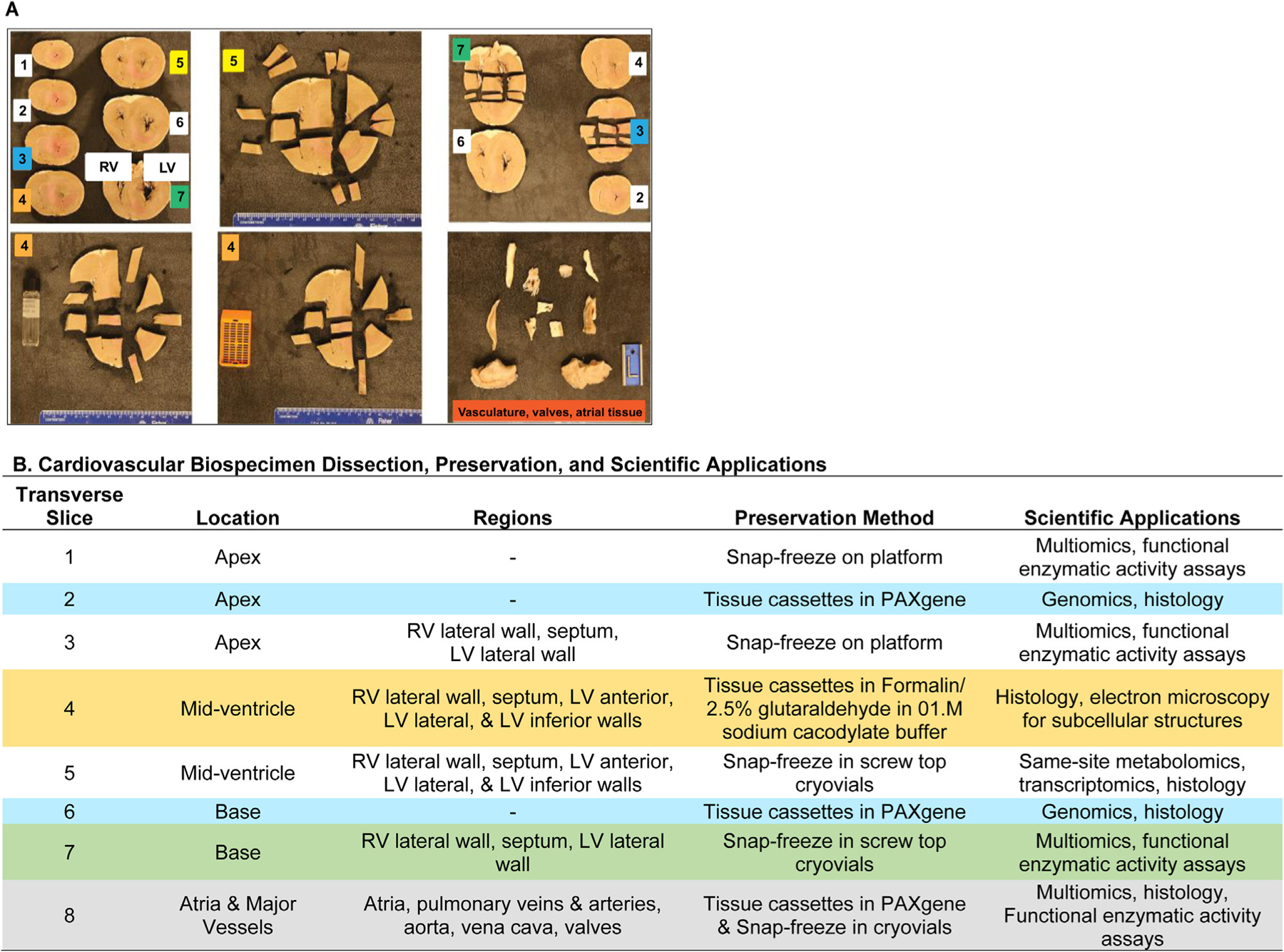
Illustrative dissection of a bovine heart for multiomics. A bovine heart was dissected, creating 7 transverse slices from the apex to 2 cm below the atrioventricular valves (A). Slices are numerically ordered from apex to base. Transverse slice 1 consists of the apex; slice 2 contains the apical most portion of the interventricular septum and left ventricle; slice 3 consists of the superior portion of the apex and ventricular tissue of both left and right ventricles; slice 4 consists of the apical-most mid-ventricular tissue; slice 5 consists of the basal-most mid-ventricular tissue; slice 6 contains the apical-most portion of the base below the atrioventricular valves; and slice 7 contains the basal-most portion of the base just below the atrioventricular valves. The base of the heart consisting of the atria and proximal major vessels was further dissected along the lines of flow (not pictured). The preservation methods and the scientific applications afforded for each transverse slice are detailed.(B).
2.2.2. PMI storage solution comparison
In bovine dissections, a comparison was performed between storage in PBS and cardioplegic solution (Belzer UW Cold Storage Solution: 320mOsm, [Na+]=29 mEq/L, [K+]=125 mEq/L, pH=7.4 at 20°C) to determine the ideal storage solution for tissue preservation.
2.3. Transcriptomics and metabolomics
RNA-seq was performed by the Boston University Microarray and Sequencing Resource and unbiased metabolomics was performed by Metabolon. (Please refer to Supplemental Materials for detailed protocol information).
2.4. Statistical analysis
2.4.1. Transcriptomics
2.4.1.1. RNA quality metrics analysis.
The amount of RNA recovered was normalized to wet weight of tissue and analyzed with respect to PMI using a repeated measure one-way analysis of variance (ANOVA) within group. A post-hoc Tukey test was used for multiple comparisons with PMI set as a fixed-effect variable. RNA Integrity Number (RIN) and 260:280 ratio were compared within group across PMI in the same manner. Comparisons of the relations of PMI on metrics of RNA quality were performed using GraphPad Prism (Version 9.2.0).
2.4.1.2. Transcriptomic analysis.
DESeq2 (Version 1.23.10)[17] was used to estimate differentially expressed genes (DEGs) between the samples.17 Principal Component Analysis (PCA) was performed using the prcomp R function with expression values transformed using the DESeq2 variance-stabilizing transformation function and z-normalized across all samples within each gene.
Differential gene expression was assessed using the likelihood ratio test and Wald test in DESeq2. To account for multiple testing, DEGs were defined using a 5% false discovery rate (FDR) using the Benjamini-Hochberg methodology [18]. We used repeated measures two-way analysis of variance (ANOVA) to identify DEGs with respect to time point (simulated PMI) or the interaction of simulated PMI with the technical variables (preservation, library preparation protocol). All analyses were performed using the R environment for statistical computing (Version 3.6.0).
2.4.2. Metabolomics
2.4.2.1. Data extraction and compound identification.
Raw metabolomic data were extracted, peak-identified, and quality control processed using Metabolon’s hardware and software with compound identification performed by comparison to Metabolon library entries of purified standards or recurrent unknown entities (based on metrics including retention time/index (RI), mass to charge ratio (m/z), and chromatographic data (including MS/MS spectral data) on all molecules available in the library. Various quality control and curation procedures were applied to ensure accurate and consistent identification of true chemical identification.
2.4.2.2. Metabolomic analysis.
Metabolite peaks were quantified using area-under-the-curve. As metabolites were processed in one day; no normalization was required except for data visualization purposes. A total of 855 compounds of known identification were present. Following log-transformation and imputation of missing values, with the minimum observed value for each compound, matched paired t-tests were used to identify bio chemicals that differed between experimental groups (each PMI time point was compared to the 0-hour time point). Statistical significance was ascribed to metabolites achieving an FDR q-value <0.05. PCA analysis was used to reduce the dimension of the data in which each principal component is a linear combination of every metabolite and principal components are uncorrelated. The number of principal components (PCs) is equal to the number of observations. Scaled data for metabolite abundance were used to calculate the log2 fold change between 0, 4, 6, and 12 hours of PMI. Based on scaled log2 fold change between metabolites between time points, a heatmap was generated with annotated hierarchical metabolite pathways using R (Version 4.0.5).
3. Results
3.1. Novel whole heart dissection and preservation protocol
Whole hearts were dissected fresh, isolating distinct anatomical areas (Fig. 2, Panel A), consistent with the established regions in the consensus statement by the Standards and Definitions Committee of the Society for Cardiovascular Pathology and the Association for European Cardiovascular Pathology [19,20]. Notably, the entirety of the heart underwent systematic dissection with same-site sampling and preservation via multiple methods (Fig. 2, Panel B), unlike previous cardiac biobank and tissue repositories (Table 1). Prior to dissection, the intact hearts were photographed and weighed. Serial transverse sections (seven total) at approximately 5–6 mm intervals were made from the apex to approximately two cm below the atrioventricular valves. The base of the heart was dissected along the lines of flow and the coronary arteries were evaluated in situ. The dissected specimen was photographed prior to sample allocation. Areas containing infarcts, acute hemorrhage, or focal pathologic change were notated by the team study pathologist and recorded but excluded from the current analyses. Only sites of grossly normal appearing tissue were used in this study for RNA and metabolite extraction. Paired, adjacent tissue samples for ribonucleic acid (RNA) and metabolomic analysis were weighed prior to PAXgene fixation (transcriptomics) or flash freezing (metabolomics). The samples were fixed in the PAXgene fixation solution for 24 hours before the samples were transferred to PAXgene stabilizing solution and stored at −80°C. The fixation rate of PAXgene is 1 mm/60 minutes; as all samples were 5–6 mm in thickness, 24 hours was sufficient time for fixation [21]. Adjacent samples from the same sites for histology were stored in 4% paraformaldehyde or 2% glutaraldehyde for 24 hours but were not analyzed for the purposes of this study. As aldehyde based fixatives penetrate greater than 8 mm of heart tissue in 24 hours, our tissue sectioning protocol of 5–6 mm was appropriate to preserve this tissue for histology [22]. All protocols were in keeping with the Established Best Practices in Postmortem Tissue Handling and Recovery guidelines set forth by the National Cancer Institute of the National Institutes of Health [23].
Table 1.
Existing cardiac biobanks/biorepositories compared to Boston University iCAMP Biobank protocol
| Alberta Human Explanted Heart Program (HELP) | Sydney Heart Bank (SHB) | University of Pennsylvania Myocardial Applied Genomics Network (MAGNet) Repository | Genotype Tissue Expression Project (GTEx) | Boston University Integrative Metabolism & Pathophysiology (BU iCAMP) Biobank protocol | |
|---|---|---|---|---|---|
| Whole hearts collected/preserved | Y | Y∗ | Y∗ | N | Y |
| Proximal large cardiac vessels | Y | N | N | N | Y |
| Other cardiac sites | LVAD core | Coronaries | U | Only LAA, LV | Y |
| Diseased cardiac phenotypes | Explants: HFrEF, CHDƗ | Explants: HFrEFƗ | Explants: HFrEF | U | HFrEF, HFpEF, all cardiac conditions |
| Normal cardiac phenotype | Y | Y | Y | Y | Y |
| Flash frozen samples | Y | Y | U | Y | Y |
| PAXgene samples | N | N | U | Y | Y |
| Aldehyde fixation | Y | Y | Y | N | Y |
| Detailed subregion chamber dissection/storage | Y | N | N | N | Y |
| Dissection protocol available for review | YƗ | N | N | Y | Y |
| Transplant/procedural tissue | YƗ | YƗ | Y | N | N |
| Postmortem cardiac tissue | N | N | N | Y | Y |
| Cardioplegic solution utilized | Y | Y | Y | N | Y |
Y, yes; N, no; U, uncertain/not described; HFrEF, heart failure with reduced ejection fraction; CHD, congenital heart disease; LVAD, left ventricular assist device; LAA, left atrial appendage; LV, left ventricle
Consists of a mixture of biopsies and whole hearts
Fresh tissue obtained rapidly with immediate processing in hospital
3.2. RNA-Seq results
Bulk, multiplexed RNA-Seq was performed using RNA isolated from the cardiac bio specimens obtained from the explanted human and postmortem bovine hearts across timepoints. The amount of RNA recovered from human or bovine PAXgene-preserved tissue was similar across all PMIs, except for the H1 right ventricular sample at 8 hour simulated PMI, which had a much lower amount of RNA recovered (80 ug RNA) than the other human right ventricular samples (505±189 ug RNA (mean ± SD)) (Fig. 3, Panel A). All human and bovine samples had a 260:280 ratio >1.8 (2.06±0.02 [mean± SD]), indicative of high RNA purity (Fig. 3, Panel B). RNA integrity was further evaluated by assessing the RIN, the ratio of 28S:18S rRNA. The mean RIN of all samples (bovine and human) was ≥5.0 (5.6±1.4 (mean±SD)); thus, all samples were deemed fit for RNA-Seq. Importantly, RIN was similar for human left ventricular samples across all PMIs. The RIN for bovine left ventricular samples was lower at 8 hours PMI compared to 0 hour (RIN bovine left ventricle-0 hours, 7.0±0.3 vs. bovine left ventricle-8 hours, 3.6±0.4; P < 0.05). (Fig. 3, Panel C).
Fig. 3.

RNA quality control metrics in human and bovine cardiac samples. Total ribonucleic acid (RNA) recovered from human left ventricle (LV) and right ventricle (RV), and bovine LV tissue normalized to tissue wet weight was evaluated across several postmortem intervals (PMI) (A). A260:280nm ratios were measured in RNA recovered from human LV and RV and bovine LV samples across several PMIs (B). RNA integrity number (RIN) for human and bovine LV samples across several PMIs was assessed (C). n=2–3/group, ∗P<.05 using repeated measures one-way analysis of variance (ANOVA) with post-hoc Tukey’s test compared to lowest PMI baseline within each group.
Evaluation of the reads by FastQC showed that the sample reads in both human and bovine samples had a mean Phred score (sequence quality) of >30 and the GC content of the reads was normally distributed, consistent with high-quality sequencing reads (Figures S1, S2). Using poly-A capture library preparation, the human cardiac samples yielded 20.4±1.9 million (mean±standard deviation) paired reads (Table 2), similar to the number observed in the GTEx studies of human left ventricular tissue [10,11,24]. An average of 20.0±2.8 million paired reads were generated from the bovine cardiac samples (Table 3). In both the human and bovine cardiac RNAseq datasets, >95% of the reads aligned to the reference genome. The average fraction of read pairs aligning to the mitochondrial genome was 33% in human and 48% in bovine cardiac samples (Tables 2, 3), which is consistent with the known high mitochondrial content of cardiac tissue [11,24].
Table 2.
Poly-A capture alignment statistics for human cardiac biospecimens
| % of Total read pairs with ≥1 alignment |
% of Non-mtDNA Uniquely aligned proper pairs |
||||||||||
|---|---|---|---|---|---|---|---|---|---|---|---|
| rRNA |
|
||||||||||
| Biological replicate | Cardiac site | PMI, hour | Total read pairs | Aligned reads, % | mtDNA reads | Ensembl | Repeat masker | Uniquely aligned proper pairs (Non-mtDNA aligned) | Assigned to 1 gene | Unassigned | Ambiguous (>1 gene) |
| H1 | LV | 4 | 22,385,800 | 97.7 | 24.6 | 0.0 | 0.2 | 15,304,463 | 84.8 | 11.2 | 4.0 |
| H2 | LV | 4 | 19,909,907 | 97.8 | 30.2 | 0.0 | 0.2 | 12,495,579 | 86.2 | 10.1 | 3.7 |
| H1 | RV | 4 | 16,228,987 | 97.6 | 23.9 | 0.0 | 0.2 | 11,201,013 | 81.2 | 15.1 | 3.7 |
| H2 | RV | 4 | 20,239,785 | 97.8 | 41.3 | 0.0 | 0.3 | 10,341,598 | 84.3 | 11.7 | 3.9 |
| H1 | LV | 8 | 17,976,822 | 97.5 | 29.2 | 0.0 | 0.3 | 11,386,073 | 81.4 | 14.9 | 3.7 |
| H2 | LV | 8 | 19,851,940 | 97.6 | 52.8 | 0.0 | 0.4 | 7,720,692 | 81.8 | 14.1 | 4.1 |
| H1 | RV | 8 | 21,363,866 | 96.8 | 12.6 | 0.0 | 1.3 | 16,586,333 | 78.9 | 18.8 | 2.4 |
| H2 | RV | 8 | 22,653,105 | 97.8 | 36.4 | 0.0 | 0.3 | 12,744,604 | 85.9 | 10.4 | 3.8 |
| H1 | LV | 12 | 20,608,209 | 97.6 | 40.2 | 0.0 | 0.4 | 10,651,907 | 77.3 | 19.3 | 3.4 |
| H2 | LV | 12 | 21,050,318 | 97.8 | 39.4 | 0.0 | 0.4 | 11,159,656 | 83.0 | 13.0 | 4.0 |
| H1 | RV | 12 | 19,853,398 | 97.6 | 24.9 | 0.0 | 0.3 | 13,450,287 | 81.6 | 14.7 | 3.7 |
| H2 | RV | 12 | 22,245,659 | 97.9 | 42.6 | 0.0 | 0.4 | 11,069,818 | 84.7 | 11.3 | 4.0 |
PMI, postmortem interval; LV, left ventricle; RV, right ventricle; mtDNA, mitochondrial DNA; n=2 human hearts/group/condition
Table 3.
Poly-A capture alignment statistics for bovine cardiac biospecimens
| % of Total read pairs with ≥1 alignment |
% of Non-mtDNA uniquely aligned proper pairs |
|||||||||
|---|---|---|---|---|---|---|---|---|---|---|
| rRNA |
|
|||||||||
| Biological replicate | PMI, hour | Total read pairs | Aligned reads, % | mtDNA reads | Ensembl | Repeat masker | Uniquely aligned proper pairs (non-mtDNA aligned) | Assigned to 1 gene | Unassigned | Ambiguous (>1 gene) |
| B1 | 0 | 21,695,675 | 97.1 | 31.6 | 0.0 | 0.7 | 13,633,581 | 70.9 | 28.2 | 0.9 |
| B2 | 0 | 18,688,199 | 97.1 | 43.6 | 0.0 | 0.8 | 9,611,367 | 64.8 | 34.4 | 0.7 |
| B3 | 0 | 25,257,748 | 97.6 | 46.3 | 0.0 | 0.5 | 12,485,539 | 66.8 | 32.4 | 0.9 |
| B1 | 6 | 16,486,026 | 97.5 | 49.4 | 0.0 | 0.5 | 7,600,051 | 69.5 | 29.7 | 0.8 |
| B2 | 6 | 20,370,200 | 97.1 | 61.0 | 0.0 | 0.7 | 7,046,281 | 66.8 | 32.5 | 0.7 |
| B3 | 6 | 20,787,188 | 96.9 | 64.8 | 0.0 | 0.5 | 6,413,389 | 64.8 | 34.4 | 0.8 |
| B1 | 8 | 24,189,098 | 97.3 | 36.2 | 0.0 | 0.5 | 14,246,915 | 65.5 | 33.6 | 0.9 |
| B2 | 8 | 19,665,443 | 97.0 | 49.1 | 0.0 | 0.8 | 9,041,827 | 64.5 | 34.8 | 0.7 |
| B3 | 8 | 16,937,918 | 97.1 | 51.0 | 0.0 | 0.5 | 7,508,435 | 68.1 | 30.9 | 0.9 |
| B1 | 12 | 20,664,057 | 95.9 | 33.1 | 0.0 | 0.6 | 12,472,364 | 66.0 | 33.0 | 1.0 |
| B2 | 12 | 17,173,781 | 96.6 | 56.5 | 0.0 | 0.7 | 6,621,510 | 58.0 | 41.4 | 0.6 |
| B3 | 12 | 18,110,216 | 97.4 | 50.2 | 0.0 | 0.5 | 8,235,646 | 66.7 | 32.4 | 0.8 |
PMI, postmortem interval; mtDNA, mitochondrial DNA; n=3 bovine hearts/group/condition
In the human and bovine cardiac samples, an average of 12.0 million and 9.6 million read pairs, respectively, aligned uniquely to non-mitochondrial sequence in proper pairs and could be assigned to a single Ensembl Gene locus. The higher fraction of unassigned read pairs in the bovine heart samples was likely driven by the incomplete annotation of the bovine reference genome [25]. The H1 right ventricular sample at 8 hour simulated PMI, which had a much lower fraction of recovered RNA, also had a lower percentage of total read pairs aligning to the genome (96.8% 97.6%±0.3% across all samples) and a greater fraction of reads that aligned to rRNA sequences (1.3% compared to 0.4%±0.3% across all samples) (Table 2), which are collectively suggestive of poor sample quality. This sample also had an unusually low fraction of reads mapping to the mitochondrial genome (13%, compared to 33.2%±10.9% across all samples).
To compare library preparation methods, rRNA reduction library preparation was also performed (Table 4). Both poly-A capture and rRNA reduction library preparation methods resulted in similar quality control measures with respect to aligned reads (>95%) and rRNA alignment. We detected few, if any, reads aligning to rRNA genes for both rRNA reduction and poly-A capture library preparation techniques. As the quality control metrics were similar between the library preparation methods, the data generated by poly-A capture library preparation and RNAseq were used for all subsequent analyses in this study.
Table 4.
rRNA reduction alignment statistics for human cardiac biospecimens
| % Of total read pairs with ≥ 1 alignment |
% of Non-mtDNA uniquely aligned Proper pairs |
||||||||||||
|---|---|---|---|---|---|---|---|---|---|---|---|---|---|
| rRNA (sense/antisense) |
|||||||||||||
| Biological replicate | PMI, hour | Total read pairs | Aligned reads, % | mtDNA reads | Ensembl | Repeat masker | Uniquely aligned proper pairs (non-mtDNA Aligned) | Assigned to 1 gene | Unassigned | Ambiguous (>1 gene) | |||
| H1 | 4 | 82,174,494 | 96.4 | 7.8 | 0.0 | 0.8 | 67,397,272 | 60.8 | 36.1 | 3.1 | |||
| H2 | 4 | 73,253,416 | 96.6 | 10.1 | 0.0 | 1.1 | 57,839,642 | 63.1 | 33.6 | 3.3 | |||
| H1 | 8 | 68,541,161 | 96.0 | 7.2 | 0.0 | 1.0 | 56,331,958 | 56.0 | 41.2 | 2.9 | |||
| H2 | 8 | 78,036,757 | 95.8 | 14.0 | 0.0 | 1.6 | 57,384,916 | 59.2 | 37.6 | 3.2 | |||
| H1 | 12 | 71,176,187 | 96.2 | 9.2 | 0.0 | 0.9 | 56,856,293 | 52.8 | 44.4 | 2.8 | |||
| H2 | 12 | 78,205,782 | 96.3 | 12.2 | 0.0 | 1.1 | 59,839,629 | 61.0 | 35.8 | 3.2 | |||
PMI, postmortem interval; mtDNA, mitochondrial DNA; n=2 human hearts/group/condition
Principal Component Analysis (PCA) was performed across all genes and all human cardiac samples (see Methods for details). First, the samples clustered separately from the H1 right ventricle sample at 8 hours along PC1, which accounted for 29% of total variance in gene expression in the experiment (Fig. 4, Panel A). The remaining samples separated strongly by donor with respect to PC2, which explained 15% of experimental variance. After adjusting the expression values for donor in a linear model, the PCA was repeated, and the human samples segregated by cardiac site and PMI, indicating that both variables are associated with differential gene expression (Fig. 4, Panel B). As the H1 right ventricle 8-hour sample was low quality, we performed a PCA excluding both right ventricle samples at 8 hour simulated PMI. The samples then separated primarily by donor with respect to PC1, which explained 23% of the experimental variance (Fig. 4, Panel C). After adjusting the expression values for donor in a linear model, PCA was repeated, and the human samples still segregated somewhat by cardiac site and PMI (Fig. 4, Panel D).
Fig. 4.

Principal component analysis (PCA) of human and bovine RNA-Seq samples across multiple cardiac sites and over varying postmortem intervals (PMI). PCA of two human heart samples (H1, H2) before (A) and after adjusting for subject as a covariate (B) shows most of the variance is explained by subject. PCA of two human heart samples (H1, H2) excluding right ventricle 8 hour PMI before (C) and after adjusting for subject as a covariate (D) shows most of the variance is still explained by subject. PCA of three bovine heart samples (B1, B2, B3) before (E) and after adjusting for subject as a covariate (F) shows that most of the variance is explained by inter-individual difference followed by PMI and cardiac site. N=2–3 samples/group/timepoint
We then compared the gene expression profile of the human left and right ventricles using a paired Wald test (to account for inter-donor variability). We found that only one gene was significantly differentially expressed (FDR q<0.25) at an 8 hour simulated PMI in the left ventricle (Table 5), and in the right ventricle, only 23 genes were significantly differentially expressed at 12 hour PMI (FDR q<0.25), indicating that very few genes are differentially expressed with respect to PMI.
Table 5.
Effect of PMI on expression-filtered DEGs in human cardiac tissue
| Left ventricle |
Right ventricle |
||||||
|---|---|---|---|---|---|---|---|
| P-value Threshold | Expected # of DEGs | 4 vs 8 hours | 4 vs 12 hours | 8 vs 12 hours | 4 vs 8 hours | 4 vs 12 hours | 8 vs 12 hours |
| .05 | 1279 | 534 | 263 | 21 | 675 | 179 | 1123 |
| .01 | 256 | 108 | 40 | 2 | 183 | 71 | 353 |
| .005 | 128 | 61 | 19 | 1 | 119 | 51 | 242 |
| .001 | 26 | 19 | 2 | 0 | 51 | 27 | 117 |
| Q-value Threshold | |||||||
| 0.25 | 1 | 0 | 0 | 41 | 23 | 178 | |
| 0.1 | 1 | 0 | 0 | 26 | 16 | 96 | |
| 0.05 | 1 | 0 | 0 | 18 | 11 | 67 | |
PMI, postmortem interval; DEGs, differentially expressed genes; n=2 human hearts/group/condition
When PCA was performed in the bovine cardiac dataset, the samples separated largely with respect to animal (Fig. 4, Panel E). However, when PCA was repeated after adjusting the expression values for animal in a linear model, the samples did not separate from one another with respect to PMI (Fig. 4, Panel F). To identify the genes that were coordinately altered with respect to PMI, we performed a repeated measures one-way ANOVA (to account for inter-animal variability) and identified a total of four DEGs associated with PMI (FDR q<0.25) (Table 6). To determine if the H1 right ventricle 8-hour sample was driving gene expression differences, we performed a repeated measures one-way ANOVA with respect to timepoint across all human ventricular samples after excluding the 8 hour sample. When adjusted for donor as a covariate, only 1 gene was associated with a PMI of 8 hours (FDR q<0.25) (Table 7). Our sensitivity analysis excluding the poor quality H1 right ventricle 8-hour sample suggests that this sample likely explained most of the differential gene expression associated with PMI.
Table 6.
Effect of PMI on the number of DEGs in bovine heart
| P-value threshold | Expected # of DEGs | Observed # of DEGs due to PMI |
|---|---|---|
| .05 | 992 | 276 |
| .01 | 198 | 93 |
| .005 | 99 | 48 |
| .001 | 20 | 13 |
| Q-value threshold | ||
| 0.25 | 4 | |
| 0.1 | 3 | |
| 0.05 | 2 | |
| 0.01 | 2 | |
One-way ANOVA across timepoints 0, 8, 12 hour PMI;
PMI, postmortem interval, DEG, differentially expressed gene
Table 7.
Effect of PMI on expression-filtered DEGs in human cardiac tissue excluding H1 right ventricle at 8 hour PMI
| Both Ventricles |
||||
|---|---|---|---|---|
| P-value threshold | Expected # of DEGs | 4 vs 8 hours | 4 vs 12 hours | 8 vs 12 hours |
| .05 | 1483 | 338 | 546 | 103 |
| .01 | 297 | 66 | 85 | 13 |
| .005 | 148 | 49 | 44 | 4 |
| .001 | 30 | 9 | 7 | 3 |
| Q-value threshold | ||||
| 0.25 | 1 | 0 | 0 | |
| 0.1 | 1 | 0 | 0 | |
| 0.05 | 1 | 0 | 0 | |
PMI, postmortem interval, DEG, differentially expressed genes; n= 2 human hearts/group/condition
3.3. Metabolomics
We performed proof-of-concept, untargeted metabolomics to evaluate metabolite stability over time with varying PMIs, as well as across different technical conditions, in both human and bovine cardiac samples. Comparable to the design of the RNA-Seq experiment, two human hearts explanted at the time of cardiac transplant (H1, H2) and three bovine hearts (B1, B2, B3) were evaluated across PMIs. Of the total 855 identified metabolites measured in the same cardiac site across several PMIs, we found that 100 metabolites decreased after 4 hours, 100 metabolites were decreased after 6 hours, and 111 metabolites were decreased after 12 hours compared to the 0 hour time point in the two human biospecimens (Table 8). In total, 503 metabolites did not change at any PMI, whereas 257 metabolites exhibited dynamic changes over the simulated PMIs. A heatmap (Fig. 5, Panel A) highlights key metabolic pathways that emerge with metabolomics across PMI and demonstrates the majority of identified metabolites remain unchanged with PMI represent multiple different metabolic pathways.
Table 8.
Effect of PMI on metabolites in human cardiac tissue
| Left ventricle |
Right ventricle | |||
|---|---|---|---|---|
| 0 vs 4 hour | 0 vs 6 hour | 0 vs 12 hour | 0 vs 4 hour | |
| Total metabolites altered (P<.05) | 104 | 103 | 111 | 35 |
| Metabolites ↑↓ | 4 | 100 | 3 | 100 | 0 | 111 | 7 | 27 |
PMI, postmortem interval; n=2 human hearts/group/condition
Fig. 5.
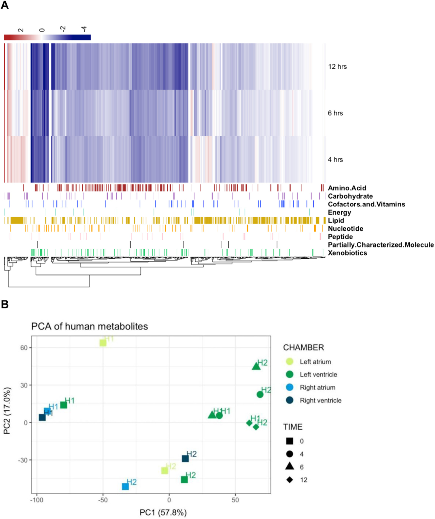
Metabolomics data revealing differentially expressed pathways in a heatmap and principal component analysis (PCA) of metabolites in human cardiac samples over multiple postmortem intervals (PMI). Heatmap displaying relative metabolite abundance in multiple metabolic pathways in human cardiac tissue across PMIs of 4, 6, and 12 hours shows that 58% (503 of 855 metabolites identified), do not significantly change across PMIs (compared to 0 hours) (A). PCA of human cardiac samples revealed that inter-individual differences and PMI were the greatest contributors to sample variation across PMIs (B).
In the metabolomic PCAs, the human samples separated into three distinct clusters along PC1, accounting for 57.8% of the variation between samples. First, H1 and H2 separated from one another into two clusters along PC1 at time 0. Additionally, both H1 and H2 time 0 clusters separated on PC1 from H1 and H2 samples at PMIs of 4, 6, and 12 hours. (Fig. 5, Panel B).
Lastly, we tested two separate conditions in samples from the three bovine hearts during the simulated PMI for our metabolomic analysis: (a) room temperature vs. 4°C storage; and (b) cardioplegic solution vs. PBS. We found altered metabolites to be comparable between room temperature and 4°C storage for samples in PBS across various PMIs. However, cardioplegic solution significantly attenuated the number of changed metabolites at greater PMIs (Table 9).
Table 9.
Effect of PMI, storage solution, and temperature on bovine cardiac tissue
| 4° C, PBS | 4° C, CP | Room temperature, PBS | ||||
|---|---|---|---|---|---|---|
| 0 vs 4 hour | 0 vs 4 hour | 0 vs 4 hour | 0 vs 6 hour | 0 vs 8 hour | 0 vs 12 hour | |
| Total metabolites altered (P<.05) | 209 | 72 | 122 | 256 | 263 | 110 |
| Metabolites ↑↓ | 6 | 203 | 21 | 51 | 18 | 104 | 31 | 225 | 20 | 243 | 24 | 86 |
PMI, postmortem interval; PBS, phosphate-buffered saline; CP, cardioplegic solution; n=3 bovine hearts/group/condition
4. Discussion
We sought to establish and optimize a novel, rigorous, systematic protocol for the postmortem dissection and preservation of whole human cardiac bio specimens for multiomics and systems biology applications. We expanded previous studies demonstrating the feasibility of RNAseq in postmortem human hearts to include metabolomics from paired samples. By identifying an optimal time window from tissue harvest to complete preservation with transition in cardioplegic solution, we found high-fidelity preservation of both RNA and metabolites, supporting the feasibility of multiomics in whole postmortem hearts.
As shown earlier (Table 1), existing cardiac biobanking/repository programs have important differences from the proposed protocol, dissection, and preservation process detailed in this study. The GTEx study, a postmortem tissue repository with RNAseq, is limited in cardiac tissue sites with only left atrial appendage and left ventricle sites obtained [14]. In contrast, the Canadian Human Explanted Heart Program (HELP) and Sydney Heart Bank offer more cardiac sites with some whole heart preservation, however, employ flash frozen and aldehyde preservation methods only (and not PAXgene) which may limit future omics applications with both biobanks utilizing explanted or donor hearts and not postmortem tissues [12,26]. Both these biobanks limit cases to HFrEF (i.e., HELP biobank has 84% dilated cardiomyopathy), and have predominantly normal referents (donor hearts who were declined due to logistical limitations that obviated cardiac transplantation) [12,26]. Our proposal, directed at postmortem tissues, is inclusive of challenging cardiac diseases (i.e., heart failure with preserved ejection fraction) as well as hearts with milder cardiac diseases and can expand human cardiac tissue access to more research centers and not restrict to only cardiac transplant centers. Our protocol encompasses tissue sites across the entire heart (including proximal great vessels) and is uniquely poised to capture and preserve postmortem cardiac tissue from a myriad of cardiac phenotypes that is more reflective of the variety of CVD present in the community, demonstrating good quality molecules extracted even from postmortem tissue. This unhindered approach to whole human heart tissue will enhance our mechanistic insight of CVDs with the capability of applying innovative systems-biology and pairing it with longitudinal clinical data, such as is available from the NHLBI cohorts.
Poly-A capture and rRNA reduction library preparations were compared to identify the optimal method for RNA-Seq with our proposed protocol. As both methods resulted in similar quality metrics, including unambiguous reference genome alignment with little rRNA region alignment, we posit that the selection of library preparation technique should be determined by the objectives of the study. In accordance with previous studies, we found that rRNA reduction increased the diversity of reads, while poly-A capture facilitated excellent coverage of coding regions [27]. Studies seeking to investigate the role of the entire transcriptome will preferentially favor the rRNA reduction approach as many noncoding RNAs are non-polyadenylated. Conversely, studies pursuing the implications of coding genes in cardiovascular diseases will favor poly-A capture, as this method provides greater RNA-Seq depth in coding regions, allowing for greater sensitivity in differences in expression.
In our investigation, the variation in the gene expression profiles between two human cardiac samples was largely attributed to differences between the donors themselves. While both hearts had ischemic disease, one was on durable mechanical support with a left ventricular assist device to augment cardiac systolic function while the other heart had aortic valvular disease and eosinophilic myocarditis. As such, these inter-individual differences may very well represent the differential effects of the VAD on the one participant[28] and myocardial inflammation on the other, but this study was not powered for such analyses. One likely outlier was noted from the 8-hour right ventricle of the H2 sample, which accounted for 29% of the variation compared to the other samples. When adjusted for donor, the samples then separate by PMI and cardiac site, which together accounted for 17% of the variation. After 8-hour right ventricle H2 sample removal, the samples still separated by PMI and site. As only a small percentage (<0.06%) of genes in certain sites were differentially expressed over multiple PMIs, our data suggests that PMI does not have a strong effect on RNA integrity. Our study revealed similar findings as the GTEx Project, and other transcriptomic studies of human cardiac tissue, suggesting stability of transcripts up to a 12-hour PMI[11,29,30] with no differences in RIN at a PMI of 12 hours compared to 0-hour measurements in human heart tissue. Even in samples with RIN values less than 5, we found good alignment quality and successfully measured gene expression, in agreement with a prior study suggesting low RIN alone does not predict poor quality sequencing [31].
A measurable effect of cardiac site on gene expression over the simulated PMIs was noted. Compared to each site’s own baseline, more DEGs were associated with the simulated PMIs in human right ventricular samples than in left ventricular samples. This finding is consistent with prior studies demonstrating differential functional pathways expressed in right vs. left ventricular tissue, and possibly owing to the thinness and lower resilience of the right ventricle [32–34]. However, in statistical comparisons excluding the human right ventricle 8-hour sample with a low RIN and RNA yield, the number of DEGs associated with PMI decreased from 41 to 1 at a q-threshold of 0.25. This suggests that the low RNA quality in this sample alone drove the right ventricle-specific differential expression. The reduction in the number of DEGs at this timepoint, in conjunction with the PCAs in Fig. 4 showing minimal association of PMI with PC1 or PC2 suggests that PMI was not a major contributor to differential gene expression. Further, this suggests that gene expression signatures in cardiac biospecimens with similar PMIs may reflect the biology pertaining to the disease state.
Our unbiased metabolomics studies revealed that over a 12-hour simulated PMI, 58% of metabolites (n = 503) remained stable in human left ventricular samples. Our PCA data suggest that both inter-individual differences and PMI affect metabolites in postmortem tissue and may require statistical adjustments in larger scale omics work in postmortem tissue. Additionally, the effect of simulated PMI on metabolite stability was attenuated when the cardiac tissue was stored in cardioplegic solution compared to PBS. While cardioplegic solution is universally used for preservation during transport in cardiac transplantation to minimize cardiac is-chemic effects [35]. studies have not yet systematically evaluated the role of cardioplegic solution in postmortem tissues for multiomics applications. Few studies have investigated the effect of PMI on cardiac metabolites in postmortem tissue, with only one study evaluating select metabolites in forensic determination of PMI but not investigating the effect of PMI on metabolite integrity [36]. In this context, our data highlight the novel utility of storage of postmortem cardiac samples in cardioplegic solution during PMI for biobanking and suggest that samples stored for up to 12 hours in cardioplegic solution will be suitable for metabolomic investigation. Based on the integrity of the RNA and metabolites harvested from human tissues at 12 hours stored in PBS and our data on cardioplegic solution in bovine hearts, human tissue stored in cardioplegic solution may be stable for longer than 12 hours, but longer PMI timepoints in postmortem tissues requires further investigation and validation.
While our study sheds light on several important technical and biological variables that affect the integrity of postmortem cardiac tissue, several limitations are present. We are likely unable to detect small differences in gene expression over time due to our sample size. However, our purpose here was not to investigate the biology of disease, but to demonstrate the feasibility of recovering high quality RNA and metabolites for multiomics. Our study was able to detect an association of 41 genes in paired, repeatedly sampled right ventricular tissue with PMI, and identify stability in metabolite expression for several hundred metabolites over multiple PMIs, suggestive that future studies with a significantly higher number of hearts will enable comparisons between disease phenotypes and likely be able to detect smaller effect sizes. Additionally, because our human biospecimens utilized explanted hearts from living patients, our study was unable to evaluate the effect of cause of death on transcriptomics and metabolomics. One study implicated cause of death in differential expression of standard reference genes;[37] however, how the cause of death affects cardiac RNA and metabolite stability and abundance[36,38] remains unclear and requires further interrogation. Thus, while PMI is important in the degradation of RNA and metabolites, a prolonged vegetative state, for example, prior to death may also contribute to the quality of acquired samples. In addition, we did not possess sufficient human cardiac tissue to replicate the bovine findings regarding enhanced RNA and metabolite quality following storage in cardioplegic solution during simulated PMI. Thus, future studies with human tissues are required. Larger samples sizes that capture sex differences through the inclusion of female participants, as well as effects specific to race and/or ethnicity are warranted. Lastly, we hope that future studies capture longitudinal phenotype data to better allow us to associate cardiac gene and metabolite signatures with specific CVDs.
Conclusions
Most CVD biomarkers are circulating, and it is currently unknown how these circulating biomarkers relate to human cardiac tissue. Herein, we demonstrate proof-of-concept for a systematic, rigorous protocol for the dissection and preservation of human cardiac tissue to facilitate highly spatially resolved multiomics interrogations. This unique protocol, when applied to a broad population of disease phenotypes in multiple human hearts, may advance our understanding of the relation of CVD risk factor burden and duration with cardiovascular structure and phenotype. Further, our protocol may enable us to validate and identify new biomarker signatures for CVD in an unprecedented and systematic manner. If applied to a large cohort study, in conjunction with longitudinal clinical data, the establishment of a human cardiac biorepository will facilitate our ability to gain an understanding of heterogeneous diseases like heart failure and discover novel therapeutic targets, as well as bring human cardiac profiling into the new age of multiomics and systems biology.
Supplementary Material
Acknowledgments
We are grateful to the Boston University Microarray and Sequencing Resource Core Facility for technical assistance in performing RNA-seq and subsequent data analysis for Boston University investigators.
Financial Support:
This research was supported by the National Heart, Lung, and Blood Institute T32 HL007224–45 (J.D.M.), K01 HL143142 (J.L.F), R01HL092577 (E.J.B.), and K23 HL135394 (M.G.F.); American Heart Association: #17FTF33670369 (D.M.G.), AF AHA_18SFRN34110082 (E.J.B); Boston University Career Development Award (D.M.G), Boston University CTSI 1UL1TR001430 (D.M.G., M.E.L., and A.C.G.), and by Grant 2021261 from the Doris Duke Charitable Foundation through the COVID-19 Fund to Retain Clinical Scientists collaborative grant program and was made possible through the support of Grant 62288 from the John Templeton Foundation (D.M.G.).
Abbreviations:
- ANOVA
Analysis of Variance
- CVD
Cardiovascular Disease
- DNA
Deoxyribonucleic acid
- DEG
Differentially Expressed Genes
- FDR
False Discovery Rate
- GTEx
Genotype-Tissue Expression Project
- PBS
Phosphate Buffered Saline
- PMI
Postmortem Interval
- PCA
Principal Components Analysis
- RNA
Ribonucleic acid
- RIN
RNA Integrity Number
- RNA-seq
RNA sequencing
Footnotes
✩✩ Disclosure: None.
Supplementary materials
Supplementary material associated with this article can be found, in the online version, at doi:10.1016/j.carpath.2022.107495.
References
- [1].Virani SS, Alonso A, Benjamin EJ, Bittencourt MS, Callaway CW, Carson AP, et al. Heart disease and stroke statistics—2020 update: a report from the american heart association. Circulation 2020:141. doi: 10.1161/cir.0000000000000757. [DOI] [PubMed] [Google Scholar]
- [2].Kannel WB. Factors of risk in the development of coronary heart disease—six-year follow-up experience. Ann Int Med 1961;55:33. doi: 10.7326/0003-4819-55-1-33. [DOI] [PubMed] [Google Scholar]
- [3].Wilson PWF, D’Agostino RB, Sullivan L, Parise H, Kannel WB. Overweight and obesity as determinants of cardiovascular risk. Arch Int Med 2002;162:1867. doi: 10.1001/archinte.162.16.1867. [DOI] [PubMed] [Google Scholar]
- [4].Khan SS, Ning H, Wilkins JT, Allen N, Carnethon M, Berry JD, et al. Association of body mass index with lifetime risk of cardiovascular disease and compression of morbidity. JAMA Cardio 2018;3:280. doi: 10.1001/jamacardio.2018.0022. [DOI] [PMC free article] [PubMed] [Google Scholar]
- [5].Rapsomaniki E, Timmis A, George J, Pujades-Rodriguez M, Shah AD, Denaxas S, et al. Blood pressure and incidence of twelve cardiovascular diseases: lifetime risks, healthy life-years lost, and age-specific associations in 1·25 million people. Lancet 2014;383:1899–911. doi: 10.1016/s0140-6736(14)60685-1. [DOI] [PMC free article] [PubMed] [Google Scholar]
- [6].Staerk L, Wang B, Preis SR, Larson MG, Lubitz SA, Ellinor PT, et al. Lifetime risk of atrial fibrillation according to optimal, borderline, or elevated levels of risk factors: cohort study based on longitudinal data from the Framingham Heart Study. BMJ 2018:k1453. doi: 10.1136/bmj.k1453. [DOI] [PMC free article] [PubMed]
- [7].Zhang Y, Pletcher MJ, Vittinghoff E, Clemons AM, Jacobs DR, Allen NB, et al. Association between cumulative low-density lipoprotein cholesterol exposure during young adulthood and middle age and risk of cardiovascular events. JAMA Cardiol 2021;6:1406–13. doi: 10.1001/jamacardio.2021.3508. [DOI] [PMC free article] [PubMed] [Google Scholar]
- [8].Vegter EL, Ovchinnikova ES, Sillje HHW, Meems LMG, van der Pol A, van der Velde AR, et al. Rodent heart failure models do not reflect the human circulating microRNA signature in heart failure. PLoS One 2017;12:e0177242. doi: 10.1371/journal.pone.0177242. [DOI] [PMC free article] [PubMed] [Google Scholar]
- [9].Schuttler D, Bapat A, Kaab S, Lee K, Tomsits P, Clauss S, et al. Animal models of atrial fibrillation. Circ Res 2020;127:91–110. doi: 10.1161/CIRCRESAHA.120.316366. [DOI] [PubMed] [Google Scholar]
- [10].Consortium GT. The Genotype-Tissue Expression (GTEx) project. Nat Genet 2013;45:580–5. doi: 10.1038/ng.2653. [DOI] [PMC free article] [PubMed] [Google Scholar]
- [11].Ferreira PG, Munoz-Aguirre M, Reverter F, Sa Godinho CP, Sousa A, Amadoz A, et al. The effects of death and post-mortem cold ischemia on human tissue transcriptomes. Nat Commun 2018;9:490. doi: 10.1038/s41467-017-02772-x. [DOI] [PMC free article] [PubMed] [Google Scholar]
- [12].Dos Remedios CG, Lal SP, Li A, McNamara J, Keogh A, Macdonald PS, et al. The Sydney heart bank: improving translational research while eliminating or reducing the use of animal models of human heart disease. Biophys Rev 2017;9:431–41. doi: 10.1007/s12551-017-0305-3. [DOI] [PMC free article] [PubMed] [Google Scholar]
- [13].Xu Y, Dong Q, Li F, Xu Y, Hu C, Wang J, et al. Identifying subpathway signatures for individualized anticancer drug response by integrating multi-omics data. J Transl Med 2019;17:255. doi: 10.1186/s12967-019-2010-4. [DOI] [PMC free article] [PubMed] [Google Scholar]
- [14].Ferreira PG, Muñoz-Aguirre M, Reverter F, Sá Godinho CP, Sousa A, Amadoz A, et al. The effects of death and post-mortem cold ischemia on human tissue transcriptomes. Nat Communicat 2018;9. doi: 10.1038/s41467-017-02772-x. [DOI] [PMC free article] [PubMed] [Google Scholar]
- [15].Andersen GB, Hager H, Hansen LL, Tost J. Improved reproducibility in genome-wide DNA methylation analysis for PAXgene-fixed samples compared with restored formalin-fixed and paraffin-embedded DNA. Anal Biochem 2015;468:50–8. doi: 10.1016/j.ab.2014.09.012. [DOI] [PubMed] [Google Scholar]
- [16].Groelz D, Sobin L, Branton P, Compton C, Wyrich R, Rainen L. Non-formalin fixative versus formalin-fixed tissue: a comparison of histology and RNA quality. Exp Mol Pathol 2013;94:188–94. doi: 10.1016/j.yexmp.2012.07.002. [DOI] [PubMed] [Google Scholar]
- [17].Love MI, Huber W, Anders S. Moderated estimation of fold change and dispersion for RNA-seq data with DESeq2. Genome Biol 2014;15:550. doi: 10.1186/s13059-014-0550-8. [DOI] [PMC free article] [PubMed] [Google Scholar]
- [18].Benjamini Y, Hochberg Y. Controlling the false discovery rate: a practical and powerful approach to multiple testing. Journal of the Royal Statistical Society Series B (Methodological) 1995;57:289–300. [Google Scholar]
- [19].Stone JR, Basso C, Baandrup UT, Bruneval P, Butany J, Gallagher PJ, et al. Recommendations for processing cardiovascular surgical pathology specimens: a consensus statement from the standards and definitions committee of the society for cardiovascular pathology and the association for European cardiovascular pathology. Cardiovasc Pathol 2012;21:2–16. doi: 10.1016/j.carpath.2011.01.001. [DOI] [PubMed] [Google Scholar]
- [20].Basso C, Aguilera B, Banner J, Cohle S, d’Amati G, de Gouveia RH, et al. Guidelines for autopsy investigation of sudden cardiac death: 2017 update from the association for european cardiovascular pathology. Virchows Arch 2017;471:691–705. doi: 10.1007/s00428-017-2221-0. [DOI] [PMC free article] [PubMed] [Google Scholar]
- [21].Howat WJ, Wilson BA. Tissue fixation and the effect of molecular fixatives on downstream staining procedures. Methods 2014;70:12–19. doi: 10.1016/j.ymeth.2014.01.022. [DOI] [PMC free article] [PubMed] [Google Scholar]
- [22].Holda MK, Klimek-Piotrowska W, Koziej M, Tyrak K, Holda J. Penetration of formaldehyde based fixatives into heart. Folia Med Cracov 2017;57:63–70. [PubMed] [Google Scholar]
- [23].OoBaB Research. Best Practices for Postmortem Recovery of Normal Human Tissue for Research Biorepositories and Biospecimen Research, National Cancer Institute, and National Institutes of Health, United States Department of Health and Human Services; 2010. [Google Scholar]
- [24].Mele M, Ferreira PG, Reverter F, DeLuca DS, Monlong J, Sammeth M, et al. Human genomics. The human transcriptome across tissues and individuals. Science 2015;348:660–5. doi: 10.1126/science.aaa0355. [DOI] [PMC free article] [PubMed] [Google Scholar]
- [25].Zimin AV, Delcher AL, Florea L, Kelley DR, Schatz MC, Puiu D, et al. A whole-genome assembly of the domestic cow, Bos taurus. Genome Biol 2009;10:R42. doi: 10.1186/gb-2009-10-4-r42. [DOI] [PMC free article] [PubMed] [Google Scholar]
- [26].Zhang H, Viveiros A, Nikhanj A, Nguyen Q, Wang K, Wang W, et al. The human explanted heart program: a translational bridge for cardiovascular medicine. Biochim Biophys Acta Mol Basis Dis 2021;1867:165995. doi: 10.1016/j.bbadis.2020.165995. [DOI] [PMC free article] [PubMed] [Google Scholar]
- [27].Zhao S, Zhang Y, Gamini R, Zhang B, von Schack D. Evaluation of two main RNA-seq approaches for gene quantification in clinical RNA sequencing: polyA+ selection versus rRNA depletion. Sci Rep 2018;8:4781. doi: 10.1038/s41598-018-23226-4. [DOI] [PMC free article] [PubMed] [Google Scholar]
- [28].Consolo F, Barberini L, Fattuoni C, Grapov D, Montisci A, Pappalardo F. Metabolomic profile of patients with left ventricular assist devices: a pilot study. Ann Cardiothorac Surg 2021;10:240–7. doi: 10.21037/acs-2020-cfmcs-117. [DOI] [PMC free article] [PubMed] [Google Scholar]
- [29].Gonzalez-Herrera L, Valenzuela A, Marchal JA, Lorente JA, Villanueva E. Studies on RNA integrity and gene expression in human myocardial tissue, pericardial fluid and blood, and its postmortem stability. Forensic Sci Int 2013;232:218–28. doi: 10.1016/j.forsciint.2013.08.001. [DOI] [PubMed] [Google Scholar]
- [30].Walker DG, Whetzel AM, Serrano G, Sue LI, Lue LF, Beach TG. Characterization of RNA isolated from eighteen different human tissues: results from a rapid human autopsy program. Cell Tissue Bank 2016;17:361–75. doi: 10.1007/s10561-016-9555-8. [DOI] [PMC free article] [PubMed] [Google Scholar]
- [31].Gupta S, Halushka MK, Hilton GM, Arking DE. Postmortem cardiac tissue maintains gene expression profile even after late harvesting. BMC Genomics 2012;13:26. doi: 10.1186/1471-2164-13-26. [DOI] [PMC free article] [PubMed] [Google Scholar]
- [32].Bernal-Ramirez J, Diaz-Vesga MC, Talamilla M, Mendez A, Quiroga C, Garza-Cervantes JA, et al. Exploring functional differences between the right and left ventricles to better understand right ventricular dysfunction. Oxid Med Cell Longev 2021;2021:9993060. doi: 10.1155/2021/9993060. [DOI] [PMC free article] [PubMed] [Google Scholar]
- [33].Friedberg MK, Redington AN. Right versus left ventricular failure: differences, similarities, and interactions. Circulation 2014;129:1033–44. doi: 10.1161/CIRCULATIONAHA.113.001375. [DOI] [PubMed] [Google Scholar]
- [34].Molina CE, Heijman J, Dobrev D. Differences in left versus right ventricular electrophysiological properties in cardiac dysfunction and arrhythmogenesis. Arrhythm Electrophysiol Rev 2016;5:14–19. doi: 10.15420/aer.2016.8.2. [DOI] [PMC free article] [PubMed] [Google Scholar]
- [35].Stoica S Two-decade analysis of cardiac storage for transplantation. Euro J Cardio-Thor Surg 2001;20:792–8. doi: 10.1016/s1010-7940(01)00847-8. [DOI] [PubMed] [Google Scholar]
- [36].Sabucedo AJ, Furton KG. Estimation of postmortem interval using the protein marker cardiac Troponin I. Forensic Sci Int 2003;134:11–16. doi: 10.1016/s0379-0738(03)00080-x. [DOI] [PubMed] [Google Scholar]
- [37].Sampaio-Silva F, Magalhaes T, Carvalho F, Dinis-Oliveira RJ, Silvestre R. Profiling of RNA degradation for estimation of post mortem [corrected] interval. PLoS One 2013;8:e56507. doi: 10.1371/journal.pone.0056507. [DOI] [PMC free article] [PubMed] [Google Scholar]
- [38].Pesko BK, Weidt S, McLaughlin M, Wescott DJ, Torrance H, Burgess K, et al. The potential of untargeted metabolomics to highlight markers for time since death. OMICS 2020;24:649–59. doi: 10.1089/omi.2020.0084. [DOI] [PMC free article] [PubMed] [Google Scholar]
Associated Data
This section collects any data citations, data availability statements, or supplementary materials included in this article.