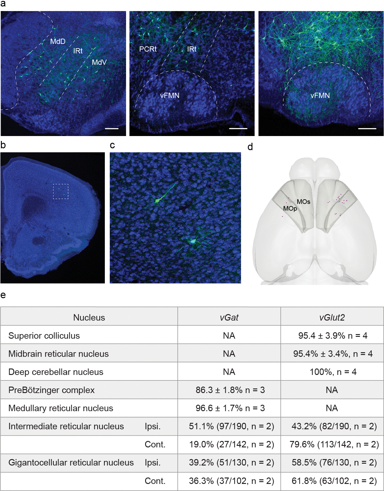
Extended Data Fig. 6. . Pre-vIRtPV neurons in the brainstem and motor cortex, and neurotransmitter characterization of pre-vIRtPV neurons.

a, Representative image of ΔG-GFP labeled pre-vIRtPVneurons in the brainstem (continued from Fig. 3c). Scale bars, 200 μm. b, Representative image of ΔG-GFP labeled pre-vIRtPV neurons in the cortex. Scale bar, 500 μm. c, Zoomed image of the boxed area in b, d, Representative 3D reconstructed image of labeled pre-vIRtPV neurons in the cortex (magenta). Shaded areas denote the primary motor (MOp) and secondary motor cortices (MOs). e, Neurotransmitter phenotype of pre-vIRtPV neurons determined by fluorescent in situ hybridization or HCR RNA-FISH.
