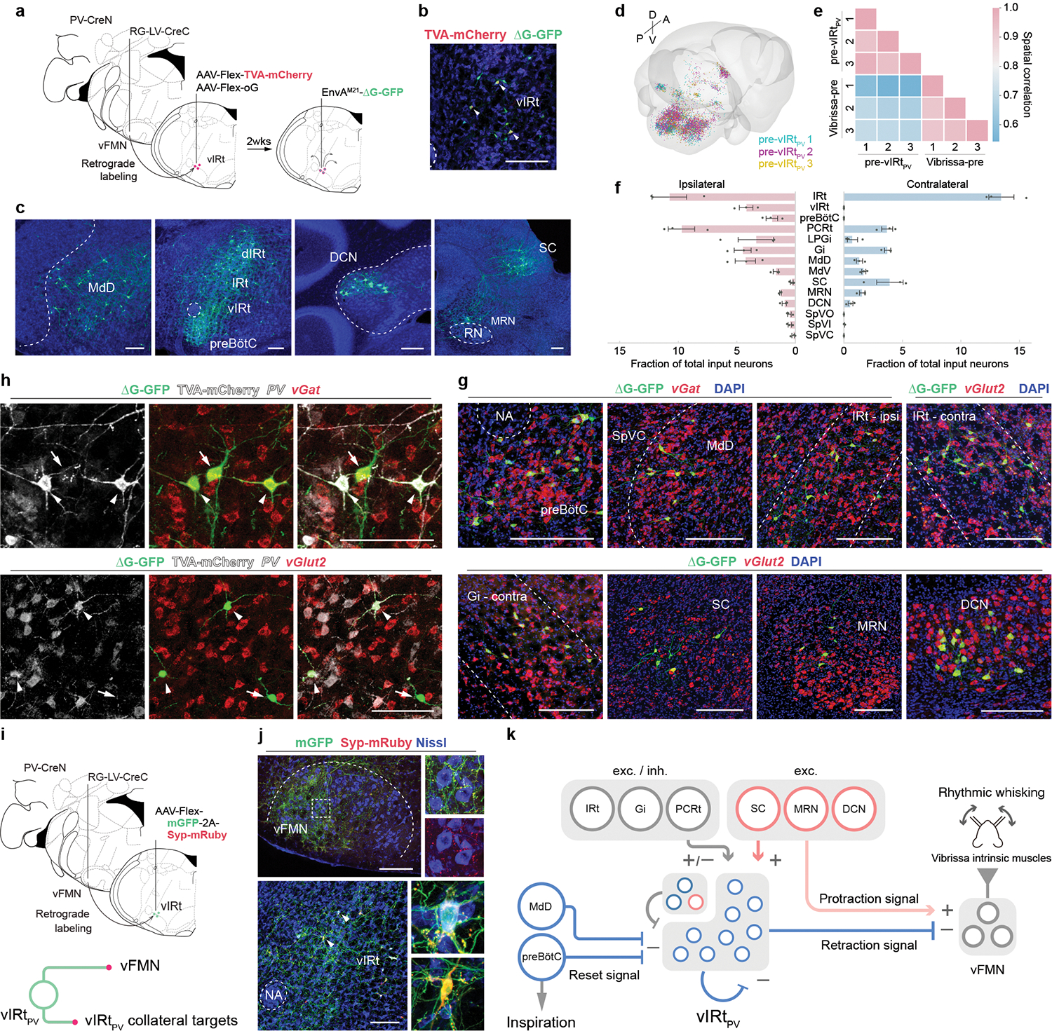
Fig. 3. Characterizing presynaptic inputs to vIRtPV neurons (pre-vIRtPV cells).

a, Schematic of monosynaptic rabies virus tracing from vIRtPV neurons. b, TVA-mCherry/GFP double-positive source cells (yellow, arrowheads), and GFP single-positive pre-vIRtPV cells in vIRt. c–f, Distribution of pre-vIRtPV neurons. c, Representative images of pre- vIRtPV neurons in the ipsilateral MdD, dIRt, IRt, DCN, preBötC, and contralateral SC, and MRN. d, Reconstructed pre-vIRtPV circuits in Allen CCF (3 mice). e, Cross-correlation analysis of pre-vIRtPV cell positions across animals; pre-vIRtPV neurons (3 mice), and vibrissa premotor neurons (3 mice) as a comparison. f, Quantification of pre-vIRtPV cell number in brain areas. Numbers are normalized by the total number of input neurons. Data are mean ± s.e.m. (n = 3 mice). g, Neurotransmitter characterization of pre- vIRtPV neurons. vGat and vGlut2 mRNA were detected by HCR RNA-FISH. See Extended Data Fig. 6 for quantification. h, Molecular characterization of pre-vIRtPV neurons within vIRt. PV, vGat, vGlut2 mRNA, and TVA-mCherry protein were detected by HCR RNA-FISH and HCR-Immunohistochemistry, respectively. Pre- vIRtPV neurons (arrows) are distinguished from source vIRtPV neurons (arrowheads) by their lack of gray-color labeled axons/dendrites. i, Top, Strategy to identify vIRtPV projections. Bottom, mGFP and Syp-mRuby label somata/axons, and axon terminals, respectively. j, Projections of vIRtPV neurons. Top, axonal projections in vFMN. Bottom, synaptic connections within vIRtPV neurons. Insets, magnified images of the boxed area and the representative vIRtPV neurons receiving mRuby-positive synaptic terminals (arrowheads). Note that strong mGFP-2A-Syp-mRuby expression causes mRuby aggregates in somata. k, Schematic summarizing the presynaptic inputs to vIRtPV. The upper left corner of vIRt indicate PV-/vGat+ (deep blue) and vGlut2+ (red) vIRt neurons. SC, MRN, and DCN provide excitatory inputs to vIRtPV neurons and presumably send simultaneous excitatory protraction signals to vFMN (translucent red). Scale bars, 200 μm (b, c, g, j), 100 μm (h). Sections were counterstained with Neurotrace blue (b, c, j) or DAPI (g, h).
