Abstract
Background
Acanthosis nigricans (AN) is a common chronic skin disorder clinically presents by velvety hyperpigmented lesions mainly at the flexural areas. Fractional photothermolysis has been reported to improve both pigmentary and textural changes by removing thin layers of skin with minimal thermal damage. Other options are the Q-switched (Qs) Nd:YAG (1064 nm) and Qs KTP (532 nm) lasers. Both can induce collagen remodeling by dermal photo-mechanical microdamage.
Aim of the Work
The aim of this study was to assess the clinical efficacy and the safety of fractional CO2 laser versus Qs Nd:YAG and KTP lasers in the treatment of acanthosis nigricans.
Methods
This randomized-controlled split neck study was conducted on 23 patients suffering from AN. For each patient, one side of the neck was randomly assigned to fractional CO2 laser and the other side to Qs Nd:YAG and KTP lasers every four weeks for four months followed by 4 monthly follow-up assessment. Acanthosis Nigricans Area and Severity Index (ANASI) score, melanin and erythema indices as well as Patient Satisfaction Scale (PSS) were used to assess improvement on each side separately.
Results
There was no statistically significant difference regarding the clinical improvement between the side treated with Fractional CO2 laser and the side treated with Qs Nd:YAG and KTP lasers (P value >0.05). In most patients, both sides showed improvement during different sessions of therapy, as regards ANASI scores, melanin indices, patient satisfaction scores, and side effects.
Conclusion
In this study, we concluded that both fractional CO2 and Q-switched lasers proved to be a safe and effective line of treatment of acanthosis nigricans.
Keywords: acanthosis nigricans, fractional CO2 laser, Q-switched Nd:YAG laser, ANASI score, Q-switched KTP laser
Introduction
Acanthosis nigricans (AN) is a chronic skin disorder that is characterized clinically by velvety darkening of the skin mainly at the back of the neck. It is most commonly associated with diabetes and insulin resistance, or hormonal disorders, certain drugs such as steroids and oral contraceptives, and rarely internal malignancy.1 Thus, acanthosis nigricans is an indicator of metabolic syndrome in many diseases such as psoriasis, lichen planus, and atopic dermatitis.2
Treatment is usually challenging. However, the condition usually improves by treating the cause as in the case of insulin resistance and weight loss. Exercise and diet help in adjusting blood glucose levels, thus improving the condition.1 Other treatment modalities include keratolytics, such as topical retinoids (eg, topical tretinoin 0.1%), topical vitamin D analogs (eg, calcipotriol 0.005%), oral retinoids, octreotide,3 dermabrasion, and chemical peels4 with variable outcome.
In addition, various types of lasers have been used for managing AN as long-pulsed alexandrite laser,5 ablative carbon dioxide laser,6 fractional 1550-nm erbium fiber,7 and fractional CO2 laser8 with variable treatment results.
Fractional CO2 laser acts by fractionated photothermolysis by making skin columns of thermal injury known as micro-thermal zones.9 It improves the condition by removing thin layers of skin with minimal thermal damage leading to collagen injury and collagen remodeling. The latter helps in improving both the texture and thickness of the skin.10 In addition, transepidermal elimination of the dermal content is a possible mechanism by which the melanin is eliminated.11
The 1064-nm Q-switched neodymium-doped yttrium aluminum garnet laser (Qs Nd:YAG) and 532-nm Q-switched potassium-titanyl-phosphate laser (Qs KTP) main mechanism on tissues is the photo-mechanical effect that promotes more effectively the synthesis of collagen type III. As collagen type III, is the predominant isotype of fetal and youth skin which is gradually replaced by collagen type I during the aging process, it may play a pivotal role in skin remodeling. In addition, it lowers the temperature and thus damages for epidermis.12
The aim of our study was to compare the efficacy and safety of Qs Nd:YAG/KTP lasers versus fractional CO2 laser in the treatment of acanthosis nigricans.
Materials and Methods
Patients
Twenty-three patients with acanthosis nigricans in otherwise healthy individuals were included in the study. Patients were recruited from the dermatology outpatient clinic in Kasr Al Ainy Hospital, Cairo University after approval by the Dermatology Research Ethics Committee of the Faculty of Medicine, Cairo University (that complies with the Declaration of Helsinki) from May 2021 to May 2022. Patients with a history of photosensitive disorders or photosensitizing drugs, endocrinal diseases, systemic steroids, uncontrolled diabetes, malignant AN, patients receiving treatment of AN, history of systemic isotretinoin use 3 months prior to this study, use of oral or injectable contraceptives or hormone replacement therapy during treatment or 12 months before, active skin infections, history of hypertrophic scars or keloids, bleeding tendency, history of liver diseases, intake of systemic chemotherapy, antiplatelets or anticoagulants, and pregnant and lactating females were excluded from the study. An informed written consent was obtained from all included patients or from guardians of those younger than 21 years.
Study Design
This is a randomized comparative split-neck study. One side of the neck was assigned blindly (using closed envelopes containing cards with fractional CO2 Rt. and fractional CO2 Lt. and the patient drew one of them blindly) to fractional CO2 laser (DEKA Smartxide DOT) every four weeks for four months with the following parameters; Power 15w, Spacing 500 µm, Dwell time 400 µs, and Stack 2, with a single pass on the affected region. The other side was assigned to Qs Nd:YAG + KTP (532 nm) (Fotona’s QX MAX®) laser every four weeks for four months with the following parameters power of 3.5–5.5 Joules/cm², 6 mm spot size pulses using Qs Nd:YAG (1064 nm). Patients developed mild delayed erythema at the end of the session. Second pass using KTP Laser: power 1Joules/cm², 3mm spot size pulses. Patients developed mild frosting at the end of the session. One hour before the session, topical anesthetic cream (Pridocaine* cream) was applied. Topical moisturizing cream was applied after the session in addition to the regular use of sunblock after complete skin healing and in between sessions. Patients were assessed all through the treatment and the follow-up period started 4 months after the last session for the emergence of any side effects.
Assessement
Acanthosis Nigricans Area and Severity Index (ANASI) Score
Patients were clinically assessed by the ANASI score system. The evaluation was done at baseline, before every session, and 1 month after the laser session. The total length of one-half of the neck (measured from a point at the junction between the chin and upper neck in full neck extension to a point at the inter-clavicular space) and the total width of one-half of the neck (measured from the point at the junction between the chin and upper neck to a point just below the nape hairline) were measured. By multiplying the resulting two numbers, we were getting the total area of half of the neck. Then, the affected area is obtained by multiplying the longest length by the longest width. A percentage was then obtained, and a value can be calculated. According to the extent of severity of pigmentation and thickness, their values were obtained. After that, both values were summed up and multiplied by the area value to get the score.8
The Melanin and Erythema Indices (MI and EI)
They were assessed using a reflectance spectrophotometer (Dermacatch, Colorix, Neuchatel, Switzerland). It is placed perpendicular to the skin with light skin pressure. In each session, three measurements were taken. The mean value of the three readings was used for each site. The area to be tested was marked and photographed for standardization at all measurement intervals; baseline, final session, and two months after the final treatment session.
Patient Satisfaction Scale (PSS)
A score is given to assess the extent of satisfaction for each side at baseline, at the final session, and 2 months after, as excellent: >75%, good: 50–75%, moderate: 25–50%, and poor: <25%.
Finally, digital photographs were taken for both sides of the neck before treatment and before every session, and 4 weeks after the last session, using Samsung ST66 16 MP Compact Digital Camera manufactured in Korea. Downtime, pain, side effects, and scabbing were reported on both sides.
Statistical Methods
Data were coded and entered using the statistical package for the Social Sciences (SPSS) version 28 (IBM Corp., Armonk, NY, USA). Data were summarized using mean, standard deviation, median, minimum, and maximum in quantitative data and using frequency (count) and relative frequency (percentage) for categorical data. Comparisons between groups were done using unpaired t-test in normally distributed quantitative variables while the non-parametric Kruskal–Wallis test and Mann–Whitney test were used for non-normally distributed quantitative variables. For comparison of serial measurements within each patient, paired t-test was used in normally distributed quantitative variables while the non-parametric Friedman test and Wilcoxon signed rank test were used for non-normally distributed quantitative variables.13
For comparing categorical data, Chi-square (χ2) test was performed. Exact test was used instead when the expected frequency is less than 5. For comparison of serial measurements within each patient, the non-parametric Marginal Homogeneity Test was used.14 Correlations between quantitative variables were done using Spearman correlation coefficient. P-values less than 0.05 were considered statistically significant.15
Results
Twenty-three patients with acanthosis nigricans were recruited and all patients completed the study. Demographic data are presented in Table 1.
Table 1.
Patients Basic Characteristics of the Studied Group
| Variables | Study Group (n=23) | ||
|---|---|---|---|
| Count | (%) | ||
| Age | Mean ±SD | 24.87±7.56 (18–42) | |
| Sex | Males | 4 | 17.4% |
| Females | 19 | 82.6% | |
| Skin type | III | 10 | 43.5% |
| IV | 13 | 56.5% | |
| Family history | No | 12 | 52.2% |
| Yes | 11 | 47.8% | |
| Diabetes | No | 21 | 91.3% |
| Yes | 2 | 8.7% | |
| Hypertension | No | 22 | 95.7% |
| Yes | 1 | 4.3% | |
| Elevated serum uric acid | No | 23 | 100% |
| Impaired lipid profile | No | 14 | 60.9% |
| Yes | 9 | 39.1% | |
Comparing Scores in Fractional CO2 Laser Side (Before and After Treatment)
The mean of ANASI Score before starting treatment in the side treated with fractional laser was (17.78 ±10.53). It was significantly decreased after treatment with a mean of (7.65 ± 8.25) (p < 0.001).
All through the laser sessions, there was a significant improvement in ANASI score in the fractional CO2 laser side after different sessions. The improvement in ANASI score started from the 2nd session and improvement remained across all the sessions (p = 0.001, <0.001, and <0.001 respectively) with minimal improvement observed upon comparing the last 2 sessions (p = 0.018) which is still significant.
The mean of the MI decreased significantly from (407.12±119.49) to (340.41±122.28) after treatment (p < 0.001) on the same side. However, EI did not show a statistically significant difference before and after treatment (p > 0.05).
Regarding patient satisfaction score, after treatment, 1 patient was considered as poor improvement (4.3%), 6 patients showed mild improvement (26.1%), 8 patients showed moderate improvement (34.8%), and 8 patients showed excellent improvement (34.8%). A significant improvement was found after treatment (p < 0.001).
Comparing Scores in Q Switched Laser Side (Before and After Treatment)
The mean of ANASI Score before starting treatment in the side treated with Q-switched laser was (18±11.49) which was significantly decreased after treatment with a mean of (7±8.69) (p<0.001). Also, there was a significant improvement in ANASI score in the Q-switched laser side after different sessions (p < 0.05). The improvement in ANASI score started from the 2nd session and improvement remained across all the sessions (p=0.001, <0.001, and <0.001 respectively) with minimal improvement observed upon comparing the last 2 sessions (p = 0.017).
The mean of MI decreased significantly from (407.16±115.71) to (335.54 ±129.4) after treatment (p < 0.001); however, EI did not show a statistically significant difference before and after treatment (p > 0.05).
Regarding patient satisfaction score, after treatment, 2 patients were considered as having poor improvement (8.7%), 3 patients showed mild improvement (13%), 10 patients showed moderate improvement (43.5%), and 8 patients showed excellent improvement (34.8%). A significant improvement was found after treatment (p < 0.001).
Comparisons of Scores on Both Sides
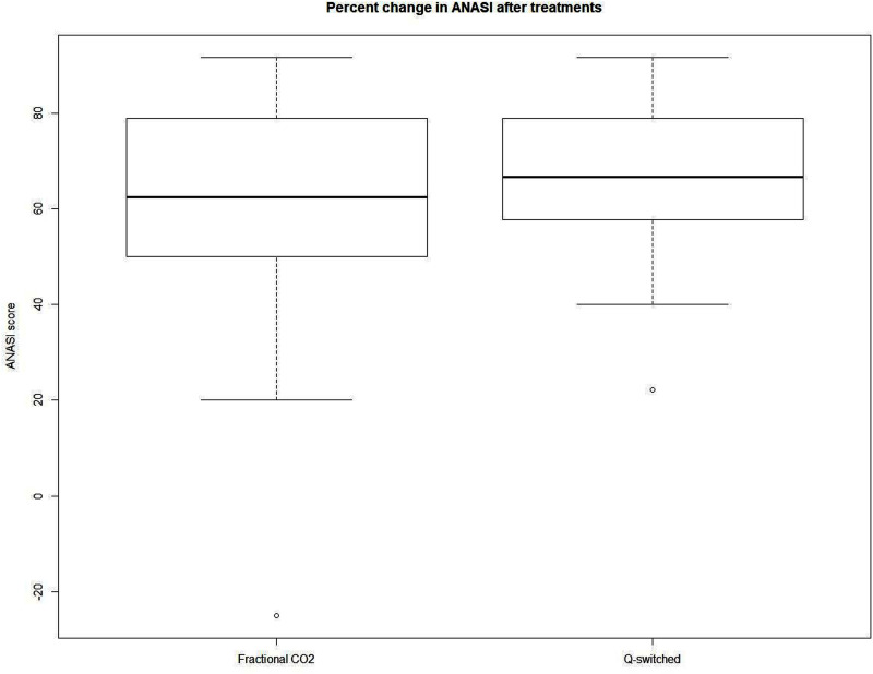
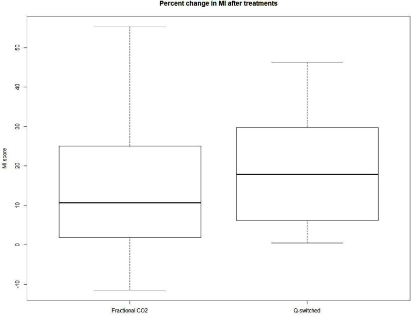
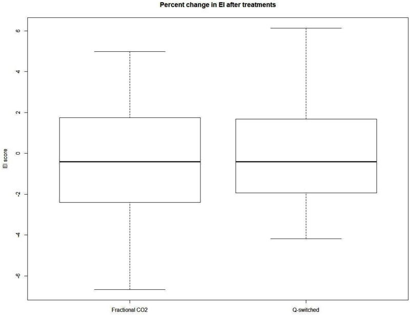
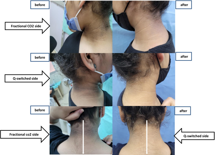
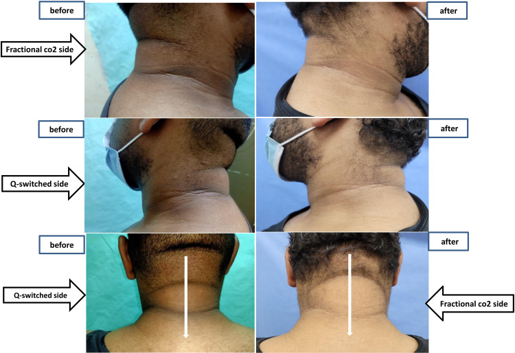
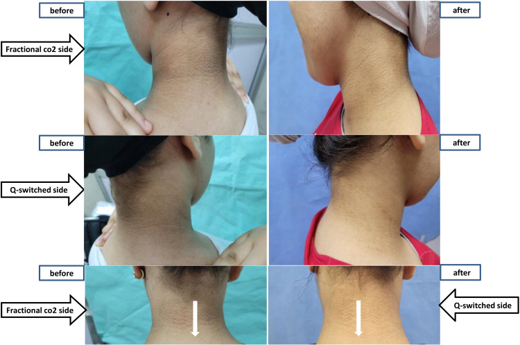
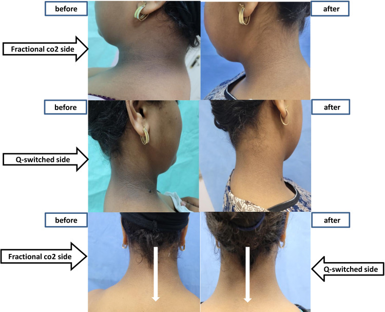
There was no statistically significant difference between both sides (Fractional CO2 laser and Q-switched laser) in all patients as regards the percentage of changes in ANASI score, MI, and EI (p > 0.05) (Figures 1–3). Photos of some of our patients are shown in Figures 4–7.
Figure 1.
Box blot illustrating percentage of change in ANASI score in both treatment sides.
Figure 2.
Box blot illustrating percentage of change in melanin index before and after treatment in both groups.
Figure 3.
Box blot illustrating percentage of change in erythema index before and after treatment in both groups.
Figure 4.
Patient 1 clinical photos before and after treatment.
Figure 5.
Patient 2 clinical photos before and after treatment.
Figure 6.
Patient 3 clinical photos before and after treatment.
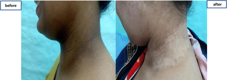
Figure 7.
Patient 4 clinical photos before and after treatment.
Correlations Between Different Parameters
A positive significant correlation was found between the change of ANASI and change of MI or patient satisfaction score after treatment on the fractional side (r = 0.529, p = 0.009 and r = 0.720, p < 0.001 respectively) and on the Q switched side (r = 0.516, p = 0.012 and r = 0.464, p = 0.026 respectively). No correlations were found between percentages of change of different scores and baseline demographic data as well as different laboratory data as regards diabetes, hypertension, impaired lipid profile, or serum uric acid (p > 0.05).
Side Effects Within Patients After Treatment
Nineteen patients of the Fractional CO2 laser (82.6%) reported transient hyperpigmentation and the other (17.4%) patients showed no side effects. Fifteen patients of Q switched laser (65.2%) reported no side effects, six patients (26.1%) reported transient hyperpigmentation and two patients (8.7%) reported hypo-pigmentation (Figure 8). All patients reported mild pain only during the session. On the fractional CO2 side, scabbing started 2–7 days after treatment, and healing occurs from 7 to 10 days after the session. However, for the Q-switched side, complete healing occurs after 7–10 days.
Figure 8.
Hypopigmentation following Q-switched laser application occurred in one patient.
Discussion
Acanthosis nigricans is a common skin disorder characterized by hyperpigmented plaques mainly on the flexural sites. The aim of treatment in AN is to treat the associated disorder and treatment of the pigmentation lesion.16
There was a significant improvement in ANASI score and MI index in our study after fractional CO2 laser; this can be explained by the fact of making skin columns of thermal injury known as micro-thermal zones and removing thin layers of skin with minimal thermal damage leading to collagen injury and collagen remodeling.10 In addition, transepidermal elimination of the dermal content is the possible mechanism by which melanin is eliminated.11 Finally, this helps in reducing both the texture and thickness of the skin thus improving the skin condition.
Similarly, a study performed by Abu Oun et al, 2022 found that fractional CO2 laser showed superior significant improvement in ANASI when compared to retinoic acid peel in the treatment of AN.6
Another study by Elsayed et al, 2021, was carried out on 20 patients with acanthosis nigricans performing 3 sessions of fractional CO2 laser on the right side of the neck and 3 sessions of glycolic acid peel on the left side of the neck. Fractional CO2 laser showed a better improvement in area index, severity and texture (ANASI Score) (p < 0.001).17
Similarly, Eldeeb et al, 2022 found that fractional CO2 laser is a better treatment option showing better improvement and fewer side effects, when compared to TCA peels, in AN.16 Leerapongnan et al, 2020 assessed the fractional laser and 0.05% tretinoin cream. He found that both improved in melanin index. However, no statistical difference was detected between both lines of treatment (p = 0.116).7
In contrast to our study, Zaki et al, 2018 found that there was no significant improvement in ANASI score after using CO2 laser on the right side of the neck as it decreased from 23.50 ± 8.85 to 19.10 ± 7.62 (P value = 0.1) with no statistical improvement. The differences between both studies may be due to good patient preparation and selection.8
Regarding the other side treated with Qs Nd:YAG laser and Qs KTP laser, there was significant improvement in ANASI score and MI. This may be due to the deep penetration with minimal epidermal damage and the good absorption by melanin, thus inducing melanosomal injury and dispersion of melanin granules in the cytoplasm in addition to collagen coagulation and collagen remodeling. As a result, new collagen synthesis is performed and that mechanism may be the cause of texture and tone improvement and rejuvenating effect on the skin. Combination of two types of Q-switched lasers is to promote better outcomes as Qs KTP targets the epidermal pigment component, however Qs Nd:YAG targets the dermal pigment component.18
In a randomized, split-faced study conducted by Yongqian et al, 2017, using the 1064-nm Qs Nd:YAG laser for skin rejuvenation in twenty-nine patients with Nevus of Ota. The participants completed 3–13 treatment sessions 3–6 months apart, the parameters were: spot diameter 3–4 mm, fluence 7–9 J/cm2, 10 Hz frequency. After the treatment, 28 patients achieved nearly complete pigmentation clearance. The degree of skin rejuvenation was positively correlated with the number of the treatment sessions, the affected skin improved in wrinkles and skin texture and pigmentation.19
Lee, 2003, used KTP laser and Nd:YAG laser, either as a monotherapy or in combination, for skin rejuvenation. The patients were divided into 3 groups, the first group of 50 patients was treated with the KTP laser alone, the second group of 50 patients was treated with the Nd:YAG laser alone, and the third group was treated with both lasers. There was a statistically significant improvement in all pigmentation, skin texture, skin tone, and rhytids in all 3 groups (p < 0.001 for all). However, KTP and Nd:YAG in combination showed better improvement than in other groups where each laser was used as a monotherapy treatment, and KTP alone showed great results when compared to Nd:YAG alone results.20
Luebberding and Armenakas, 2012, reported that treatment with the fractional, non-ablative, 1064-nm Qs Nd:YAG laser device significantly improves fine wrinkles, leading to an 11.3% improvement over baseline.21
Agarwal and Velaskar, 2020, used the fractional 1064 Qs Nd:YAG laser on 252 Indian patients with skin types III–VI for skin rejuvenation. Patients were subjected to 6 sessions with two weeks intervals with the following parameters: 5 mm spot size, fluence ranging from 1.2 J to 2 J/cm2, 2 passes were done with laser on face and neck every session. After complete laser toning for 3 sessions, there was a great improvement in pigmentation, toning, and texture of the skin.22
De Filippis et al, 2019, proved that 1064 nm Qs Nd:YAG laser treatment is a good option for skin rejuvenation.23 Moreover, Gálvez et al, 2020, used non-ablative fractional high-power 1064-nm Qs Nd:YAG laser for face and neck rejuvenation on 16 patients who were subjected to 3 sessions with maximum energy (2400 mJ/pulse). He reported a reduction in signs of skin aging with an improvement of 30–40% on the Global Esthetic Improvement Scale.24
On comparing both types of lasers (fractional CO2 laser and Qs Nd:YAG and KTP lasers), our study showed a significant improvement in ANASI and MI after both lines of treatment with no statistical difference; moreover, both lines of treatment showed no statistical significance in patients’ satisfaction score showing both fractional CO2 laser and Qs Nd:YAG and KTP lasers are effective lines of treatment.
Moreover, patients reported equal downtime l on both sides (average from 7 to 10 days) which are matching the reported downtime following similar procedures. During both procedures, pain was tolerated by patients as topical anesthesia was used prior to the procedure. During the 4 month follow-up period, no recurrences have been detected.25,26
Conclusion
Finally, we concluded that both fractional CO2 and Q-switched Nd:YAG+ KTP lasers proved to be safe and effective in the treatment of acanthosis nigricans, so both treatment modalities can be considered as a potentially useful treatment, although further studies are necessary to confirm these findings as the limitations of this study are the small sample size and the short follow-up time of 4 months.
Data Sharing Statement
The authors have the master table including the participants full data with all the required identifications and are willing to share it with the editor of the journal upon request. The specific data include the patients names, sex, age, skin type…etc. The data is available upon request any time. You can contact the corresponding author: Aya Mohamed Fahim, email: ayafahim2011@cu.edu.eg.
Ethical Committee Approval
This study was approved by the ethical committee of Cairo University in April 2021 (that complies with the Declaration of Helsinki).
Clinical Trial Approval
This study was approved by the clinical trial with ID NCT04893304.
Disclosure
The authors report no conflicts of interest in this work.
References
- 1.Patel NU, Roach C, Alinia H, Huang WW, Feldman SR. Current treatment options for acanthosis nigricans. Clin Cosmet Investig Dermatol. 2018;11:407. doi: 10.2147/CCID.S137527 [DOI] [PMC free article] [PubMed] [Google Scholar]
- 2.Daye M, Temiz SA, Işık B, Durduran Y. Relationship between acanthosis nigricans, acrochordon and metabolic syndrome in patients with lichen planus. Int J Clin Pract. 2021;75(10):e14687. doi: 10.1111/ijcp.14687 [DOI] [PubMed] [Google Scholar]
- 3.Phiske MM. An approach to acanthosis nigricans. Indian Dermatol Online J. 2014;5(3):239–249. doi: 10.4103/2229-5178.137765 [DOI] [PMC free article] [PubMed] [Google Scholar]
- 4.Das A, Datta D, Kassir M, et al. Acanthosis nigricans: a review. J Cosmet Dermatol. 2020;19(8):1857–1865. doi: 10.1111/jocd.13544 [DOI] [PubMed] [Google Scholar]
- 5.Ehsani A, Noormohammadpour P, Goodarzi A, et al. Comparison of long-pulsed alexandrite laser and topical tretinoin-ammonium lactate in axillary acanthosis nigricans: a case series of patients in a before-after trial. Caspian J Intern Med. 2016;7(4):290–293. [PMC free article] [PubMed] [Google Scholar]
- 6.Abu Oun AA, Ahmed NA, Hafiz HS. Comparative study between fractional carbon dioxide laser versus retinoic acid chemical peel in the treatment of acanthosis nigricans. J Cosmet Dermatol. 2022;21(3):1023–1030. doi: 10.1111/jocd.14224 [DOI] [PubMed] [Google Scholar]
- 7.Leerapongnan P, Jurairattanaporn N, Kanokrungsee S, Udompataikul M. Comparison of the effectiveness of fractional 1550-nm erbium fiber laser and 0.05% tretinoin cream in the treatment of acanthosis nigricans: a prospective, randomized, controlled trial. Lasers Med Sci. 2020;35(5):1153–1158. doi: 10.1007/s10103-019-02944-9 [DOI] [PubMed] [Google Scholar]
- 8.Zaki NS, Hilal RF, Essam RM. Comparative study using fractional carbon dioxide laser versus glycolic acid peel in treatment of pseudo-acanthosis nigricans. Lasers Med Sci. 2018;33(7):1485–1491. doi: 10.1007/s10103-018-2505-x [DOI] [PubMed] [Google Scholar]
- 9.Ciocon DH, Engelman DE, Hussain M, Goldberg DJ. A split-face comparison of two ablative fractional carbon dioxide lasers for the treatment of photodamaged facial skin. Dermatol Surg. 2011;37(6):784–790. doi: 10.1111/j.1524-4725.2011.01964.x [DOI] [PubMed] [Google Scholar]
- 10.Bogdan AI, Kaufman J. Fractional photothermolysis-an update. Lasers Med Sci. 2011;25(1):137–144. doi: 10.1007/s10103-009-0734-8 [DOI] [PubMed] [Google Scholar]
- 11.Katz TM, Goldberg LH, Firoz BF, Friedman PM. Fractional photothermolysis for the treatment of postinflammatory hyperpigmentation. Dermatol Surg. 2009;35(11):1844–1848. doi: 10.1111/j.1524-4725.2009.01303.x [DOI] [PubMed] [Google Scholar]
- 12.de Filippis A, D’Agostino A, Pirozzi AVA, Tufano MA, Schiraldi C, Baroni A. Q-switched Nd-YAG laser alone and in combination with innovative hyaluronic acid gels improve keratinocytes wound healing in vitro. Lasers Med Sci. 2021;36(5):1047–1057. doi: 10.1007/s10103-020-03145-5 [DOI] [PMC free article] [PubMed] [Google Scholar]
- 13.Chan YH. Biostatistics102: quantitative data – parametric & non-parametric tests. Singapore Med J. 2003a;44(8):391–396. [PubMed] [Google Scholar]
- 14.Chan YH. Biostatistics 103: qualitative data – tests of independence. Singapore Med J. 2003b;44(10):498–503. [PubMed] [Google Scholar]
- 15.Chan YH. Biostatistics 104: correlational analysis. Singapore Med J. 2003c;44(12):614–619. [PubMed] [Google Scholar]
- 16.Eldeeb F, Wahid RM, Alakad R. Fractional carbon dioxide laser versus trichloroacetic acid peel in the treatment of pseudo‐acanthosis nigricans. J Cosmet Dermatol. 2022;21(1):247–253. doi: 10.1111/jocd.14088 [DOI] [PubMed] [Google Scholar]
- 17.Elsayed GEM, Mohammed SEE, Mohammed SEE. Fractional carbon dioxide laser versus glycolic acid peel in treatment of pseudo-acanthosis nigricans. Egypt J Hosp Med. 2021;85(2):4173–4178. doi: 10.21608/ejhm.2021.207816 [DOI] [Google Scholar]
- 18.Beigvand HH, Razzaghi M, Rostami-Nejad M, et al. Assessment of laser effects on skin rejuvenation. J Lasers Med Sci. 2020;11(2):212. doi: 10.34172/jlms.2020.35 [DOI] [PMC free article] [PubMed] [Google Scholar]
- 19.Yongqian C, Li L, Jianhai B, et al. A split-face comparison of Q-switched Nd: YAG 1064-nm laser for facial rejuvenation in nevus of ota patients. Lasers Med Sci. 2017;32(4):765–769. doi: 10.1007/s10103-017-2161-6 [DOI] [PubMed] [Google Scholar]
- 20.Lee MWC. Combination 532-nm and 1064-nm lasers for noninvasive skin rejuvenation and toning. Arch Dermatol. 2003;139(10):1265–1276. doi: 10.1001/archderm.139.10.1265 [DOI] [PubMed] [Google Scholar]
- 21.Luebberding S, Alexiades-Armenakas MR. Fractional, nonablative Q-switched 1064-nm neodymium YAG laser to rejuvenate photoaged skin: a pilot case series. J Drugs Dermatol. 2012;11:1300–1304. [PubMed] [Google Scholar]
- 22.Agarwal M, Velaskar S. Laser skin rejuvenation with fractional 1064 Q‐switched Nd: YAG In 252 patients: an Indian experience. J Cosmet Dermatol. 2020;19(2):382–387. doi: 10.1111/jocd.13050 [DOI] [PubMed] [Google Scholar]
- 23.De Filippis A, Perfetto B, Guerrera LP, Oliviero G, Baroni A. Q-switched 1064 nm Nd-Yag nanosecond laser effects on skin barrier function and on molecular rejuvenation markers in keratinocyte-fibroblasts interaction. Lasers Med Sci. 2019;34(3):595–605. doi: 10.1007/s10103-018-2635-1 [DOI] [PubMed] [Google Scholar]
- 24.Gálvez FU, Trelles MA, Martín-Sánchez S, Maiz-Jimenez M. Face and neck rejuvenation using an improved non- ablative fractional high power 1064-nm Q-switched Nd: yAGLaser: clinical results in 16 women. J Cosmet Laser Ther. 2020;22(2):70–76. doi: 10.1080/14764172.2020.1726962 [DOI] [PubMed] [Google Scholar]
- 25.Mario A, Michael S, Fernando U. Safe and effective one-session fractional skin resurfacing using a carbon dioxide laser device in super-pulse mode: a clinical and histologic study. Aesth Plast Surg. 2011;35:31–42. doi: 10.1007/s00266-010-9553-3 [DOI] [PubMed] [Google Scholar]
- 26.Steven P, Mario S, Gaia F, et al. Fractional Q-switched 1064 nm laser for treatment of atrophic scars in Asian skin. Medicina. 2022;58:1190. doi: 10.3390/medicina58091190 [DOI] [PMC free article] [PubMed] [Google Scholar]
Associated Data
This section collects any data citations, data availability statements, or supplementary materials included in this article.
Data Availability Statement
The authors have the master table including the participants full data with all the required identifications and are willing to share it with the editor of the journal upon request. The specific data include the patients names, sex, age, skin type…etc. The data is available upon request any time. You can contact the corresponding author: Aya Mohamed Fahim, email: ayafahim2011@cu.edu.eg.


