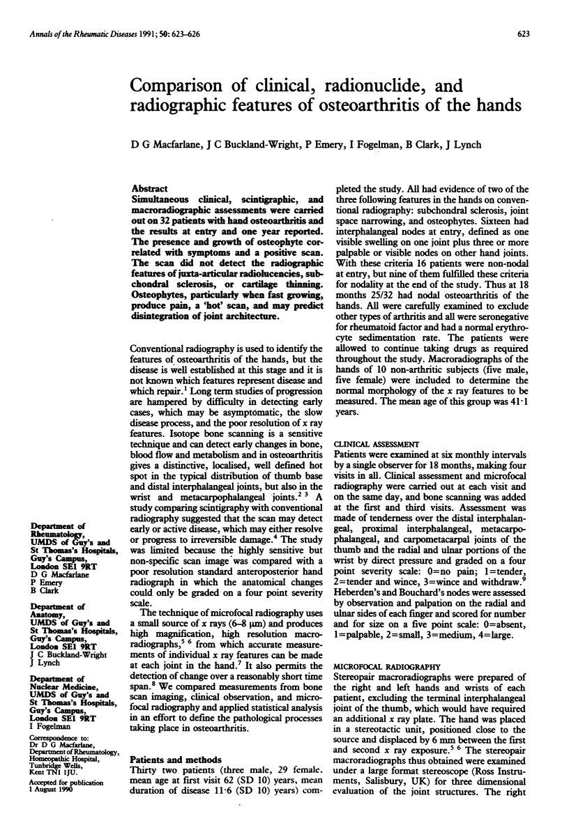
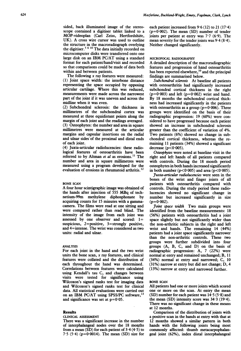
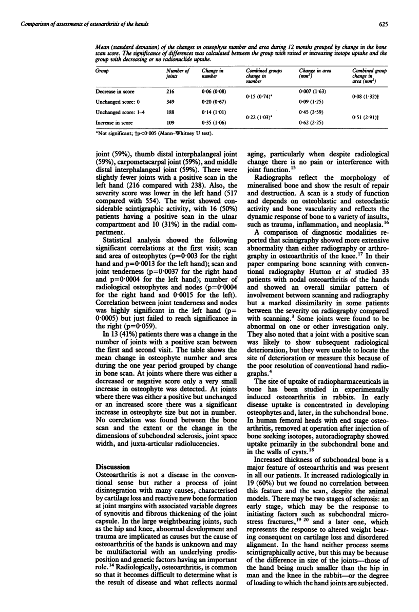
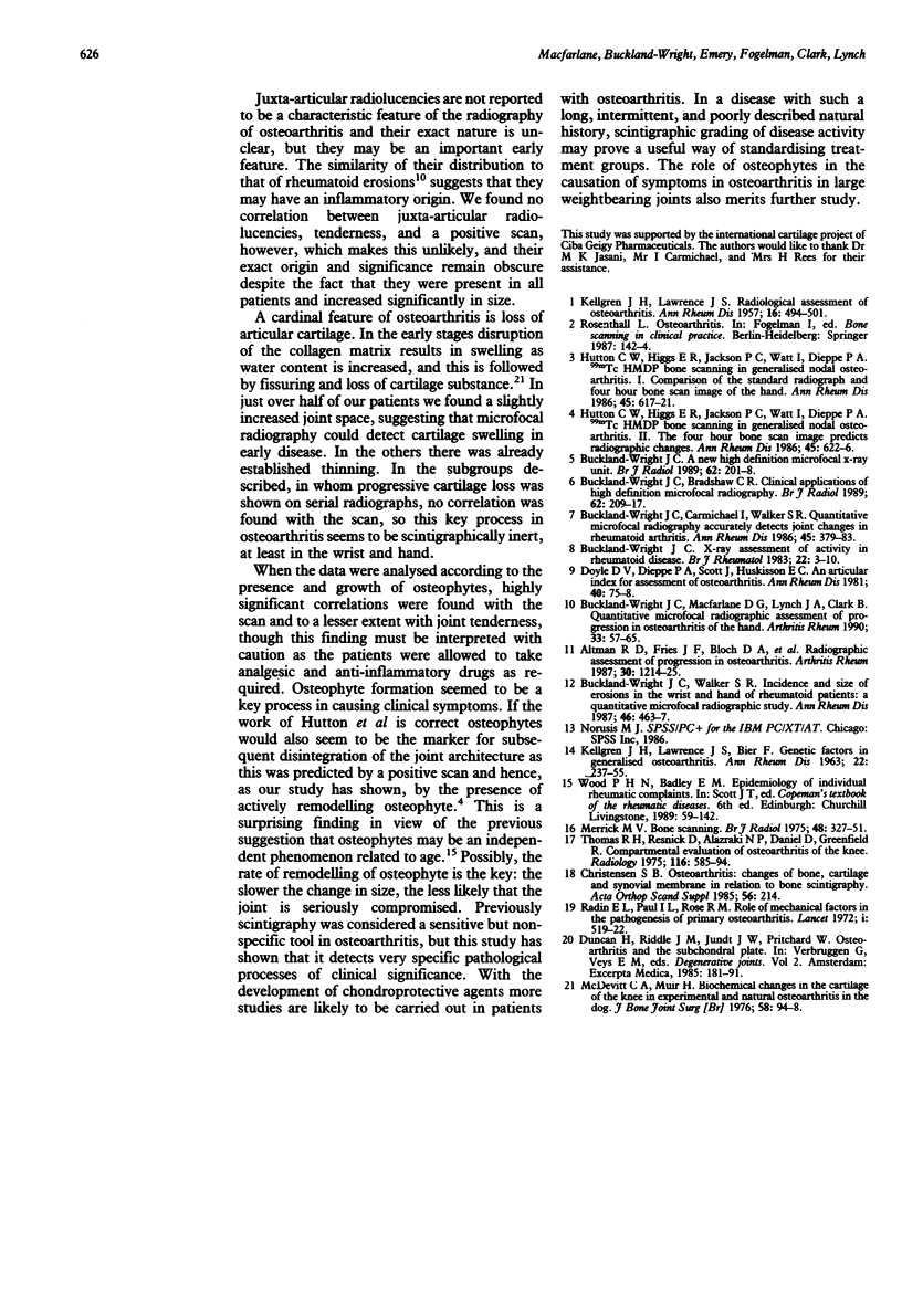
Abstract
Simultaneous clinical, scintigraphic, and macroradiographic assessments were carried out on 32 patients with hand osteoarthritis and the results at entry and one year reported. The presence and growth of osteophyte correlated with symptoms and a positive scan. The scan did not detect the radiographic features of juxta-articular radiolucencies, subchondral sclerosis, or cartilage thinning. Osteophytes, particularly when fast growing, produce pain, a 'hot' scan, and may predict disintegration of joint architecture.
Full text
PDF


Selected References
These references are in PubMed. This may not be the complete list of references from this article.
- Altman R. D., Fries J. F., Bloch D. A., Carstens J., Cooke T. D., Genant H., Gofton P., Groth H., McShane D. J., Murphy W. A. Radiographic assessment of progression in osteoarthritis. Arthritis Rheum. 1987 Nov;30(11):1214–1225. doi: 10.1002/art.1780301103. [DOI] [PubMed] [Google Scholar]
- Buckland-Wright J. C. A new high-definition microfocal X-ray unit. Br J Radiol. 1989 Mar;62(735):201–208. doi: 10.1259/0007-1285-62-735-201. [DOI] [PubMed] [Google Scholar]
- Buckland-Wright J. C., Bradshaw C. R. Clinical applications of high-definition microfocal radiography. Br J Radiol. 1989 Mar;62(735):209–217. doi: 10.1259/0007-1285-62-735-209. [DOI] [PubMed] [Google Scholar]
- Buckland-Wright J. C., Carmichael I., Walker S. R. Quantitative microfocal radiography accurately detects joint changes in rheumatoid arthritis. Ann Rheum Dis. 1986 May;45(5):379–383. doi: 10.1136/ard.45.5.379. [DOI] [PMC free article] [PubMed] [Google Scholar]
- Buckland-Wright J. C., Macfarlane D. G., Lynch J. A., Clark B. Quantitative microfocal radiographic assessment of progression in osteoarthritis of the hand. Arthritis Rheum. 1990 Jan;33(1):57–65. doi: 10.1002/art.1780330107. [DOI] [PubMed] [Google Scholar]
- Buckland-Wright J. C., Walker S. R. Incidence and size of erosions in the wrist and hand of rheumatoid patients: a quantitative microfocal radiographic study. Ann Rheum Dis. 1987 Jun;46(6):463–467. doi: 10.1136/ard.46.6.463. [DOI] [PMC free article] [PubMed] [Google Scholar]
- Buckland-Wright J. C. X-ray assessment of activity in rheumatoid disease. Br J Rheumatol. 1983 Feb;22(1):3–10. doi: 10.1093/rheumatology/22.1.3. [DOI] [PubMed] [Google Scholar]
- Hutton C. W., Higgs E. R., Jackson P. C., Watt I., Dieppe P. A. 99mTc HMDP bone scanning in generalised nodal osteoarthritis. I. Comparison of the standard radiograph and four hour bone scan image of the hand. Ann Rheum Dis. 1986 Aug;45(8):617–621. doi: 10.1136/ard.45.8.617. [DOI] [PMC free article] [PubMed] [Google Scholar]
- KELLGREN J. H., LAWRENCE J. S., BIER F. GENETIC FACTORS IN GENERALIZED OSTEO-ARTHROSIS. Ann Rheum Dis. 1963 Jul;22:237–255. doi: 10.1136/ard.22.4.237. [DOI] [PMC free article] [PubMed] [Google Scholar]
- Merrick M. V. Review article-Bone scanning. Br J Radiol. 1975 May;48(569):327–351. doi: 10.1259/0007-1285-48-569-327. [DOI] [PubMed] [Google Scholar]
- Radin E. L., Paul I. L., Rose R. M. Role of mechanical factors in pathogenesis of primary osteoarthritis. Lancet. 1972 Mar 4;1(7749):519–522. doi: 10.1016/s0140-6736(72)90179-1. [DOI] [PubMed] [Google Scholar]


