Abstract
The application of 3D printing in bone grafts is gaining in importance and is becoming more and more popular. The choice of the method has a direct impact on the preparation of the patient for surgery, the probability of rejection of the transplant, and many other complications. The aim of the article is to discuss methods of bone grafting and to compare these methods. This review of literature is based on a selective literature search of the PubMed and Web of Science databases from 2001 to 2022 using the search terms “bone graft”, “bone transplant”, and “3D printing”. In addition, we also reviewed non-medical literature related to materials used for 3D printing. There are several methods of bone grafting, such as a demineralized bone matrix, cancellous allograft, nonvascular cortical allograft, osteoarticular allograft, osteochondral allograft, vascularized allograft, and an autogenic transplant using a bone substitute. Currently, autogenous grafting, which involves removing the patient’s bone from an area of low aesthetic importance, is referred to as the gold standard. 3D printing enables using a variety of materials. 3D technology is being applied to bone tissue engineering much more often. It allows for the treatment of bone defects thanks to the creation of a porous scaffold with adequate mechanical strength and favorable macro- and microstructures. Bone tissue engineering is an innovative approach that can be used to repair multiple bone defects in the process of transplantation. In this process, biomaterials are a very important factor in supporting regenerative cells and the regeneration of tissue. We have years of research ahead of us; however, it is certain that 3D printing is the future of transplant medicine.
Keywords: 3D printing, bone grafts, FDM, scaffold, cells
1. Introduction
Extensive bone loss caused by high-energy fractures or pathological fractures require bone transplantation. The first documented bone transplant took place in 1686 by a Dutch surgeon, Job van Meekeren, when he used dog cranium (xenograft) to repair a soldier’s skull defect [1]. Today, more than two million bone transplants are performed worldwide each year. The treatment of bone defects of critical size associated with tumors or resulting from trauma remains an unmet clinical need [2,3,4]. Current treatments using xenografts, autografts, or allografts have multiple serious limitations, including limited supply, donor site morbidity, and disease transmission. There is also a risk of foreign body rejection [4,5,6].
Autograft is considered to be the best method, but it is also associated with disorders at the site of collection and poor availability. Allografts (tissues obtained from human cadavers or living donors) and xenografts (tissue that is derived from a species that is different from the recipient of the specimen) carry the risk of excessive immune reaction and, consequently, rejection of the transplant [2].
Reconstruction of the defect in bone tissue as a surgical procedure is a time-consuming and technically difficult process [7]. The intended effect is not always achieved, and the patient is exposed to residual pain, non-union, or a treatment-resistant infection. Then, a decision may be made to perform a secondary amputation [8,9,10,11]. Spatial printing (3D printing) is the process of producing physical objects based on a computer model [12,13,14]. It is a subset of manufacturing called additive manufacturing (AM).
Three-dimensional (3D) additive manufacturing has recently been widely used in a great many medical fields. Among them, orthopedic oncology is the one using it most actively. Bones and their defects and tumors are modeled for surgical planning, personalized surgical tools for personalized surgical instruments, and implant manufacturing, which are currently the most typical applications [15,16,17,18].
The beginnings of 3D printing date back to the 1990s. Huge interest in this method results from the detailed control of the entire process, the possibility of creating non-standard shapes, and the production of structures with specific physical properties. Due to lower costs, shorter time, and a wide possibility of adjusting parameters with a precise layer-by-layer method, it is possible to recreate structures on a micro- and macroscopic scale. These methods have also found their application in medicine [19,20,21,22]. Three-dimensional printing technology has revolutionized the medical field in recent years, allowing the creation of custom personalized implants, prosthetics, and surgical instruments [18,20,23,24]. Some of the key applications of 3D printing in medicine include:
Personalized implants: 3D printing technology can be used to create personalized implants tailored to the specific needs of individual patients. For example, cranial implants can be created to match the exact shape and size of a patient’s skull, providing a more secure and comfortable fit [25,26].
Prosthetics: 3D printing allows for the creation of custom-fit prosthetics that are lighter, stronger, and more comfortable than traditional prosthetics [27,28].
Surgical planning and training: Surgeons can use 3D printing to create replicas of a patient’s anatomy, allowing them to plan and practice complex procedures before performing them on a patient [29,30,31,32].
Surgical tools: 3D printing can be used to create customized surgical instruments, such as scissors, forceps, and retractors. These custom tools can help improve the accuracy and efficiency of surgeries [33,34].
Tissue engineering: 3D printing is being used to create functional tissue and organs for use in transplants. Researchers are using 3D printing to create structures that mimic the complex architecture of human tissues, with the goal of eventually being able to print functional replacements for damaged or diseased organs [35,36,37,38]. Bioprinting is currently in its early stages, but due to the significant effort put into research real progress is being made [39,40,41]. The technology still has a long way to go, but its possible application will greatly influence the field of transplantology.
Training and simulation tools: Various devices for students and trainees that are helpful in the process of education. These can include laparoscopy simulators, models of bones, organs, joints, medical equipment, tools, or even entire body parts constructed from various materials with different mechanical properties and colors [42,43,44]. These are easy to replace, modify, and duplicate, and are a great solution for institutions that are not permitted to use actual human specimens.
In the field of orthopedics, it is used inter alia in bone tissue engineering as a substitute for the previously mentioned human bone tissue transplants. This innovative method aims to solve problems related to tissue source, immune rejection, and disease transmission [8], as well as the reconstruction of normal bone structure—compact and spongy bone requires the use of bone scaffolds, cells, and growth factors [45]. The scaffolding method is the most promising and currently most widely studied. The bone scaffold should contain the appropriate density and size of the pores for the proper angiogenesis process [46]. For this purpose, the most common methods are: chemical foaming, solvent casting, and freeze drying [10]. It is possible to adjust the appropriate amount of minerals, such as calcium phosphate, magnesium or silicon. The conducted studies indicate that the presence of these components incorporated into macroporous bone scaffolds has a positive effect on the rate of bone tissue formation and indicates their use in early wound healing [47,48,49].
The porosity of the implanted scaffold holds a critical role in the process of osteogenesis. It improves osteogenesis by protein adsorption and the generation of capillary forces. These forces help to attach cells on the surface of the implant [50]. Moreover, porous structures have significantly increased surface area compared with non-porous structures, which allows for the faster permeation of chemicals, such as drugs and nutrients, aiding in vascularization and metabolite removal [51,52]. Macro–mesoporous composites containing PEEK and mesoporous diopside as bone implants are characterized by in vitro mineralization and cytocompatibility, as well as vascularization potential and osteogenesis in vivo. This is very important in the process of fluid circulation and helps cell migration towards the center of the implant [53,54]. Additionally, porous structures can absorb antibiotics and other substances introduced before implantation [55]. By controlling the porosity of the structure, it is also possible to impact the degradation rate of the scaffold. In the ideal case, the rates of bone regeneration and implant degradation should be equal. For example, mesoporous silica shows beneficial properties as a part of an antibacterial strategy during implantation [56], and can impact the degradation rate of the implant [57]. Nevertheless, too much porosity can negatively impact the mechanical properties and robustness of the implanted structure [58]. This is important, as only strong and stiff materials should be considered for implantation, as they need to provide structural support for the newly formed bone tissues [59,60].
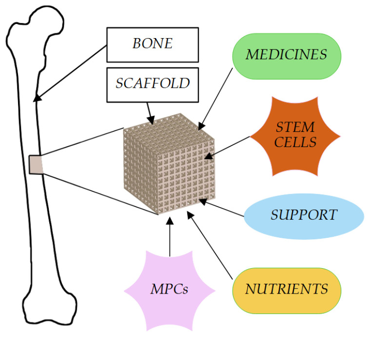
Scaffolds can be filled with medicines, which are then released locally at the site of scaffold implantation [61] (Figure 1). There is a wide range of materials used in bone tissue engineering, e.g., gypsum, ceramics, resin, and plastics [62,63,64,65,66]. Additionally, the bio-ink ensures the proper regeneration and reconstruction of bone tissue. The resulting material largely corresponds to the morphological features of normal human tissue [67]. The advantage of the entire process is the ability to adjust all parameters to the patient’s needs and, consequently, to improve the condition of life [45].
Figure 1.
Usage of microporous scaffolds for supporting bone formation.
2. Bone Grafts
Bone transplantation is a medical procedure aimed at filling the defects in the recipient’s bone by implanting the donor’s bone. Several methods of this procedure have been described [68,69]. The first is demineralized bone matrix (DBM), which consists of taking the bone matrix from a donor and treating it with chemicals—acids—and then demineralizing it. Furthermore, additional substances are used, such as ethylene oxide, which kills pathogens but affects the acceptance of the transplant by the recipient’s organism, meaning that it is practically not used. Currently, gamma irradiation is more commonly used because, in addition to neutralizing pathogens such as HIV or HCV viruses, it has less impact on graft acceptance; however, it weakens the structural integrity of the graft material. Currently, DBM is used to fill cavernous defects and to fill in non-fused bones [70]. Similar to DBM, a cancellous allograft is used, but they differ in the method of fixation [71]. Unlike the demineralized bone matrix, it does not undergo demineralization but is deeply frozen. This increases the possibility of contracting HIV; however, the chance of it is still low. Another method of bone transplantation is the nonvascular cortical allograft. This bone has a much higher density than the trabecular bone, making it more difficult to eliminate pathogens, and therefore there is a risk of HIV infection from the donor. This material is freeze-dried, which reduces its immunogenicity but also reduces its strength. The healing process of such a bone is also significantly extended as the patient’s cells slowly absorb and turn the transplanted bone into its own; however, often it does not fully disintegrate and there is necrosis of the grafted bone, followed by inflammation [72]. During the first six months after the process of implantation, the nonvascularized cortical grafts become progressively weaker and later resorb. Nevertheless, the area regains structural mechanical strength within twelve months [73]. There are also osteoarticular and osteochondral allografts, of which only in the osteochondral allografts the material is not frozen in order to enable the survival of chondrocytes, which are very sensitive to this process. They are used in joint resurfacing procedures. The osteoarticular allograft is used in arthroplasts, thanks to the fact that a large fragment of bone can be transplanted, which will ensure the efficiency of the joint. This type of allograft is characterized by a deep freeze in order to reduce immunogenicity [74,75]. One further type of allograft is vascularized allograft, but this requires the administration of immunosuppressive drugs to the patient, such as cyclosporine, after surgery. This procedure is performed only in serious cases, such as in large bone defects [72]. This method is associated with many health complications for the patient because they must subsequently take lifelong immunosuppressive drugs that can cause various types of neuropathies, myopathies, and nephropathies. Therefore, the so-called autogenic transplant, i.e., the removal of the patient’s bone from a place of low aesthetic importance, is increasingly used [72]. Although the viability of osteoinductive proteins and osteogenic cells is diminished after such transplantation, it is referred to as the “gold standard” due to the lower risk of infection, transplant rejection, or arthrodesis in the patient. Most often it is taken from the iliac plate (posterior iliac crest); however, it is a very invasive procedure, causing additional complications for the patient, including pain, infection, nerve damage, iatrogenic fracture, incisional hernia, and ending with hematoma [72]. The other sites from which the transplant is performed are the proximal part of the tibia, the distal end of the radius, the distal part of the tibia, and the greater trochanter [72]. Approximately three cubic centimeters of cancellous or corticocancellous graft can be obtained from the distal end of the radius for applications in the hand and upper extremity surgery [40,76,77,78,79]. The greater trochanter can also be used as a useful source of bone graft for surgery in the ipsilateral lower extremity [80]. A cancellous autograft is the best material for filling a bone defect smaller than 6 cm caused by, e.g., cancer or acute bone fracture, allowing it to heal properly without defects. Another example is the nonvascularized cortical autograft, which is used to increase the bone’s structural strength, treating defects up to 6 cm. It is usually performed by cutting out large amounts of the anterior or posterior iliac crest. This way, a large number of osteoprogenitor cells is obtained. Nonvascularized cortical grafts have less mechanical strength several months after transplantation because they are associated with a revascularization process that resorbs parts of the bone to form new blood vessels within the fracture—a process that takes much longer in the cortex than in the trabecular bone. Therefore, they are not recommended for filling cavities larger than 6 cm. Vascularized autografts heal much faster due to the fact that there is no revascularization process in them. It was observed by Waitayawinyu et al. [81] that clinical outcomes for scaphoid non-unions with avascular necrosis improved. Union rates of 93% were observed in those patients who received vascularized grafts. Merrell et al. [82] found in the process of meta-analysis that a vascularized graft may be beneficial for those patients who suffer scaphoid avascular necrosis. Additionally, patients after implantation of vascularized bone graft showed a union rate of 88%. Simultaneously, the recipients of screw-and-wedge fixation achieved a union only 47% of the time. Typically, 90% of osteocytes present in these grafts are able to survive the transplant and bring their blood supply [72]. This type of graft material is most often taken from the ribs, fibula, and scapula, and is mainly used to treat osteonecrosis, non-unions of the scaphoid, and Kienböck’s disease (necrosis of the lunate or osteonecrosis of the lunate) [74,81,82]. In the event that the bone defect is greater than 6 cm, the so-called induced membrane technique can be used. It consists of two key steps: The first is the removal of necrotic bone tissue at the site of the damage, and then the polymethylmethacrylate cement spacer (with or without antibiotics) is applied [83,84,85]. After a few weeks, the second stage begins, which consists of removing the spacer and filling the bone defect with an autogenous cancellous bone graft. Then, the surgeon sutures the wound together with the graft and the membrane, which protects the newly transplanted tissue against too-fast resorption [86]. The use of bone substitutes is increasingly noticed, and they are used with two main advantages in mind: an ability to integrate with the regenerating bone and a strength similar to the [87,88] cortical bone. The most commonly used are ceramics or hydroxyapatite cement, which the patient’s bone uses up and replaces with bone tissue [89,90,91]. Recent studies show that patients with transplanted bone substitutes experience less pain, have fewer complications, and are more agile than patients with other types of transplants [92,93]. In addition to transplants, patients also receive special types of drugs—bone morphogenetic proteins. They stimulate cell division, matrix synthesis, and tissue differentiation. In addition, they also stimulate osteoblasts, osteoclasts, and osteoprogenitor cells to the process of osteogenesis. The strongest activities, and the best-researched and available for treatment, are BMP-2 and BMP-7; however, despite their beneficial bone-forming properties, they increase the risk of cancer and diseases of the genito-urinary system [74].
3. Three-Dimensional Printing and Computer-Aided Design
Three-dimensional (3D) printing, a part of additive manufacturing (AM) and rapid prototyping methods, is a process of creating a 3D object from layer-by-layer-joined material. This makes it an opposite technique to traditional machining, where material is removed from a block to form the desired shape. It is a versatile technique, allowing the fabrication of complex parts from a variety of types of materials. These include polymers, ceramics, metals, and composites. The method can be customized to create various shapes and dense or macro/microporous architecture [94,95,96,97,98,99,100]. Three-dimensional printed objects are being used in many industries. The typical applications include the manufacturing of complex geometries, such as turbine blades, jewelry, molds, implants, prosthetics, mechanical parts, and it is even used in the construction of buildings, tissue engineering, etc. [101,102,103,104,105]. Mentions of the 3D printing process can be found in the late 19th century. These first methods were used when photosculpture and geomorphology technologies were developed. Significant progress was made between the 1980s and 2010s. At that time, a number of 3D printing techniques were developed, including Stereolithography, Fused Deposition Modeling (FDM), ink-jet 3D printing, adhesive-droplet- and powder-bed-based AM, Selective Laser Sintering (SLS), Selective Laser Melting, Continuous Liquid Interface Production, Digital Laser Processing, etc. [106,107,108,109,110,111,112,113,114,115,116,117].
Stereolithography is the first 3D printing process. It uses photocurable polymers that are solidified by a UV laser beam a delivered through a tunable optical system with moving mirrors. The method produces high-level detail but is slow. Digital Light Processing is similar to Stereolithography, as both use light to cure a resin. The difference is that in this case the light passes through a liquid crystal display screen, and the whole layer can be produced at once. This makes the method significantly faster. PolyJet is a method that also uses photocurable resins, but the head of the machine can deliver microdrops of materials of different properties to different spots in the print. Selective Laser Sintering and Multi-Jet Fusion use different approaches to binding polymer powders. The first one uses heat delivered by a laser, while the second one binds the powder using a binding agent. Fused Deposition Modeling uses a heated nozzle to melt material and deposit it on the bed of the machine, and it can print using various materials in the form of spools, granules, or liquids.
Technologies that allow printing from metal powders include Direct Metal Laser Sintering, Electron Beam Melting, Selective Metal Melting, and Selective Laser Sintering. They all heat the material in certain points to induce its fusion. DMLS and SLS use a laser beam but only sinter the powder, which in turn results in a print that should be later heated in an external oven to ensure the proper binding of granules. EBM and SLM methods produce objects made from melted powder. The first method uses an electron beam focused by a series of coils, while the second one is based on a high-power and focused laser. The resulting prints require less postprocessing and are stronger, more uniform, and more robust. Comparison of the methods is presented in Table 1.
Table 1.
Advantages and disadvantages of various 3D printing technologies.
| Type | Technology | Materials | Advantages | Disadvantages | Resolution |
|---|---|---|---|---|---|
| Polymer printing | Stereolithography (SLA) [118,119] | UV-curable resins and photopolymers | High level of detail, smooth finish, and tight tolerances | Slow and can be brittle | 30 to 140 microns |
| Digital Light Processing (DLP) [118,119] | UV-curable resins and photopolymers | High level of detail, smooth finish, and tight tolerances | Fast, suitable for low-volume production, and can be brittle | 35 to 100 microns | |
| Selective Laser Sintering (SLS) [107,120,121] | Nylon-based polymer powders and ceramics | Strong parts and no support structure required | Rough surface finish and slow | 80 microns | |
| Multi-Jet Fusion (MJF) [122,123] | Polymer powders | Strength and fast speed | Rough surface | 1200 dpi and 22 microns | |
| PolyJet [124,125] | Multiple materials | Multiple materials in one print, and colorful prints | Mediocre rigidity | 14 microns | |
| Fused Deposition Modeling (FDM) [119,125] | Multiple polymers with additives | Cost effective, quick, simple, cheap, and many materials | Rough surface finish, slow, and mediocre precision | 10 microns vertical; 100 microns horizontal | |
| Metal printing | Direct Metal Laser Sintering (DMLS) [126,127] | Alloy powders | Strong; dense parts | Often requires post processing via sintering and normalizing in a furnace; expensive; rough surface | 40 microns |
| Electron Beam Melting (EBM) [128,129] | Metal powders | Very strong parts, high speed, energy efficiency, and low distortion | Very expensive | 50 microns | |
| Selective Metal Melting (SLM) [130,131] | Metal powders | Very strong parts; usage of single-component metals | Often requires postprocessing via normalizing in a furnace; expensive | 30 microns | |
| Selective Laser Sintering (SLS) [121,130] | Alumide (aluminum plus polyamide) | Strong parts; no support structure required | Rough surface finish; slow | 80 microns | |
| Bioimplant production | Bioprinting [132,133,134,135] | Gels containing living cells and collagen, gelatin, hyaluronan, etc. | Can produce element or complete organs | Young technology | 100 microns |
| Construction 3D printing | Extruding [136,137,138] | Concrete, clay, and soil | Rapid construction of buildings in various shapes, requiring less labor and resulting in less construction waste | Very expensive, large, and complex machines | 6000 microns |
Parts created by the use of different technologies have vastly different properties and uses (Table 2). Additionally, printed models can be used as a step in the process of creating an object by casting. Forms can be printed directly, or the material can be melted by hot material poured into the mold, leaving a cavity in the shape of the desired object.
Table 2.
Uses of various 3D printing technologies.
| Technology | Anatomical Models | Implants | Prosthetics | Dentistry | Industrial Production |
|---|---|---|---|---|---|
| Stereolithography (SLA) | yes | ||||
| Digital Light Processing (DLP) | yes | yes | |||
| Selective Laser Sintering (SLS) polymer | yes | yes | |||
| Multi-Jet Fusion (MJF) | yes | ||||
| PolyJet | yes | ||||
| Fused Deposition Modeling (FDM) | yes | yes | yes | ||
| Direct Metal Laser Sintering (DMLS) | yes | ||||
| Electron Beam Melting (EBM) | yes | yes | yes | yes | |
| Selective Metal Melting (SLM) | yes | yes | yes | ||
| Selective Laser Sintering (SLS) metal | yes | yes | yes |
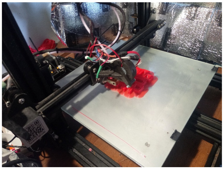
Although multiple printing technologies exist, FDM printing is by far the most popular due to its ease of use, the low price of various printing materials with different properties, and the availability of devices and parts (Figure 2). These materials include easy to use and cheap polylactic acid (PLA) and acrylonitrile butadiene styrene (ABS), tough and resistant Polyethylene Terephthalate (PET) and nylon, flexible and shock-absorbing thermoplastic polyurethane (TPU) and thermoplastic elastomer (TPE), water-soluble polyvinyl alcohol (PVA) and other materials doped with carbon for electric conductivity, Kevlar for additional strength, and even metal or wood particles for various looks and applications. Some printers can print using various filaments during the same process using multiple nozzles, making the technology very flexible [139].
Figure 2.
Typical FDM printer during operation. The heated nozzle deposits the material (red) and places it layer by layer on the print bed in the required shape.
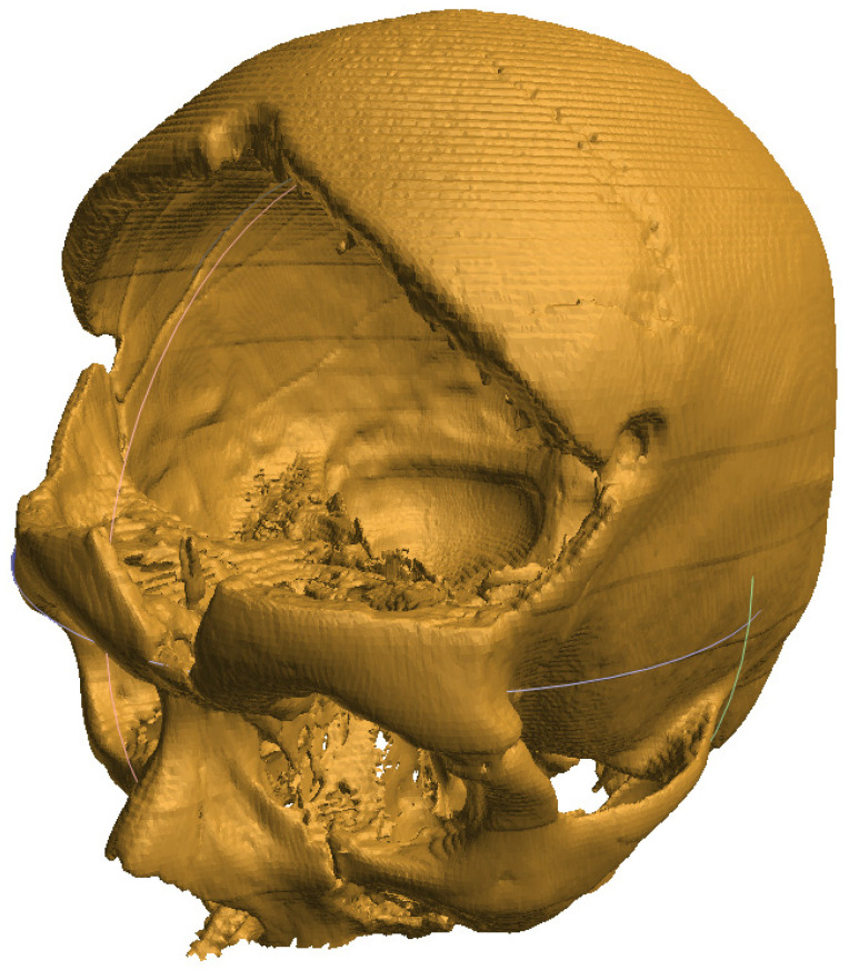
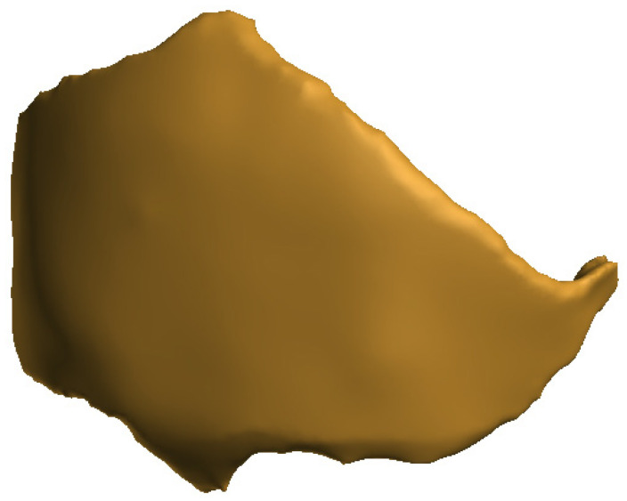
Three-dimensional technology enables the production of implants, tools, instruments, and devices. Now, it is also successfully used in bone tissue engineering to replace critical bone defects [5]. A high-performance scaffold underpins the success of a bone tissue engineering strategy, which is a crucial part of the process. The resulting scaffold should be designed with multiple characteristics in mind, including a desirable shape; physical, chemical, structural, and biological features for regenerating complex bone tissues; and enhanced biological performance [49,140]. Magnetic resonance imaging (MRI) and computed tomography (CT) [141,142] provide images of specific defects in an individual patient, and the images can in turn be further used for 3D printing the defective object [11] (Figure 3 and Figure 4).
Figure 3.
Image of a skull after removal of damaged bone following an accident, obtained by processing a set of tomography images.
Figure 4.
A model of an implant designed to replace the missing bone tissue.
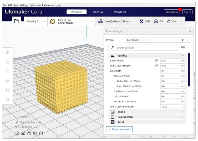
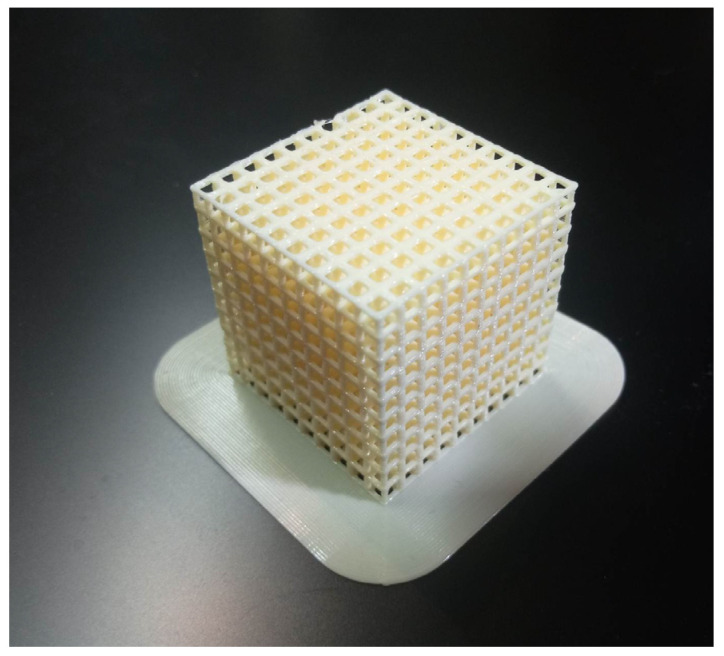
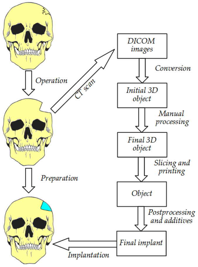
The process of implant preparation can be divided into several steps. The initial stage is collecting data on the defective bone using CT and MRI [141,142]. Through programs such as 3D doctors, 3D slicer, etc., the data are converted into 3D CAD. This is later used as a base for implant creation, as the resulting object must fit exactly in the area in question. This requires the use of graphic design tools and can vary between cases. Currently, the process of model creation is simplified and can be performed using easy to use and free software that creates models that need very little processing before slicing and printing [16]. Nevertheless, the automated algorithms can sometimes struggle when processing complex, low-quality, or noisy images. The skull, especially when damaged in the face area, can be quite challenging and requires a significant amount of manual postprocessing [143]. After the process of computer-aided design is complete, the precise scaffolding design is created [144,145]. During production, scaffolding requires a precise shape, size, and mechanical and biological properties to improve the reliability of patient outcomes after surgery [11,146]. Later, the digital CAD data are processed for 3D printing in an appropriate format and printed with the required material [30] (Figure 5 and Figure 6). Finally, the printed scaffold is planted with cells and tissues and then implanted into the patient’s body during surgery (Figure 7).
Figure 5.
A model of a 30 mm × 30 mm × 30 mm lattice during preprocessing in Ultimaker Cura. The software allows the user to adjust printing parameters for the desired effect.
Figure 6.
A model of a 30 mm × 30 mm × 30 mm lattice after printing on an FDM printer using PLA.
Figure 7.
The steps required before final implantation of the prepared structure.
Bone tissue engineering with the use of 3D printing provides new insights into the treatment of bone defects thanks to the production of a porous scaffold with appropriate mechanical strength and favorable macro-and microstructures. Tissue function is restored by the appropriate use of cells placed on the scaffolds produced from a combination of biomaterials. The process of healing and osteointegration is heavily influenced by the design of 3D structures, constituting the template for cell transplantation. Multiple parameters must be taken into consideration to ensure the proper performance of scaffolds. These include pore volume, pore size, chemical properties, and mechanical strength. The biomaterials used for scaffold formations need to be structurally stable, fast-curing, biomimetic, biocompatible, and resistant, and have mechanical properties similar to bone [147]. Bones have varying density and elasticity depending on the area and type of the missing piece, and the method of biostructure production must also take that into consideration [3]. For the production of porous bone scaffolds, widely used methods include chemical foaming, freeze drying, solvent casting, and foaming gel [45,47,148,149]. These technologies use materials such as gypsum powder, plastics, resins, aluminide, ceramics, sand, metal, Polyether ether ketone (PEEK), and graphene to create the required part [45]. Appropriate macro- and microstructures are key features in bone tissue engineering scaffolds [49]. Patterned macropores in scaffolds influence cell penetration and cell distribution and, most importantly, enable the transportation of gases and nutrients into the deeper layer of scaffolds, hence maintaining cell viability at a high level [62]. Bioceramic powders, non-hydrogel polymers, natural/synthetic hydrogels, and composites of various materials can be used to formulate printing “inks” for 3D printing. Biodegradable and biocompatible polyesters, such as poly (L-lactic acid) (PLLA), poly-β-hydroxybutyrate (PHB), poly (vinyl alcohol) (PVA), polyurethane elastomers, poly (D, L-lactic acid) (PDLLA), poly (3-hydroxybutyrate-co-3-hydroxyvalerate) (PHBV), polycaprolactone (PCL), poly (lactic-co-glycolic acid) (PLGA) and polyurethanes, can be processed into the form of wires, granules, and even powders. This enables the 3D printing of polymer scaffolds using melting extrusion at high temperature and sintering or dissolving in organic/water solvents in order to enable 3D printing based on micro-extrusion at room/low temperature [62]. The usage of hydrogels allows the trapping of proteins or cells inside the mesh, as well as control over the release of materials as per requirements. In addition, hydrogels due to their absorbable nature and excellent integration capabilities into the surrounding tissues do not need a complex process of surgical removal and also reduce the risk of an inflammatory reaction. Because of their high water-holding capacity (similar to that of soft tissues), hydrogels can support cells better than other 3D scaffolds [150].
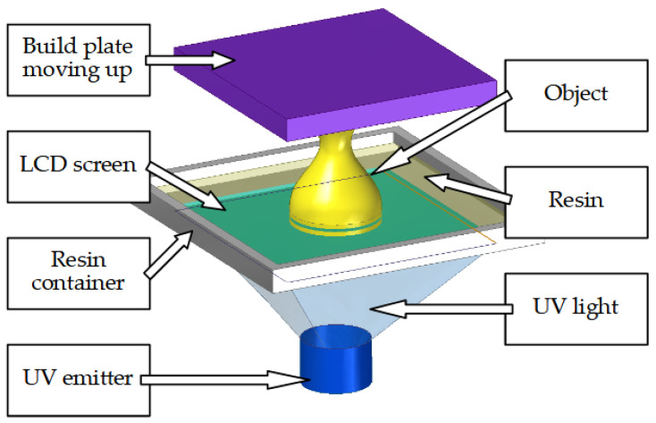
Stereolithography (SLA) is one of the earliest 3D printing techniques used in bone engineering [9]. In this method, the object is created from photosensitive fluid resin. Hardening occurs when resin is exposed to precisely controlled light beams. The process occurs layer by layer and the machine requires few moving parts as the light source is often in the form of an LCD screen and the object is rapidly created one full layer at a time (Figure 8). The method offers high accuracy in the micro- and nanometric scales. It allows the creation of high-resolution complex shapes with an internal architecture. SLA has low biodegradability and biocompatibility. Photo-cross-linkable poly (propylene fumarate) (PPF) is commonly used in SLA [9,151]; however, many other materials such as poly (caprolactone) have also been used.
Figure 8.
Main components of an SLA printer.
However, difficulties have been encountered, one of which is SLA’s inability to print micron-sized scaffolds due to the limitation of the layer thickness and too much cure, which can cause the resin to polymerize in the lower layers. The second difficulty is that there are too few bone engineering materials compatible with SLA due to reduced viscosity, stability, and refractive index. There is also a risk of a cytotoxic effect on enveloped cells due to their radiation by ultraviolet light. Additionally, improper printing parameters can result in the resin not bonding properly. Third, it has also been shown that the size of the light pixels in some SLA processes also limits in-plane microstructure creation [9]. The models created using this method have mediocre mechanical stability and can deteriorate over time.
Fused Deposition Modeling (FDM) is a 3D printing process based on a continuous filament extrusion approach. It allows the fabrication of complex, three-dimensional geometries. The process works by the extrusion of small beads or polymer filament through a small, motorized nozzle in a molten form (Figure 9). The material then hardens post-printing to form a solid construct [9,152]. With this method, the size of the pores in the scaffolds, the morphology, and the joints can be controlled. As a result, the FDM process allows the creation of complex 3D structures that are unattainable in traditional methods such as lithography and micro-machining. Thermoplastic materials, such as PCL, poly(lactic acid) (PLA), and PLGA, are often also combined with other biomaterials, and have been used with FDM to create biocompatible, tissue-engineered scaffolds with a low melting temperature [9]. FDM scaffolds showed favorable mechanical and biochemical properties in the bone regeneration process. The performance of PCL scaffolds in the compression and biocompatibility module has also been demonstrated [153]. On the other hand, PLA scaffolds with different pore sizes showed the appropriate mechanical properties and the distribution of bone marrow stromal cells. The resolution of the FDM printers largely depends on the diameter of the extrusion nozzle. These typically range from 0.3 to 0.8 mm, but in some cases even 0.1 mm or 1 mm can be used [4]. Some studies also show that objects obtained by using FDM printing are sterilized as a result of the high temperatures present in the nozzle [154]. This is a significant positive factor that should be considered when choosing the technology for implant creation.
Figure 9.
Process of creating an object using FDM.
Despite its obvious advantages, FDM printing is not without problems. The main limitation of this method is that this technique only produces biological scaffolds with organic shapes and regular structures due to its resolution compared with SLA, as the viscosity of the polymer melts limits the achievable print resolution [9]. Additionally, using a very small nozzle significantly increases printing time and requires really precise calibration and high-quality material, as tiny dust particles can easily clog the opening. Another disadvantage is that popular FDM machines do not support the simultaneous integration of temperature-sensitive cells or biological factors due to the relatively high temperatures used, although it is possible to modify existing designs for such purposes [155]. The overall conversion, processing, and printing time can be long depending on multiple parameters, such as object complexity, nozzle size, the speed of the printer, etc. In some cases, prints may range from several hours to a full day. The method also produces non-uniform objects, as the layers are arranged horizontally. This results in an object that can effectively handle splitting forces perpendicular to the median and frontal planes, while also being weak to splitting across the horizontal plane. This can be slightly remedied by heating the object in a controlled manner to achieve better bonding between the layers. Such an operation can be performed in an open-air environment or after filling the cavities with a powdery medium that is resistant to temperatures, for example table salt or very fine sand. This ensures the dimensional stability of the scaffold when the material is close to its glassing or melting point [156]. After the process, the interlayer bonding is significantly better, leading to increased tensile strength. Additionally, FDM prints often require support material while printing overhangs or at an angle. This material later needs to be removed, either by mechanical processing or by dissolving if using water-soluble materials. The process of design, printing, and processing requires significant technical expertise and experience. Computer-aided design (CAD) tools may be easy to learn but need time to master. The cases can wildly differ from patient to patient, and the designer needs to develop a significant amount of skills and knowledge in medicine, material science, and the practical aspects of printing technologies. Comparison between various grafting techniques is presented in Table 3.
Table 3.
Comparison between various grafting techniques.
| Method | Pros | Cons | |
|---|---|---|---|
| Autograft | Cancellous | Biocompatibility Best for defects smaller than 6 cm Fastest healing |
Poor availability Disorders at the site of collection |
| Allograft | Demineralized bone matrix graft | Higher availability Sterilization process |
Lower acceptance Lower structural integrity Risk of rejection |
| Cancellous | Availability Ease of application No prior harvesting required |
Possibility of infection Low initial strength Risk of rejection |
|
| Nonvascular cortical | High initial density of bone Mechanical properties |
Weakening of graft after a time Risk of rejection |
|
| Vascularized | Can be used in serious cases | Requires lifelong immunosuppressive drugs High risk of rejection |
|
| Synthetic | Ceramic | Rapid resorption and osteointegration Tailored shape and composition |
Mediocre mechanical properties Material degradation |
| 3D printing using SLA | Precisely designed shapes High precision High speed Cheap materials |
Lower biocompatibility Degradation over time Mediocre mechanical properties |
|
| 3D printing using FDM | Precisely designed shapes Wide range of materials Sterilization during the process High biocompatibility Dimensional stability Cheap materials |
Lower print speed than SLA Lower resolution than SLA Non-uniform tensile strength |
Additive manufacturing by 3D printing offers many possibilities, both for current clinical practice and for the future of the medical industry due to complete freedom of shape design based on patient-specific data. Rapid development in the field of material science will undoubtedly lead to discovering multiple new biocompatible polymers for use in many branches of healthcare. These include, among others, orthopedics, dentistry, prosthetics, and medical simulation. Medical tool production can also be significantly improved by using rapid prototyping. Bioprinting development will allow the production of tissue that can be used to replace the elements of body parts lost in accidents or that are removed during surgery due to various pathologies, fractures, and deformities. This innovative form of therapy will significantly decrease the waiting time of the patients for the procedure and minimize the risk connected to performing allograft and autograft transplantations. In some cases, the usage of 3D printing will benefit patients with conditions that were not curable before. Nevertheless, new technologies also come with new challenges, and this case is no different. The risk involved in all the procedures needs to be assessed, and new guidelines should be put in place to avoid danger to the patients. In the future, we can expect a significant growth in interest regarding the applications of 3D printing in medicine.
4. Conclusions
Bone transplants are associated with the risk of too-turbulent immune reaction, which may even lead to the rejection of the transplant, as well as inflammatory reaction. The preparation for this treatment is long and very complicated in terms of technical difficulty. It is not always possible to perform a bone transplant, which may be associated with chronic pain and chronic inflammation, resulting in amputation. Three-dimensional printing gives provides control over the entire process, including the possibility of creating non-standard shapes and the production of structures with specific physical properties.
During bone transplantation, the patient can be infected by HIV or HCV. Gamma rays are most commonly used to neutralize these viruses; however, unfortunately, they weaken the grafted material. Three-dimensional printing minimizes the chances of contamination.
The possibility of adjusting the porosity of the grafted fragment through 3D printing allows for the acceleration of the formation of new bone tissue, which has a positive effect on wound healing.
Hydrogels used in 3D printing show excellent integration with surrounding tissues, avoiding the complexity of surgical removal and a reduction in the possibility of an inflammatory reaction. Bone tissue engineering is an innovative approach that can be directly used to repair bone defects during the process of transplantation. During the construction of bone tissue implants, biomaterials play an important role in supporting the regeneration of cells and tissue. We have years of research ahead of us; however, it is certain that 3D printing is the future of transplant medicine.
Author Contributions
Conceptualization, J.B. and A.B. (Adam Brachetand); methodology, A.B. (Adam Brachetand); investigation, R.K. and M.M.; resources, R.K., M.M. and J.B.; data curation, J.B.; writing—original draft preparation, A.B. (Aleksandra Bełżek), D.F., Z.G. and D.T.; writing—review and editing, K.K., R.K. and M.M.; visualization, R.K. and M.M.; supervision, J.B.; project administration, R.K. and M.M.; funding acquisition, J.B. All authors have read and agreed to the published version of the manuscript.
Institutional Review Board Statement
Not applicable.
Informed Consent Statement
Not applicable.
Data Availability Statement
No new data was created.
Conflicts of Interest
The authors declare no conflict of interest.
Funding Statement
This research was funded by the Medical University of Lublin, grant number DS 201.
Footnotes
Disclaimer/Publisher’s Note: The statements, opinions and data contained in all publications are solely those of the individual author(s) and contributor(s) and not of MDPI and/or the editor(s). MDPI and/or the editor(s) disclaim responsibility for any injury to people or property resulting from any ideas, methods, instructions or products referred to in the content.
References
- 1.Williams A., Szabo R.M. Bone Transplantation. Orthopedics. 2004;27:488–495. doi: 10.3928/0147-7447-20040501-17. quiz 496–497. [DOI] [PubMed] [Google Scholar]
- 2.Bose S., Sarkar N. Natural Medicinal Compounds in Bone Tissue Engineering. Trends Biotechnol. 2020;38:404–417. doi: 10.1016/j.tibtech.2019.11.005. [DOI] [PMC free article] [PubMed] [Google Scholar]
- 3.Koons G.L., Diba M., Mikos A.G. Materials Design for Bone-Tissue Engineering. Nat. Rev. Mater. 2020;5:584–603. doi: 10.1038/s41578-020-0204-2. [DOI] [Google Scholar]
- 4.Mirkhalaf M., Men Y., Wang R., No Y., Zreiqat H. Personalized 3D Printed Bone Scaffolds: A Review. Acta Biomater. 2023;156:110–124. doi: 10.1016/j.actbio.2022.04.014. [DOI] [PubMed] [Google Scholar]
- 5.Campana V., Milano G., Pagano E., Barba M., Cicione C., Salonna G., Lattanzi W., Logroscino G. Bone Substitutes in Orthopaedic Surgery: From Basic Science to Clinical Practice. J. Mater. Sci. Mater. Med. 2014;25:2445–2461. doi: 10.1007/s10856-014-5240-2. [DOI] [PMC free article] [PubMed] [Google Scholar]
- 6.Schulze M., Gosheger G., Bockholt S., De Vaal M., Budny T., Tönnemann M., Pützler J., Bövingloh A.S., Rischen R., Hofbauer V., et al. Complex Bone Tumors of the Trunk—The Role of 3D Printing and Navigation in Tumor Orthopedics: A Case Series and Review of the Literature. JPM. 2021;11:517. doi: 10.3390/jpm11060517. [DOI] [PMC free article] [PubMed] [Google Scholar]
- 7.Karpiński R., Jaworski Ł., Zubrzycki J. Structural Analysis of Articular Cartilage of the Hip Joint Using Finite Element Method. Adv. Sci. Technol. Res. J. 2016;10:240–246. doi: 10.12913/22998624/64064. [DOI] [Google Scholar]
- 8.Keating J.F., Simpson A.H.R.W., Robinson C.M. The Management of Fractures with Bone Loss. J. Bone Jt. Surg. Br. 2005;87:142–150. doi: 10.1302/0301-620X.87B2.15874. [DOI] [PubMed] [Google Scholar]
- 9.Zhang L., Yang G., Johnson B.N., Jia X. Three-Dimensional (3D) Printed Scaffold and Material Selection for Bone Repair. Acta Biomater. 2019;84:16–33. doi: 10.1016/j.actbio.2018.11.039. [DOI] [PubMed] [Google Scholar]
- 10.Athanasiou V.T., Papachristou D.J., Panagopoulos A., Saridis A., Scopa C.D., Megas P. Histological Comparison of Autograft, Allograft-DBM, Xenograft, and Synthetic Grafts in a Trabecular Bone Defect: An Experimental Study in Rabbits. Med. Sci. Monit. 2010;16:BR24–BR31. [PubMed] [Google Scholar]
- 11.Dimitriou R., Jones E., McGonagle D., Giannoudis P.V. Bone Regeneration: Current Concepts and Future Directions. BMC Med. 2011;9:66. doi: 10.1186/1741-7015-9-66. [DOI] [PMC free article] [PubMed] [Google Scholar]
- 12.Karpiński R., Jaworski Ł., Zubrzycki J. The Design and Structural Analysis of the Endoprosthesis of the Shoulder Joint. ITM Web Conf. 2017;15:07015. doi: 10.1051/itmconf/20171507015. [DOI] [Google Scholar]
- 13.Karpiński R., Jaworski Ł., Szala M., Mańko M. Influence of Patient Position and Implant Material on the Stress Distribution in an Artificial Intervertebral Disc of the Lumbar Vertebrae. ITM Web Conf. 2017;15:07006. doi: 10.1051/itmconf/20171507006. [DOI] [Google Scholar]
- 14.Karpiński R., Jaworski Ł., Jonak J., Krakowski P. Stress Distribution in the Knee Joint in Relation to Tibiofemoral Angle Using the Finite Element Method. MATEC Web Conf. 2019;252:07007. doi: 10.1051/matecconf/201925207007. [DOI] [Google Scholar]
- 15.Park J.W., Kang H.G. Application of 3-Dimensional Printing Implants for Bone Tumors. Clin. Exp. Pediatr. 2022;65:476–482. doi: 10.3345/cep.2021.01326. [DOI] [PMC free article] [PubMed] [Google Scholar]
- 16.Marro A., Bandukwala T., Mak W. Three-Dimensional Printing and Medical Imaging: A Review of the Methods and Applications. Curr. Probl. Diagn. Radiol. 2016;45:2–9. doi: 10.1067/j.cpradiol.2015.07.009. [DOI] [PubMed] [Google Scholar]
- 17.Xue N., Ding X., Huang R., Jiang R., Huang H., Pan X., Min W., Chen J., Duan J.-A., Liu P., et al. Bone Tissue Engineering in the Treatment of Bone Defects. Pharmaceuticals. 2022;15:879. doi: 10.3390/ph15070879. [DOI] [PMC free article] [PubMed] [Google Scholar]
- 18.Abdullah K.A., Reed W. 3D Printing in Medical Imaging and Healthcare Services. J. Med. Radiat. Sci. 2018;65:237–239. doi: 10.1002/jmrs.292. [DOI] [PMC free article] [PubMed] [Google Scholar]
- 19.Liaw C.-Y., Guvendiren M. Current and Emerging Applications of 3D Printing in Medicine. Biofabrication. 2017;9:024102. doi: 10.1088/1758-5090/aa7279. [DOI] [PubMed] [Google Scholar]
- 20.Aimar A., Palermo A., Innocenti B. The Role of 3D Printing in Medical Applications: A State of the Art. J. Healthc. Eng. 2019;2019:1–10. doi: 10.1155/2019/5340616. [DOI] [PMC free article] [PubMed] [Google Scholar]
- 21.Mohammed A.A., Algahtani M.S., Ahmad M.Z., Ahmad J., Kotta S. 3D Printing in Medicine: Technology Overview and Drug Delivery Applications. Ann. 3d Print. Med. 2021;4:100037. doi: 10.1016/j.stlm.2021.100037. [DOI] [Google Scholar]
- 22.Xiao J., Gao Y. The Manufacture of 3D Printing of Medical Grade TPU. Prog. Addit. Manuf. 2017;2:117–123. doi: 10.1007/s40964-017-0023-1. [DOI] [Google Scholar]
- 23.Rybicki F.J., Grant G.T., editors. 3D Printing in Medicine. Springer International Publishing; Cham, Switzerland: 2017. [Google Scholar]
- 24.Morrison R.J., Hollister S.J., Niedner M.F., Mahani M.G., Park A.H., Mehta D.K., Ohye R.G., Green G.E. Mitigation of Tracheobronchomalacia with 3D-Printed Personalized Medical Devices in Pediatric Patients. Sci. Transl. Med. 2015;7 doi: 10.1126/scitranslmed.3010825. [DOI] [PMC free article] [PubMed] [Google Scholar]
- 25.Nagarajan N., Dupret-Bories A., Karabulut E., Zorlutuna P., Vrana N.E. Enabling Personalized Implant and Controllable Biosystem Development through 3D Printing. Biotechnol. Adv. 2018;36:521–533. doi: 10.1016/j.biotechadv.2018.02.004. [DOI] [PubMed] [Google Scholar]
- 26.Ackland D.C., Robinson D., Redhead M., Lee P.V.S., Moskaljuk A., Dimitroulis G. A Personalized 3D-Printed Prosthetic Joint Replacement for the Human Temporomandibular Joint: From Implant Design to Implantation. J. Mech. Behav. Biomed. Mater. 2017;69:404–411. doi: 10.1016/j.jmbbm.2017.01.048. [DOI] [PubMed] [Google Scholar]
- 27.ten Kate J., Smit G., Breedveld P. 3D-Printed Upper Limb Prostheses: A Review. Disabil. Rehabil. Assist. Technol. 2017;12:300–314. doi: 10.1080/17483107.2016.1253117. [DOI] [PubMed] [Google Scholar]
- 28.Tanaka K.S., Lightdale-Miric N. Advances in 3D-Printed Pediatric Prostheses for Upper Extremity Differences. J. Bone Jt. Surg. 2016;98:1320–1326. doi: 10.2106/JBJS.15.01212. [DOI] [PubMed] [Google Scholar]
- 29.Mobbs R.J., Coughlan M., Thompson R., Sutterlin C.E., Phan K. The Utility of 3D Printing for Surgical Planning and Patient-Specific Implant Design for Complex Spinal Pathologies: Case Report. SPI. 2017;26:513–518. doi: 10.3171/2016.9.SPINE16371. [DOI] [PubMed] [Google Scholar]
- 30.Tejo-Otero A., Buj-Corral I., Fenollosa-Artés F. 3D Printing in Medicine for Preoperative Surgical Planning: A Review. Ann. Biomed. Eng. 2020;48:536–555. doi: 10.1007/s10439-019-02411-0. [DOI] [PubMed] [Google Scholar]
- 31.Zoabi A., Redenski I., Oren D., Kasem A., Zigron A., Daoud S., Moskovich L., Kablan F., Srouji S. 3D Printing and Virtual Surgical Planning in Oral and Maxillofacial Surgery. JCM. 2022;11:2385. doi: 10.3390/jcm11092385. [DOI] [PMC free article] [PubMed] [Google Scholar]
- 32.Wake N., Rude T., Kang S.K., Stifelman M.D., Borin J.F., Sodickson D.K., Huang W.C., Chandarana H. 3D Printed Renal Cancer Models Derived from MRI Data: Application in Pre-Surgical Planning. Abdom. Radiol. 2017;42:1501–1509. doi: 10.1007/s00261-016-1022-2. [DOI] [PMC free article] [PubMed] [Google Scholar]
- 33.Colan J., Davila A., Zhu Y., Aoyama T., Hasegawa Y. OpenRST: An Open Platform for Customizable 3D Printed Cable-Driven Robotic Surgical Tools. IEEE Access. 2023;11:6092–6105. doi: 10.1109/ACCESS.2023.3236821. [DOI] [Google Scholar]
- 34.Pugliese L., Marconi S., Negrello E., Mauri V., Peri A., Gallo V., Auricchio F., Pietrabissa A. The Clinical Use of 3D Printing in Surgery. Updates Surg. 2018;70:381–388. doi: 10.1007/s13304-018-0586-5. [DOI] [PubMed] [Google Scholar]
- 35.Richards D.J., Tan Y., Jia J., Yao H., Mei Y. 3D Printing for Tissue Engineering. Isr. J. Chem. 2013;53:805–814. doi: 10.1002/ijch.201300086. [DOI] [PMC free article] [PubMed] [Google Scholar]
- 36.Almela T., Al-Sahaf S., Brook I.M., Khoshroo K., Rasoulianboroujeni M., Fahimipour F., Tahriri M., Dashtimoghadam E., Bolt R., Tayebi L., et al. 3D Printed Tissue Engineered Model for Bone Invasion of Oral Cancer. Tissue Cell. 2018;52:71–77. doi: 10.1016/j.tice.2018.03.009. [DOI] [PubMed] [Google Scholar]
- 37.Zhu W., Ma X., Gou M., Mei D., Zhang K., Chen S. 3D Printing of Functional Biomaterials for Tissue Engineering. Curr. Opin. Biotechnol. 2016;40:103–112. doi: 10.1016/j.copbio.2016.03.014. [DOI] [PubMed] [Google Scholar]
- 38.Randhawa A., Dutta S.D., Ganguly K., Patel D.K., Patil T.V., Lim K. Recent Advances in 3D Printing of Photocurable Polymers: Types, Mechanism, and Tissue Engineering Application. Macromol. Biosci. 2023;23:2200278. doi: 10.1002/mabi.202200278. [DOI] [PubMed] [Google Scholar]
- 39.Cohen D.L., Malone E., Lipson H., Bonassar L.J. Direct Freeform Fabrication of Seeded Hydrogels in Arbitrary Geometries. Tissue Eng. 2006;12:1325–1335. doi: 10.1089/ten.2006.12.1325. [DOI] [PubMed] [Google Scholar]
- 40.Schuringa M., Fechner M. Cancellous Bone Grafting from the Distal Radius. Eur. J. Plast. Surg. 2002;25:319–322. doi: 10.1007/s00238-002-0378-4. [DOI] [Google Scholar]
- 41.Marques D.M.C., Silva J.C., Serro A.P., Cabral J.M.S., Sanjuan-Alberte P., Ferreira F.C. 3D Bioprinting of Novel κ-Carrageenan Bioinks: An Algae-Derived Polysaccharide. Bioengineering. 2022;9:109. doi: 10.3390/bioengineering9030109. [DOI] [PMC free article] [PubMed] [Google Scholar]
- 42.Garcia J., Yang Z., Mongrain R., Leask R.L., Lachapelle K. 3D Printing Materials and Their Use in Medical Education: A Review of Current Technology and Trends for the Future. BMJ Simul. Technol. Enhanc. Learn. 2018;4:27–40. doi: 10.1136/bmjstel-2017-000234. [DOI] [PMC free article] [PubMed] [Google Scholar]
- 43.Mafeld S., Nesbitt C., McCaslin J., Bagnall A., Davey P., Bose P., Williams R. Three-Dimensional (3D) Printed Endovascular Simulation Models: A Feasibility Study. Ann. Transl. Med. 2017;5:42. doi: 10.21037/atm.2017.01.16. [DOI] [PMC free article] [PubMed] [Google Scholar]
- 44.Lichtenberger J.P., Tatum P.S., Gada S., Wyn M., Ho V.B., Liacouras P. Using 3D Printing (Additive Manufacturing) to Produce Low-Cost Simulation Models for Medical Training. Mil. Med. 2018;183:73–77. doi: 10.1093/milmed/usx142. [DOI] [PubMed] [Google Scholar]
- 45.Haleem A., Javaid M., Khan R.H., Suman R. 3D Printing Applications in Bone Tissue Engineering. J. Clin. Orthop. Trauma. 2020;11:S118–S124. doi: 10.1016/j.jcot.2019.12.002. [DOI] [PMC free article] [PubMed] [Google Scholar]
- 46.Li R., Chen K.L., Wang Y., Liu Y.S., Zhou Y.S., Sun Y.C. Establishment of a 3D printing system for bone tissue engineering scaffold fabrication and the evaluation of its controllability over macro and micro structure precision. J. Peking Univ. Health Sci. 2019;51:115–119. doi: 10.19723/j.issn.1671-167X.2019.01.021. [DOI] [PMC free article] [PubMed] [Google Scholar]
- 47.Bose S., Tarafder S., Bandyopadhyay A. Effect of Chemistry on Osteogenesis and Angiogenesis towards Bone Tissue Engineering Using 3D Printed Scaffolds. Ann. Biomed. Eng. 2017;45:261–272. doi: 10.1007/s10439-016-1646-y. [DOI] [PMC free article] [PubMed] [Google Scholar]
- 48.Tarafder S., Balla V.K., Davies N.M., Bandyopadhyay A., Bose S. Microwave-Sintered 3D Printed Tricalcium Phosphate Scaffolds for Bone Tissue Engineering: 3D Printed TCP Scaffolds for Bone Tissue Engineering. J. Tissue Eng. Regen. Med. 2013;7:631–641. doi: 10.1002/term.555. [DOI] [PMC free article] [PubMed] [Google Scholar]
- 49.Tarafder S., Davies N.M., Bandyopadhyay A., Bose S. 3D Printed Tricalcium Phosphate Bone Tissue Engineering Scaffolds: Effect of SrO and MgO Doping on in Vivo Osteogenesis in a Rat Distal Femoral Defect Model. Biomater. Sci. 2013;1:1250. doi: 10.1039/c3bm60132c. [DOI] [PMC free article] [PubMed] [Google Scholar]
- 50.Zhang K., Fan Y., Dunne N., Li X. Effect of Microporosity on Scaffolds for Bone Tissue Engineering. Regen. Biomater. 2018;5:115–124. doi: 10.1093/rb/rby001. [DOI] [PMC free article] [PubMed] [Google Scholar]
- 51.Perez R.A., Mestres G. Role of Pore Size and Morphology in Musculo-Skeletal Tissue Regeneration. Mater. Sci. Eng. C. 2016;61:922–939. doi: 10.1016/j.msec.2015.12.087. [DOI] [PubMed] [Google Scholar]
- 52.Pérez R.A., Won J.-E., Knowles J.C., Kim H.-W. Naturally and Synthetic Smart Composite Biomaterials for Tissue Regeneration. Adv. Drug Deliv. Rev. 2013;65:471–496. doi: 10.1016/j.addr.2012.03.009. [DOI] [PubMed] [Google Scholar]
- 53.Rouahi M., Gallet O., Champion E., Dentzer J., Hardouin P., Anselme K. Influence of Hydroxyapatite Microstructure on Human Bone Cell Response. J. Biomed. Mater. Res. 2006;78A:222–235. doi: 10.1002/jbm.a.30682. [DOI] [PubMed] [Google Scholar]
- 54.Sachot N., Mateos-Timoneda M.A., Planell J.A., Velders A.H., Lewandowska M., Engel E., Castaño O. Towards 4th Generation Biomaterials: A Covalent Hybrid Polymer–Ormoglass Architecture. Nanoscale. 2015;7:15349–15361. doi: 10.1039/C5NR04275E. [DOI] [PubMed] [Google Scholar]
- 55.Liu M., Huang F., Hung C.-T., Wang L., Bi W., Liu Y., Li W. An Implantable Antibacterial Drug-Carrier: Mesoporous Silica Coatings with Size-Tunable Vertical Mesochannels. Nano Res. 2022;15:4243–4250. doi: 10.1007/s12274-021-4055-y. [DOI] [Google Scholar]
- 56.Vandamme K., Thevissen K., Mesquita M.F., Coropciuc R., Agbaje J., Thevissen P., da Silva W.J., Vleugels J., De Cremer K., Gerits E., et al. Implant Functionalization with Mesoporous Silica: A Promising Antibacterial Strategy, but Does Such an Implant Osseointegrate? Clin. Exp Dent. Res. 2021;7:502–511. doi: 10.1002/cre2.389. [DOI] [PMC free article] [PubMed] [Google Scholar]
- 57.Yang Y., Guo X., He C., Gao C., Shuai C. Regulating Degradation Behavior by Incorporating Mesoporous Silica for Mg Bone Implants. ACS Biomater. Sci. Eng. 2018;4:1046–1054. doi: 10.1021/acsbiomaterials.8b00020. [DOI] [PubMed] [Google Scholar]
- 58.Franco J., Hunger P., Launey M.E., Tomsia A.P., Saiz E. Direct Write Assembly of Calcium Phosphate Scaffolds Using a Water-Based Hydrogel. Acta Biomater. 2010;6:218–228. doi: 10.1016/j.actbio.2009.06.031. [DOI] [PMC free article] [PubMed] [Google Scholar]
- 59.Shao X., Goh J.C.H., Hutmacher D.W., Lee E.H., Zigang G. Repair of Large Articular Osteochondral Defects Using Hybrid Scaffolds and Bone Marrow-Derived Mesenchymal Stem Cells in a Rabbit Model. Tissue Eng. 2006;12:1539–1551. doi: 10.1089/ten.2006.12.1539. [DOI] [PubMed] [Google Scholar]
- 60.Woodard J.R., Hilldore A.J., Lan S.K., Park C.J., Morgan A.W., Eurell J.A.C., Clark S.G., Wheeler M.B., Jamison R.D., Wagoner Johnson A.J. The Mechanical Properties and Osteoconductivity of Hydroxyapatite Bone Scaffolds with Multi-Scale Porosity. Biomaterials. 2007;28:45–54. doi: 10.1016/j.biomaterials.2006.08.021. [DOI] [PubMed] [Google Scholar]
- 61.van der Heide D., Cidonio G., Stoddart M.J., D’Este M. 3D Printing of Inorganic-Biopolymer Composites for Bone Regeneration. Biofabrication. 2022;14:042003. doi: 10.1088/1758-5090/ac8cb2. [DOI] [PubMed] [Google Scholar]
- 62.Wang C., Huang W., Zhou Y., He L., He Z., Chen Z., He X., Tian S., Liao J., Lu B., et al. 3D Printing of Bone Tissue Engineering Scaffolds. Bioact Mater. 2020;5:82–91. doi: 10.1016/j.bioactmat.2020.01.004. [DOI] [PMC free article] [PubMed] [Google Scholar]
- 63.Karpiński R., Szabelski J., Maksymiuk J. Effect of Physiological Fluids Contamination on Selected Mechanical Properties of Acrylate Bone Cement. Materials. 2019;12:3963. doi: 10.3390/ma12233963. [DOI] [PMC free article] [PubMed] [Google Scholar]
- 64.Karpiński R., Szabelski J., Maksymiuk J. Seasoning Polymethyl Methacrylate (PMMA) Bone Cements with Incorrect Mix Ratio. Materials. 2019;12:3073. doi: 10.3390/ma12193073. [DOI] [PMC free article] [PubMed] [Google Scholar]
- 65.Wang M., Hu Y., Zi Y., Huang W. Functionalized Hybridization of Bismuth Nanostructures for Highly Improved Nanophotonics. APL Mater. 2022;10:050901. doi: 10.1063/5.0091341. [DOI] [Google Scholar]
- 66.Wang M., Zhang Z., Li Y., Men X. An Eco-Friendly One-Step Method to Fabricate Superhydrophobic Nanoparticles with Hierarchical Architectures. Chem. Eng. J. 2017;327:530–538. doi: 10.1016/j.cej.2017.06.143. [DOI] [Google Scholar]
- 67.Gungor-Ozkerim P.S., Inci I., Zhang Y.S., Khademhosseini A., Dokmeci M.R. Bioinks for 3D Bioprinting: An Overview. Biomater. Sci. 2018;6:915–946. doi: 10.1039/C7BM00765E. [DOI] [PMC free article] [PubMed] [Google Scholar]
- 68.Roberts T.T., Rosenbaum A.J. Bone Grafts, Bone Substitutes and Orthobiologics: The Bridge between Basic Science and Clinical Advancements in Fracture Healing. Organogenesis. 2012;8:114–124. doi: 10.4161/org.23306. [DOI] [PMC free article] [PubMed] [Google Scholar]
- 69.Wisanuyotin T., Paholpak P., Sirichativapee W., Kosuwon W. Allograft versus Autograft for Reconstruction after Resection of Primary Bone Tumors: A Comparative Study of Long-Term Clinical Outcomes and Risk Factors for Failure of Reconstruction. Sci. Rep. 2022;12:14346. doi: 10.1038/s41598-022-18772-x. [DOI] [PMC free article] [PubMed] [Google Scholar]
- 70.Kim B.-J., Kim S.-H., Lee H., Lee S.-H., Kim W.-H., Jin S.-W. Demineralized Bone Matrix (DBM) as a Bone Void Filler in Lumbar Interbody Fusion: A Prospective Pilot Study of Simultaneous DBM and Autologous Bone Grafts. J. Korean Neurosurg. Soc. 2017;60:225–231. doi: 10.3340/jkns.2017.0101.006. [DOI] [PMC free article] [PubMed] [Google Scholar]
- 71.Prall W.C., Kusmenkov T., Schmidt B., Fürmetz J., Haasters F., Naendrup J.H., Böcker W., Shafizadeh S., Mayr H.O., Pfeiffer T.R. Cancellous Allogenic and Autologous Bone Grafting Ensure Comparable Tunnel Filling Results in Two-Staged Revision ACL Surgery. Arch. Orthop. Trauma Surg. 2020;140:1211–1219. doi: 10.1007/s00402-020-03421-7. [DOI] [PMC free article] [PubMed] [Google Scholar]
- 72.Myeroff C., Archdeacon M. Autogenous Bone Graft: Donor Sites and Techniques. J. Bone Jt. Surg. Am. 2011;93:2227–2236. doi: 10.2106/JBJS.J.01513. [DOI] [PubMed] [Google Scholar]
- 73.Finkemeier C.G. Bone-Grafting and Bone-Graft Substitutes. J. Bone Jt. Surg. Am. 2002;84:454–464. doi: 10.2106/00004623-200203000-00020. [DOI] [PubMed] [Google Scholar]
- 74.Klifto C.S., Gandi S.D., Sapienza A. Bone Graft Options in Upper-Extremity Surgery. J. Hand Surg. Am. 2018;43:755–761.e2. doi: 10.1016/j.jhsa.2018.03.055. [DOI] [PubMed] [Google Scholar]
- 75.De Long W.G., Einhorn T.A., Koval K., McKee M., Smith W., Sanders R., Watson T. Bone Grafts and Bone Graft Substitutes in Orthopaedic Trauma Surgery: A Critical Analysis. J. Bone Jt. Surg. 2007;89:649–658. doi: 10.2106/00004623-200703000-00026. [DOI] [PubMed] [Google Scholar]
- 76.Patel J.C., Watson K., Joseph E., Garcia J., Wollstein R. Long-Term Complications of Distal Radius Bone Grafts. J. Hand Surg. 2003;28:784–788. doi: 10.1016/S0363-5023(03)00364-2. [DOI] [PubMed] [Google Scholar]
- 77.Horne L.T., Murray P.M., Saha S., Sidhar K. Effects of Distal Radius Bone Graft Harvest on the Axial Compressive Strength of the Radius. J. Hand Surg. Am. 2010;35:262–266. doi: 10.1016/j.jhsa.2009.10.034. [DOI] [PubMed] [Google Scholar]
- 78.Bruno R.J., Cohen M.S., Berzins A., Sumner D.R. Bone Graft Harvesting from the Distal Radius, Olecranon, and Iliac Crest: A Quantitative Analysis. J. Hand Surg. 2001;26:135–141. doi: 10.1053/jhsu.2001.20971. [DOI] [PubMed] [Google Scholar]
- 79.Jarrett P., Kinzel V., Stoffel K. A Biomechanical Comparison of Scaphoid Fixation with Bone Grafting Using Iliac Bone or Distal Radius Bone. J. Hand Surg. Am. 2007;32:1367–1373. doi: 10.1016/j.jhsa.2007.06.009. [DOI] [PubMed] [Google Scholar]
- 80.Geideman W., Early J.S., Brodsky J. Clinical Results of Harvesting Autogenous Cancellous Graft from the Ipsilateral Proximal Tibia for Use in Foot and Ankle Surgery. Foot Ankle Int. 2004;25:451–455. doi: 10.1177/107110070402500702. [DOI] [PubMed] [Google Scholar]
- 81.Waitayawinyu T., McCallister W.V., Katolik L.I., Schlenker J.D., Trumble T.E. Outcome after Vascularized Bone Grafting of Scaphoid Nonunions with Avascular Necrosis. J. Hand Surg. Am. 2009;34:387–394. doi: 10.1016/j.jhsa.2008.11.023. [DOI] [PubMed] [Google Scholar]
- 82.Merrell G.A., Wolfe S.W., Slade J.F. Treatment of Scaphoid Nonunions: Quantitative Meta-Analysis of the Literature. J. Hand Surg. Am. 2002;27:685–691. doi: 10.1053/jhsu.2002.34372. [DOI] [PubMed] [Google Scholar]
- 83.Karpinski R., Szabelski J., Maksymiuk J. Analysis of the Properties of Bone Cement with Respect to Its Manufacturing and Typical Service Lifetime Conditions. MATEC Web Conf. 2018;244:01004. doi: 10.1051/matecconf/201824401004. [DOI] [Google Scholar]
- 84.Machrowska A., Karpiński R., Jonak J., Szabelski J., Krakowski P. Numerical Prediction of the Component-Ratio-Dependent Compressive Strength of Bone Cement. Appl. Comput. Sci. 2020;16:87–101. doi: 10.35784/acs-2020-24. [DOI] [Google Scholar]
- 85.Szabelski J., Karpiński R., Krakowski P., Jonak J. The Impact of Contaminating Poly (Methyl Methacrylate) (PMMA) Bone Cements on Their Compressive Strength. Materials. 2021;14:2555. doi: 10.3390/ma14102555. [DOI] [PMC free article] [PubMed] [Google Scholar]
- 86.Pereira R., Perry W.C., Crisologo P.A., Liette M.D., Hall B., Hafez Hassn S.G., Masadeh S. Membrane-Induced Technique for the Management of Combined Soft Tissue and Osseous Defects. Clin. Podiatr. Med. Surg. 2021;38:99–110. doi: 10.1016/j.cpm.2020.09.005. [DOI] [PubMed] [Google Scholar]
- 87.Karpiński R., Szabelski J., Krakowski P., Jonak J. Effect of Physiological Saline Solution Contamination on Selected Mechanical Properties of Seasoned Acrylic Bone Cements of Medium and High Viscosity. Materials. 2021;14:110. doi: 10.3390/ma14010110. [DOI] [PMC free article] [PubMed] [Google Scholar]
- 88.Szabelski J., Karpiński R., Krakowski P., Jojczuk M., Jonak J., Nogalski A. Analysis of the Effect of Component Ratio Imbalances on Selected Mechanical Properties of Seasoned, Medium Viscosity Bone Cements. Materials. 2022;15:5577. doi: 10.3390/ma15165577. [DOI] [PMC free article] [PubMed] [Google Scholar]
- 89.Machrowska A., Szabelski J., Karpiński R., Krakowski P., Jonak J., Jonak K. Use of Deep Learning Networks and Statistical Modeling to Predict Changes in Mechanical Parameters of Contaminated Bone Cements. Materials. 2020;13:5419. doi: 10.3390/ma13235419. [DOI] [PMC free article] [PubMed] [Google Scholar]
- 90.Karpiński R., Szabelski J., Krakowski P., Jojczuk M., Jonak J., Nogalski A. Evaluation of the Effect of Selected Physiological Fluid Contaminants on the Mechanical Properties of Selected Medium-Viscosity PMMA Bone Cements. Materials. 2022;15:2197. doi: 10.3390/ma15062197. [DOI] [PMC free article] [PubMed] [Google Scholar]
- 91.Litak J., Czyzewski W., Szymoniuk M., Pastuszak B., Litak J., Litak G., Grochowski C., Rahnama-Hezavah M., Kamieniak P. Hydroxyapatite Use in Spine Surgery—Molecular and Clinical Aspect. Materials. 2022;15:2906. doi: 10.3390/ma15082906. [DOI] [PMC free article] [PubMed] [Google Scholar]
- 92.Buser Z., Brodke D.S., Youssef J.A., Meisel H.-J., Myhre S.L., Hashimoto R., Park J.-B., Tim Yoon S., Wang J.C. Synthetic Bone Graft versus Autograft or Allograft for Spinal Fusion: A Systematic Review. J. Neurosurg. Spine. 2016;25:509–516. doi: 10.3171/2016.1.SPINE151005. [DOI] [PubMed] [Google Scholar]
- 93.Bhatt R.A., Rozental T.D. Bone Graft Substitutes. Hand Clin. 2012;28:457–468. doi: 10.1016/j.hcl.2012.08.001. [DOI] [PubMed] [Google Scholar]
- 94.Ligon S.C., Liska R., Stampfl J., Gurr M., Mülhaupt R. Polymers for 3D Printing and Customized Additive Manufacturing. Chem. Rev. 2017;117:10212–10290. doi: 10.1021/acs.chemrev.7b00074. [DOI] [PMC free article] [PubMed] [Google Scholar]
- 95.Sun K., Wei T.-S., Ahn B.Y., Seo J.Y., Dillon S.J., Lewis J.A. 3D Printing of Interdigitated Li-Ion Microbattery Architectures. Adv. Mater. 2013;25:4539–4543. doi: 10.1002/adma.201301036. [DOI] [PubMed] [Google Scholar]
- 96.Ladd C., So J.-H., Muth J., Dickey M.D. 3D Printing of Free Standing Liquid Metal Microstructures. Adv. Mater. 2013;25:5081–5085. doi: 10.1002/adma.201301400. [DOI] [PubMed] [Google Scholar]
- 97.Scheithauer U., Slawik T., Schwarzer E., Richter H.J., Moritz T., Michaelis A. Additive Manufacturing of Metal-Ceramic-Composites by Thermoplastic 3D-Printing (3DTP) J. Ceram. Sci. Tech. 2015 doi: 10.1111/ijac.12306. [DOI] [Google Scholar]
- 98.Hwang S., Reyes E.I., Moon K., Rumpf R.C., Kim N.S. Thermo-Mechanical Characterization of Metal/Polymer Composite Filaments and Printing Parameter Study for Fused Deposition Modeling in the 3D Printing Process. J. Elec Mater. 2015;44:771–777. doi: 10.1007/s11664-014-3425-6. [DOI] [Google Scholar]
- 99.Carrico J.D., Traeden N.W., Aureli M., Leang K.K. Fused Filament 3D Printing of Ionic Polymer-Metal Composites (IPMCs) Smart Mater. Struct. 2015;24:125021. doi: 10.1088/0964-1726/24/12/125021. [DOI] [Google Scholar]
- 100.Ngo T.D., Kashani A., Imbalzano G., Nguyen K.T.Q., Hui D. Additive Manufacturing (3D Printing): A Review of Materials, Methods, Applications and Challenges. Compos. B Eng. 2018;143:172–196. doi: 10.1016/j.compositesb.2018.02.012. [DOI] [Google Scholar]
- 101.Lipian M., Kulak M., Stepien M. Fast Track Integration of Computational Methods with Experiments in Small Wind Turbine Development. Energies. 2019;12:1625. doi: 10.3390/en12091625. [DOI] [Google Scholar]
- 102.Pasricha A., Greeninger R. Exploration of 3D Printing to Create Zero-Waste Sustainable Fashion Notions and Jewelry. Fash Text. 2018;5:30. doi: 10.1186/s40691-018-0152-2. [DOI] [Google Scholar]
- 103.Altaf K., Qayyum J., Rani A., Ahmad F., Megat-Yusoff P., Baharom M., Aziz A., Jahanzaib M., German R. Performance Analysis of Enhanced 3D Printed Polymer Molds for Metal Injection Molding Process. Metals. 2018;8:433. doi: 10.3390/met8060433. [DOI] [Google Scholar]
- 104.Kelly C.N., Miller A.T., Hollister S.J., Guldberg R.E., Gall K. Design and Structure-Function Characterization of 3D Printed Synthetic Porous Biomaterials for Tissue Engineering. Adv. Health Mater. 2018;7:e1701095. doi: 10.1002/adhm.201701095. [DOI] [PubMed] [Google Scholar]
- 105.Tay Y.W.D., Panda B., Paul S.C., Noor Mohamed N.A., Tan M.J., Leong K.F. 3D Printing Trends in Building and Construction Industry: A Review. Virtual Phys. Prototyp. 2017;12:261–276. doi: 10.1080/17452759.2017.1326724. [DOI] [Google Scholar]
- 106.Chia H.N., Wu B.M. Recent Advances in 3D Printing of Biomaterials. J. Biol. Eng. 2015;9:4. doi: 10.1186/s13036-015-0001-4. [DOI] [PMC free article] [PubMed] [Google Scholar]
- 107.Fina F., Goyanes A., Gaisford S., Basit A.W. Selective Laser Sintering (SLS) 3D Printing of Medicines. Int. J. Pharm. 2017;529:285–293. doi: 10.1016/j.ijpharm.2017.06.082. [DOI] [PubMed] [Google Scholar]
- 108.Joe Lopes A., MacDonald E., Wicker R.B. Integrating Stereolithography and Direct Print Technologies for 3D Structural Electronics Fabrication. Rapid Prototyp. J. 2012;18:129–143. doi: 10.1108/13552541211212113. [DOI] [Google Scholar]
- 109.Yap C.Y., Chua C.K., Dong Z.L., Liu Z.H., Zhang D.Q., Loh L.E., Sing S.L. Review of Selective Laser Melting: Materials and Applications. Appl. Phys. Rev. 2015;2:041101. doi: 10.1063/1.4935926. [DOI] [Google Scholar]
- 110.Frazier W.E. Metal Additive Manufacturing: A Review. J. Mater. Eng. Perform. 2014;23:1917–1928. doi: 10.1007/s11665-014-0958-z. [DOI] [Google Scholar]
- 111.Tumbleston J.R., Shirvanyants D., Ermoshkin N., Janusziewicz R., Johnson A.R., Kelly D., Chen K., Pinschmidt R., Rolland J.P., Ermoshkin A., et al. Continuous Liquid Interface Production of 3D Objects. Science. 2015;347:1349–1352. doi: 10.1126/science.aaa2397. [DOI] [PubMed] [Google Scholar]
- 112.Jiménez M., Romero L., Domínguez I.A., Espinosa M.d.M., Domínguez M. Additive Manufacturing Technologies: An Overview about 3D Printing Methods and Future Prospects. Complexity. 2019;2019:1–30. doi: 10.1155/2019/9656938. [DOI] [Google Scholar]
- 113.Manoj Prabhakar M., Saravanan A.K., Haiter Lenin A., Jerinleno I., Mayandi K., Sethu Ramalingam P. A Short Review on 3D Printing Methods, Process Parameters and Materials. Mater. Today Proc. 2021;45:6108–6114. doi: 10.1016/j.matpr.2020.10.225. [DOI] [Google Scholar]
- 114.Awad R.H., Habash S.A., Hansen C.J. 3D Printing Applications in Cardiovascular Medicine. Elsevier; Amsterdam, The Netherlands: 2018. 3D Printing Methods; pp. 11–32. [Google Scholar]
- 115.Fritzler K.B., Prinz V.Y. 3D Printing Methods for Micro- and Nanostructures. Phys. Uspekhi. 2019;62:54–69. doi: 10.3367/UFNe.2017.11.038239. [DOI] [Google Scholar]
- 116.Jeong Y.-G., Lee W.-S., Lee K.-B. Accuracy Evaluation of Dental Models Manufactured by CAD/CAM Milling Method and 3D Printing Method. J. Adv. Prosthodont. 2018;10:245. doi: 10.4047/jap.2018.10.3.245. [DOI] [PMC free article] [PubMed] [Google Scholar]
- 117.Victor A. Lifton; Gregory Lifton; Steve Simon Options for Additive Rapid Prototyping Methods (3D Printing) in MEMS Technology. Rapid Prototyp. J. 2014;20:403–412. doi: 10.1108/RPJ-04-2013-0038. [DOI] [Google Scholar]
- 118.Maines E.M., Porwal M.K., Ellison C.J., Reineke T.M. Sustainable Advances in SLA/DLP 3D Printing Materials and Processes. Green Chem. 2021;23:6863–6897. doi: 10.1039/D1GC01489G. [DOI] [Google Scholar]
- 119.Kafle A., Luis E., Silwal R., Pan H.M., Shrestha P.L., Bastola A.K. 3D/4D Printing of Polymers: Fused Deposition Modelling (FDM), Selective Laser Sintering (SLS), and Stereolithography (SLA) Polymers. 2021;13:3101. doi: 10.3390/polym13183101. [DOI] [PMC free article] [PubMed] [Google Scholar]
- 120.Gueche Y.A., Sanchez-Ballester N.M., Cailleaux S., Bataille B., Soulairol I. Selective Laser Sintering (SLS), a New Chapter in the Production of Solid Oral Forms (SOFs) by 3D Printing. Pharmaceutics. 2021;13:1212. doi: 10.3390/pharmaceutics13081212. [DOI] [PMC free article] [PubMed] [Google Scholar]
- 121.Charoo N.A., Barakh Ali S.F., Mohamed E.M., Kuttolamadom M.A., Ozkan T., Khan M.A., Rahman Z. Selective Laser Sintering 3D Printing–An Overview of the Technology and Pharmaceutical Applications. Drug Dev. Ind. Pharm. 2020;46:869–877. doi: 10.1080/03639045.2020.1764027. [DOI] [PubMed] [Google Scholar]
- 122.Cai C., Tey W.S., Chen J., Zhu W., Liu X., Liu T., Zhao L., Zhou K. Comparative Study on 3D Printing of Polyamide 12 by Selective Laser Sintering and Multi Jet Fusion. J. Mater. Process. Technol. 2021;288:116882. doi: 10.1016/j.jmatprotec.2020.116882. [DOI] [Google Scholar]
- 123.O’Connor H.J., Dickson A.N., Dowling D.P. Evaluation of the Mechanical Performance of Polymer Parts Fabricated Using a Production Scale Multi Jet Fusion Printing Process. Addit. Manufact. 2018;22:381–387. doi: 10.1016/j.addma.2018.05.035. [DOI] [Google Scholar]
- 124.Tee Y.L., Peng C., Pille P., Leary M., Tran P. PolyJet 3D Printing of Composite Materials: Experimental and Modelling Approach. JOM. 2020;72:1105–1117. doi: 10.1007/s11837-020-04014-w. [DOI] [Google Scholar]
- 125.Nguyen T.T., Kim J. 4D-Printing–Fused Deposition Modeling Printing and PolyJet Printing with Shape Memory Polymers Composite. Fibers Polym. 2020;21:2364–2372. doi: 10.1007/s12221-020-9882-z. [DOI] [Google Scholar]
- 126.Pradhan S.R., Singh R., Banwait S.S. On 3D Printing of Dental Crowns with Direct Metal Laser Sintering for Canine. J. Mech. Sci. Technol. 2022;36:4197–4203. doi: 10.1007/s12206-022-0737-y. [DOI] [Google Scholar]
- 127.Nancharaiah T. A Review Paper on Metal 3D Printing Technology. In: Patnaik A., Kozeschnik E., Kukshal V., editors. Advances in Materials Processing and Manufacturing Applications. Springer; Singapore: 2021. pp. 251–259. Lecture Notes in Mechanical Engineering. [Google Scholar]
- 128.Popov V.V., Muller-Kamskii G., Kovalevsky A., Dzhenzhera G., Strokin E., Kolomiets A., Ramon J. Design and 3D-Printing of Titanium Bone Implants: Brief Review of Approach and Clinical Cases. Biomed. Eng. Lett. 2018;8:337–344. doi: 10.1007/s13534-018-0080-5. [DOI] [PMC free article] [PubMed] [Google Scholar]
- 129.Liu P., Yang Y., Liu R., Shu H., Gong J., Yang Y., Sun Q., Wu X., Cai M. A Study on the Mechanical Characteristics of the EBM-Printed Ti-6Al-4V LCP Plates in Vitro. J. Orthop. Surg. Res. 2014;9:106. doi: 10.1186/s13018-014-0106-3. [DOI] [PMC free article] [PubMed] [Google Scholar]
- 130.Padmakumar M. Additive Manufacturing of Tungsten Carbide Hardmetal Parts by Selective Laser Melting (SLM), Selective Laser Sintering (SLS) and Binder Jet 3D Printing (BJ3DP) Techniques. Lasers Manuf. Mater. Process. 2020;7:338–371. doi: 10.1007/s40516-020-00124-0. [DOI] [Google Scholar]
- 131.Subbiah R., Bensingh J., Kader A., Nayak S. Influence of Printing Parameters on Structures, Mechanical Properties and Surface Characterization of Aluminium Alloy Manufactured Using Selective Laser Melting. Int. J. Adv. Manuf. Technol. 2020;106:5137–5147. doi: 10.1007/s00170-020-04929-3. [DOI] [Google Scholar]
- 132.Chen Z., Zhao D., Liu B., Nian G., Li X., Yin J., Qu S., Yang W. 3D Printing of Multifunctional Hydrogels. Adv. Funct. Mater. 2019;29:1900971. doi: 10.1002/adfm.201900971. [DOI] [Google Scholar]
- 133.Xu H.-Q., Liu J.-C., Zhang Z.-Y., Xu C.-X. A Review on Cell Damage, Viability, and Functionality during 3D Bioprinting. Military Med. Res. 2022;9:70. doi: 10.1186/s40779-022-00429-5. [DOI] [PMC free article] [PubMed] [Google Scholar]
- 134.Lee J.M., Ng W.L., Yeong W.Y. Resolution and Shape in Bioprinting: Strategizing towards Complex Tissue and Organ Printing. Appl. Phys. Rev. 2019;6:011307. doi: 10.1063/1.5053909. [DOI] [Google Scholar]
- 135.Miri A.K., Mirzaee I., Hassan S., Mesbah Oskui S., Nieto D., Khademhosseini A., Zhang Y.S. Effective Bioprinting Resolution in Tissue Model Fabrication. Lab Chip. 2019;19:2019–2037. doi: 10.1039/C8LC01037D. [DOI] [PMC free article] [PubMed] [Google Scholar]
- 136.Zhang J., Wang J., Dong S., Yu X., Han B. A Review of the Current Progress and Application of 3D Printed Concrete. Compos. Part A Appl. Sci. Manuf. 2019;125:105533. doi: 10.1016/j.compositesa.2019.105533. [DOI] [Google Scholar]
- 137.Mohan M.K., Rahul A.V., De Schutter G., Van Tittelboom K. Extrusion-Based Concrete 3D Printing from a Material Perspective: A State-of-the-Art Review. Cem. Concr. Compos. 2021;115:103855. doi: 10.1016/j.cemconcomp.2020.103855. [DOI] [Google Scholar]
- 138.Duballet R., Baverel O., Dirrenberger J. Classification of Building Systems for Concrete 3D Printing. Autom. Constr. 2017;83:247–258. doi: 10.1016/j.autcon.2017.08.018. [DOI] [Google Scholar]
- 139.Pugliese R., Beltrami B., Regondi S., Lunetta C. Polymeric Biomaterials for 3D Printing in Medicine: An Overview. Ann. 3d Print. Med. 2021;2:100011. doi: 10.1016/j.stlm.2021.100011. [DOI] [Google Scholar]
- 140.Liu X., Fang Z., Feng J., Yang S., Ren Y. Application of Computer-Aided Design and 3D-Printed Template for Accurate Bone Augmentation in the Aesthetic Region of Anterior Teeth. BMC Oral. Health. 2023;23:13. doi: 10.1186/s12903-023-02707-7. [DOI] [PMC free article] [PubMed] [Google Scholar]
- 141.Krakowski P., Karpiński R., Jonak J., Maciejewski R. Evaluation of Diagnostic Accuracy of Physical Examination and MRI for Ligament and Meniscus Injuries. J. Phys. Conf. Ser. 2021;1736:012027. doi: 10.1088/1742-6596/1736/1/012027. [DOI] [Google Scholar]
- 142.Krakowski P., Karpiński R., Maciejewski R., Jonak J. Evaluation of the Diagnostic Accuracy of MRI in Detection of Knee Cartilage Lesions Using Receiver Operating Characteristic Curves. J. Phys. Conf. Ser. 2021;1736:012028. doi: 10.1088/1742-6596/1736/1/012028. [DOI] [Google Scholar]
- 143.Ebert L.C., Franckenberg S., Sieberth T., Schweitzer W., Thali M., Ford J., Decker S. A Review of Visualization Techniques of Post-Mortem Computed Tomography Data for Forensic Death Investigations. Int. J. Legal. Med. 2021;135:1855–1867. doi: 10.1007/s00414-021-02581-4. [DOI] [PMC free article] [PubMed] [Google Scholar]
- 144.Torres K., Staśkiewicz G., Śnieżyński M., Drop A., Maciejewski R. Application of Rapid Prototyping Techniques for Modelling of Anatomical Structures in Medical Training and Education. Folia Morphol. 2011;70:1–4. [PubMed] [Google Scholar]
- 145.Czyżewski W., Jachimczyk J., Hoffman Z., Szymoniuk M., Litak J., Maciejewski M., Kura K., Rola R., Torres K. Low-Cost Cranioplasty—A Systematic Review of 3D Printing in Medicine. Materials. 2022;15:4731. doi: 10.3390/ma15144731. [DOI] [PMC free article] [PubMed] [Google Scholar]
- 146.Lu W., Shi Y., Xie Z. Novel Structural Designs of 3D-Printed Osteogenic Graft for Rapid Angiogenesis. Biodes. Manuf. 2023;6:51–73. doi: 10.1007/s42242-022-00212-4. [DOI] [Google Scholar]
- 147.Whitehouse M., Blom A. The Use of Ceramics as Bone Substitutes in Revision Hip Arthroplasty. Materials. 2009;2:1895–1907. doi: 10.3390/ma2041895. [DOI] [Google Scholar]
- 148.Vaishya R., Vijay V., Vaish A., Agarwal A.K. Computed Tomography Based 3D Printed Patient Specific Blocks for Total Knee Replacement. J. Clin. Orthop. Trauma. 2018;9:254–259. doi: 10.1016/j.jcot.2018.07.013. [DOI] [PMC free article] [PubMed] [Google Scholar]
- 149.De Witte T.-M., Fratila-Apachitei L.E., Zadpoor A.A., Peppas N.A. Bone Tissue Engineering via Growth Factor Delivery: From Scaffolds to Complex Matrices. Regen. Biomater. 2018;5:197–211. doi: 10.1093/rb/rby013. [DOI] [PMC free article] [PubMed] [Google Scholar]
- 150.Qasim M., Chae D.S., Lee N.Y. Advancements and Frontiers in Nano-Based 3D and 4D Scaffolds for Bone and Cartilage Tissue Engineering. Int. J. Nanomed. 2019;14:4333–4351. doi: 10.2147/IJN.S209431. [DOI] [PMC free article] [PubMed] [Google Scholar]
- 151.Lee K.-W., Wang S., Fox B.C., Ritman E.L., Yaszemski M.J., Lu L. Poly(Propylene Fumarate) Bone Tissue Engineering Scaffold Fabrication Using Stereolithography: Effects of Resin Formulations and Laser Parameters. Biomacromolecules. 2007;8:1077–1084. doi: 10.1021/bm060834v. [DOI] [PubMed] [Google Scholar]
- 152.Zein I., Hutmacher D.W., Tan K.C., Teoh S.H. Fused Deposition Modeling of Novel Scaffold Architectures for Tissue Engineering Applications. Biomaterials. 2002;23:1169–1185. doi: 10.1016/S0142-9612(01)00232-0. [DOI] [PubMed] [Google Scholar]
- 153.Avanzi I.R., Parisi J.R., Souza A., Cruz M.A., Martignago C.C.S., Ribeiro D.A., Braga A.R.C., Renno A.C. 3D-printed Hydroxyapatite Scaffolds for Bone Tissue Engineering: A Systematic Review in Experimental Animal Studies. J. Biomed. Mater. Res. 2023;111:203–219. doi: 10.1002/jbm.b.35134. [DOI] [PubMed] [Google Scholar]
- 154.Skelley N.W., Hagerty M.P., Stannard J.T., Feltz K.P., Ma R. Sterility of 3D-Printed Orthopedic Implants Using Fused Deposition Modeling. Orthopedics. 2020;43:46–51. doi: 10.3928/01477447-20191031-07. [DOI] [PubMed] [Google Scholar]
- 155.Koch F., Thaden O., Tröndle K., Zengerle R., Zimmermann S., Koltay P. Open-Source Hybrid 3D-Bioprinter for Simultaneous Printing of Thermoplastics and Hydrogels. HardwareX. 2021;10:e00230. doi: 10.1016/j.ohx.2021.e00230. [DOI] [PMC free article] [PubMed] [Google Scholar]
- 156.Szust A., Adamski G. Using Thermal Annealing and Salt Remelting to Increase Tensile Properties of 3D FDM Prints. Eng. Fail. Anal. 2022;132:105932. doi: 10.1016/j.engfailanal.2021.105932. [DOI] [Google Scholar]
Associated Data
This section collects any data citations, data availability statements, or supplementary materials included in this article.
Data Availability Statement
No new data was created.


