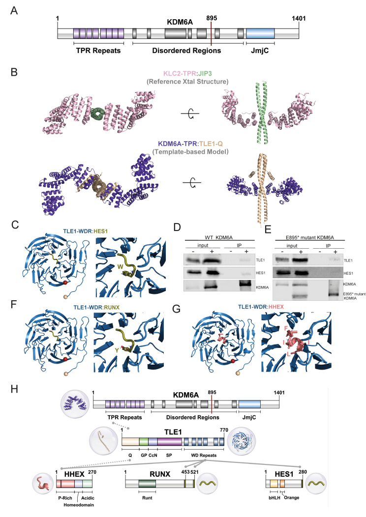
Figure 5.
KDM6A TPR motif related interactions. (A) Domain illustration of KDM6A. (B) Modeling of KDM6A-TPR:TLE1-Q. The reference structure, containing KLC2-TPR (pink) and JIP3 (light green), is given in the first row (PDB ID: 6EJN). Our KDM6A-TPR (purple) and TLE1-Q (wheat) interaction model is shown in the second row. (C,F,G) Refined structures for TLE1-WDR containing interactions, together with the interacting motif closeups. N-ter (wheat) and C-ter (red) are represented by spheres. (D,E) Immunoprecipitation using anti-FLAG affinity gel in HEK293T cells expressing wild-type FLAG-tagged KDM6A (D) and E895* mutant FLAG-tagged KDM6A (E). Western blot images show the bands detected for WT-KDM6A, truncated KDM6A, TLE1, and HES1. ‘−’ and ‘+’ denote the untransfected and transfected HEK293T cells, respectively, for the input and IP samples. (H) Overall representation of interactions of co-repressors with KDM6A and TLE1 at domain level. Gray lines represent interacting regions between proteins, the source of the interaction is grouped as literature information and structural evidence, shown with dotted and solid lines, respectively. Three-dimensional structures are also provided for each interacting region.

