Abstract
The Chemokine-like factor (CKLF)-like MARVEL transmembrane domain-containing (CMTM) family, comprising nine members, is involved in the tumorigenesis and progression of various cancers. However, the expression profiles and clinical significance of CMTM family members in hepatocellular carcinoma (HCC) are not fully clarified. In this study, the RNA-sequencing and clinical data were downloaded from The Cancer Genome Atlas (TCGA) databases. The Kaplan–Meier method and the Cox proportional hazards regression analysis were used to evaluate the prognostic significance of CMTM family members. Single-sample gene set enrichment analysis (ssGSEA) and ESTIMATE algorithms were employed to explore the relationship between CMTM family genes and the tumor microenvironment in HCC. Finally, the prognostic CMTM family gene expression was further validated by quantitative real-time polymerase chain reaction (qRT-PCR) and immunohistochemical (IHC) staining in clinical HCC tissue specimens. The results indicated that, compared with normal tissues, the expression of CKLF, CMTM1, CMTM3, CMTM4, CMTM7, and CMTM8 were significantly upregulated in HCC, while the expression of CMTM2, CMTM5, and CMTM6 were significantly downregulated in HCC. Univariate and multivariate Cox regression analysis demonstrated that CKLF was an independent prognostic biomarker for the overall survival (OS) of HCC patients. In HCC, the expression of CKLF was found to be correlated with immune cell infiltration, immune-related functions, and immune checkpoint genes. The qRT-PCR and IHC confirmed that CKLF was highly expressed in HCC. Overall, this research suggested that CKLF is involved in immune cell infiltration and may serve as a critical prognostic biomarker, which provides new light on the therapeutics for HCC.
Keywords: CMTM family member, hepatocellular carcinoma, immune infiltration, prognosis, survival
1. Introduction
Liver cancer is one of the most common malignancies of the digestive system and the fourth cause of death due to cancer worldwide [1]. Hepatocellular carcinoma (HCC) is the main type of primary liver cancer, accounting for approximately 75–85% of all cases [2]. Hepatectomy, radiofrequency ablation (RFA), liver transplantation, transcatheter arterial chemoembolization (TACE), and targeted therapy are the main treatments for HCC, but the 5-year overall survival rate of HCC patients remains poor [3,4]. In addition, HCC is usually asymptomatic in its early stages, leading to a poor prognosis for most patients who are already in the middle and advanced stages at the time of diagnosis [5]. Therefore, it is urgent to identify robust diagnostic biomarkers and effective therapeutic targets to improve the prognosis and curative effect of HCC patients.
Chemokine-like factor (CKLF)-like MARVEL transmembrane domain-containing (CMTM) is a novel gene family which consist of CKLF and CMTM1-8 [6]. The MARVEL domain of the CMTM family comprises four transmembrane helices and is involved in vesicle trafficking and membrane linking [7]. The structural features of the encoded proteins of the CMTM family are intermediate between the classical chemokines and the transmembrane 4-superfamily (TM4SF) [8]. CMTM family members have been associated with tumor cell proliferation, apoptosis, invasion, and migration [9]. Furthermore, angiogenesis and the recruitment of immune cells are also linked to CMTM family members [10,11]. Additionally, previous studies have revealed that the expression and prognostic roles of CMTM family members are quite different in various tumors [12]. For instance, overexpression of CKLF was detected in HCC and was associated with poor survival in HCC patients [13]. Research reported that high expression of CMTM1 in glioblastoma enhanced aggressive tumor behavior, resulting in a worse prognosis [14]. Increased expression of CMTM3 in pancreatic carcinoma was correlated with lower pathological grade, higher recurrence/distant metastasis rate, and poorer survival time [15]. Additionally, abnormal expression of CMTM4 and CMTM6, which serve as a regulator of programmed death-ligand 1 (PD-L1), has been reported to be associated with survival and they may be a new target for prognostic biomarkers and immunotherapy [16,17,18]. Similarly, previous studies found that CKLF had broad chemotactic activity on many cells, including lymphocytes, macrophages, and neutrophils, and was involved in promoting the proliferation and differentiation of human bone marrow cells [19]. In addition, CKLF expression was increased in monocytes and activated CD4+ and CD8+ lymphocytes [20]. These studies suggest that some CMTM family genes (CKLF, CMTM4, and CMTM6) might not only predict prognosis, but also be considered as a potential immune target, which indicated its high prospect of clinical application.
In this study, the expression and prognostic value of CMTM family members in HCC were comprehensively investigated through public resources and multiple bioinformatics analyses. Furthermore, we discuss the correlation of CMTM family member expression with immune cell infiltration, immune-related functions, and immune checkpoint genes in HCC. Finally, we conducted quantitative real-time polymerase chain reaction (qRT-PCR) and immunohistochemistry (IHC) experiments to validate the prognostic CMTM family gene expression in HCC. Collectively, the purpose of this experiment was to determine whether the CMTM gene could be a new potential prognostic biomarker for HCC and hopefully contribute to the screening of a prognostic indicator that could be used to predict survival and guide immunotherapy in HCC patients.
2. Materials and Methods
2.1. Data Sources
This study obtained the RNA-sequence data of 374 HCC tumor tissues and 50 normal tissues from The Cancer Genome Atlas (TCGA) (https://portal.gdc.cancer.gov, accessed on 20 April 2022) [21]. RNA-sequence data in fragments per kilobase million (FPKM) format were converted to transcripts per million (TPM) formats, and then log2 transformed for analysis. Meanwhile, the clinical data were also acquired from the TCGA database, including age, gender, histological grade, tumor (T) stage, node (N) stage, metastasis (M) stage, survival time, survival status, and so on.
2.2. Differential Expression Data of CMTM Family Members in HCC
The differential expression of CMTM family between HCC tissues and paired normal tissues was identified using the limma package [22] in R 4.0.2 software. CMTM family gene expression in tumor and normal tissues was analyzed by Wilcoxon rank sum test. The results were plotted as pairwise boxplot and heatmap, which were visualized by the ggplot2 [23] and pheatmap packages [24].
2.3. Clinicopathological Characteristics Analysis of CMTM Family Members in HCC
UALCAN (http://ualcan.path.uab.edu, accessed on 25 April 2022) database was used to investigate the association between the mRNA expression of CMTM family members and clinicopathological features (cancer stage and tumor grade) in patients with HCC [25]. Additionally, TCGA database was used to complement the association between the mRNA expression of CMTM family members and clinicopathological characteristics of HCC patients, such as age, gender, AFP, and vascular invasion status. Expression differences were verified by Student’s t-test or Wilcoxon signed-rank test, and p < 0.05 was considered statistically significant.
2.4. Survival and Prognostic Analysis of CMTM Family Members in HCC
The diagnostic receiver operating characteristic (ROC) curves and the time-dependent receiver operating characteristic (ROC) curve were performed using the “pROC” and “timeROC” R packages [26] to evaluate the predictive value of CMTM family members expression for diagnosis and prognosis of HCC. An AUC of 0.5–0.7 was indicative of low diagnostic accuracy, an AUC of 0.7–0.9 was indicative of moderate diagnostic accuracy, and an AUC higher than 0.9 was indicative of high diagnostic accuracy.
Kaplan–Meier plotters (http://kmplot.com/analysis/, accessed on 25 April 2022) were used to evaluate the prognostic significance of the expression of CMTM family genes regarding OS (overall survival) and progression-free survival (PFS) [27]. Based on the median expression value of CMTM family members, HCC patients were divided into high- and low-expression groups and verified by Kaplan–Meier survival curves and log-rank tests. The number-at-risk cases, log-rank p-value, and hazard ratio (HR) with 95% confidence intervals (CIs) were presented in every survival curve plotting.
Cox proportional hazards regression analyses was used to assess the potential of CMTM family members as independent prognostic factors in patients diagnosed with HCC. First, the relationship between CMTM members and clinicopathological parameters (including clinical stage, grade gender, and age) and survival of HCC patients was evaluated using univariate Cox proportional hazards regression analysis. Subsequently, clinical characteristics with a p-value < 0.05, including CMTM expression, were included for multivariate analysis. Similarly, HCC clinical samples were performed to validate whether CKLF was an independent predictive factor for the prognosis of HCC patients by the same method mentioned above. A p < 0.05 was considered statistically significant.
2.5. The Investigation of CMTM Family Members with Tumor Microenvironment and Immune Checkpoint Genes in HCC
The GSVA packages [28] in R language was applied to estimate the correlation between the expression of the CMTM family members and 24 types of immune cell infiltration. The single-sample gene set enrichment analysis (ssGSEA) algorithm was used to assess 16 types of immune cell infiltration and 13 immune functions between the high and low expressions of CMTM family members [29]. Afterwards, we used the ESTIMATE algorithm to calculate the ImmuneScore, StromalScore, and ESTIMATEScore in HCC samples, which analyzes the tumor microenvironment (TME) status of HCC [30]. In addition, we obtained a heatmap presenting the correlation between the immune checkpoint genes and CMTM family members [31]. We also compared the expression of immune checkpoint genes between the high and low expressions of CMTM family members, and the results were presented as a box chart.
2.6. Gene Set Enrichment Analysis (GSEA)
To investigate the molecular mechanisms by which CMTM family members were involved in the development and progression of HCC, we explored the signal pathways related to CMTM members in HCC via GSEA (version 4.0.1; http://software.broadinstitute.org/gsea/index.jsp, accessed on 1 May 2022) [32]. The c2.cp.kegg.v7.5.symbols.gmt was utilized as the reference gene set. The results with a false discovery rate (FDR) < 0.05 and p < 0.05 were considered statistically significant.
2.7. Patients and Tumor Tissues
The HCC tissues and adjacent normal tissues of 41 patients with HCC were collected from the Second Affiliated Hospital of Nanchang University. Patients with HCC confirmed by histopathological diagnosis and without adjuvant anticancer treatment such as TACE, radiotherapy, and chemotherapy prior to surgery were selected for the study. The experiments were approved by the Ethics Committee of the Second Affiliated Hospital of Nanchang University and conducted following the Declaration of Helsinki. All participants signed an informed consent form.
2.8. qRT-PCR
HCC tissue samples were extracted for total RNA according to the product manual of TRIzol Reagent (Invitrogen). RNA was inversely transcribed into complementary DNA (cDNA) using PrimeScript™ RT Kit (TaKaRa, Kusatsu, Japan; RR047A). TB Green® Premix Ex Taq™ II kit (TaKaRa, RR820A) was used to conduct qRT-PCR. Additionally, all PCRs were amplified by the CFX96 real-time PCR detection system with the following reaction conditions: an initial step of 94.0 °C for 30 s, followed by 40 cycles of 94.0 °C for 4 s, 58.0 °C for 15 s, and 72 °C for 15 s. The results were analyzed by 2−ΔΔCt. The primer sequences of genes are provided in Table 1.
Table 1.
Sequences of qRT-PCR primers.
| Gene Name | Sequence (5′-3′) |
|---|---|
| CKLF | F: CAGCAGTATGCTGTCTTGCCGA; R: TTTTTCATGCACAGGCTTTTTCTGG |
| GADPH | F: GGAGCGAGATCCCTCCAAAAT; R: GGCTGTTGTCATACTTCTCATGG |
qRT-PCR, quantitative real-time reverse transcription polymerase chain reaction. F, forward primer. R, reverse primer.
2.9. Immunohistochemistry Staining
IHC analysis of CKLF procedures was performed as described previously [33]. Antibodies used were as follows: rabbit antibody CKLF (1:200 primary antibody dilution, abs138894, Absin, Shanghai, China).
2.10. Statistical Analysis
All statistical analyses were performed in R software version 4.0.2 and GraphPad Prism software version 7.0 and SPSS software version 22.0. Paired t-test, Wilcoxon rank sum test, or one-way ANOVA was used to analyze the differences between groups. Spearman correlation analysis was applied to evaluated correlations. Survival rates were assessed using Kaplan–Meier (K-M) curves and the log-rank test. Univariate and multivariate analyses were conducted by Cox proportional hazards regression model. Variables with prognostic significance from univariate analysis were incorporate into subsequent multivariate analysis, and a p < 0.05 was considered statistically significant.
3. Result
3.1. The Expression of CMTM Family Members in HCC
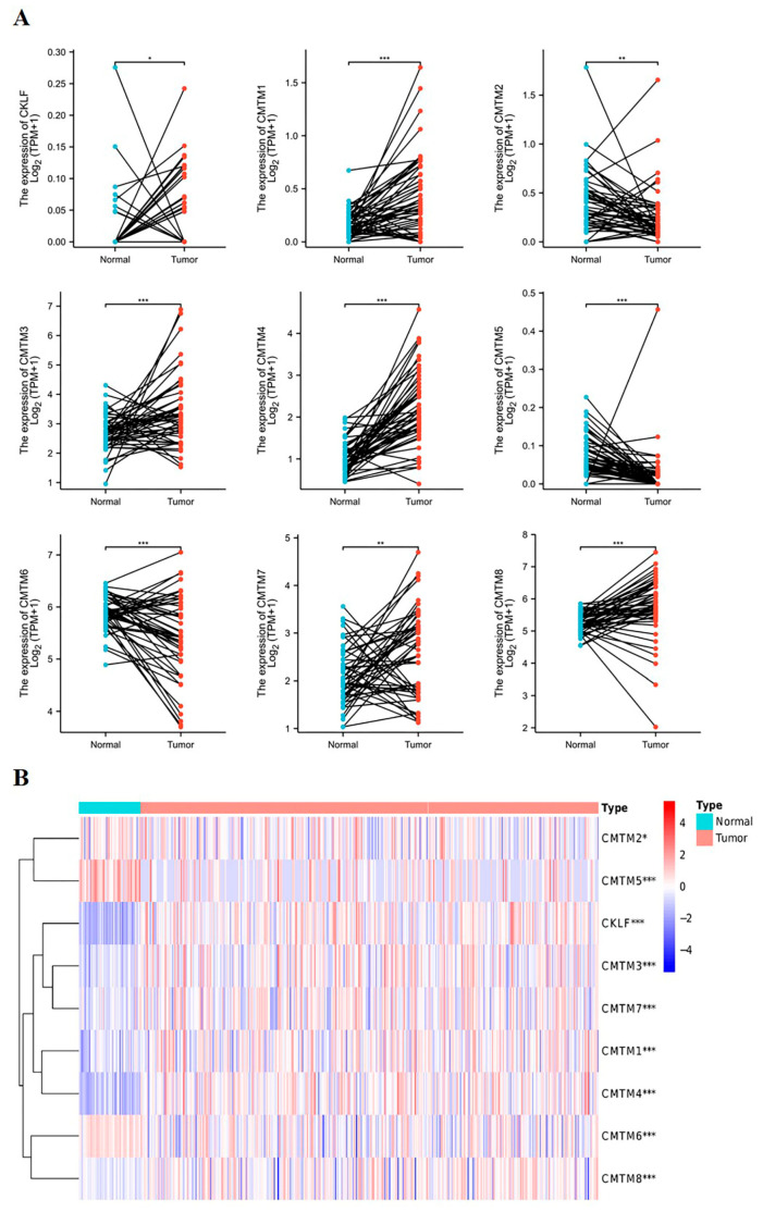
First, we used the TCGA database to analyze the expression of CMTM family members in paired HCC tissues and normal tissues. The paired data (50 case) results found that the mRNA expression of CKLF, CMTM1, CMTM3, CMTM4, CMTM7, and CMTM8 was significantly increased in HCC tissues compared with normal tissues. However, the mRNA expression of CMTM2, CMTM5, and CMTM6 was evidently decreased in HCC tissues compared to normal tissues (Figure 1A). The heatmap displayed the differential expression of CMTM family members between the HCC tissues and normal tissues (Figure 1B).
Figure 1.
The expression profile of CMTM family members in HCC tissues based on TCGA database. The pairwise boxplot shows the mRNA expression of CMTM family in 50 paired HCC tissues and normal tissues (A). The heatmap shows the expression of CMTM family members between HCC tissues and normal tissues (B). The darkness of color indicates the expression level of the gene (red indicates high and blue low). * p < 0.05, ** p < 0.01, *** p < 0.001. HCC, hepatocellular carcinoma. CMTM, Chemokine-like factor-like MARVEL transmembrane domain-containing family.
3.2. Relationship between the mRNA Expression of CMTM Family Members and Clinicopathological Parameters of HCC Patients
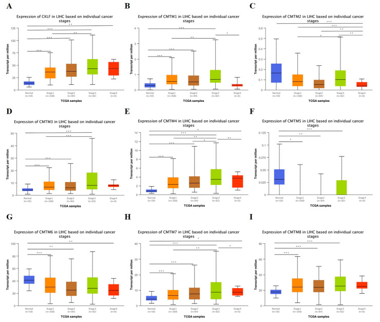
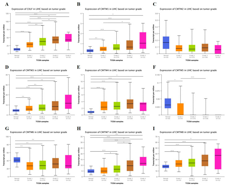
We analyzed the relationship between the mRNA expression of CMTM family members and clinicopathological parameters (cancer stage and tumor grade) of HCC patients based on the UALCAN database. As presented in Figure 2, the mRNA expression of CKLF, CMTM1, CMTM3, CMTM4, and CMTM7 was correlated with the cancer stage, revealing that patients with more advanced cancer stages tended to have higher mRNA expression of the CMTM family. As for the relationship between mRNA expression of CMTM family members and tumor grade, as shown in Figure 3, the results indicated that mRNA expressions of CKLF, CMTM1, CMTM3, and CMTM7 was significantly related to tumor grade. As tumor grade increased, the mRNA expression of CKLF, CMTM1, CMTM3, and CMTM7 tended to be higher. Following that, we used the TCGA database to explore the relationship between the expression of CMTM family members and clinicopathological features of HCC, including age, gender, APF, and vascular invasion status. The results showed that the expressions of CMTM3 and CMTM7 were significantly lower in HCC patients over 60 years of age (Supplementary Figure S1). We found that the expressions of CMTM1, CMTM3, and CMTM4 were significantly higher in the females compared to males (Supplementary Figure S2). Additionally, the patients with a high expression of CMTM3, CMTM4, CMTM6, CMTM7, and CMTM8 were associated with a higher AFP level (>400 ng/mL vs. ≤400 ng/mL) (Supplementary Figure S3). However, the expressions of CMTM family members were not associated with vascular invasion status in HCC patients (Supplementary Figure S4).
Figure 2.
Association of mRNA expression of CMTM family members with cancer stage of HCC patients in UALCAN database (A–I). * p < 0.05, ** p < 0.01, *** p < 0.001. HCC, hepatocellular carcinoma. CMTM, Chemokine-like factor-like MARVEL transmembrane domain-containing family.
Figure 3.
Association of mRNA expression of CMTM family members with tumor grade of HCC patients in UALCAN database (A–I). * p < 0.05, ** p < 0.01, *** p < 0.001. HCC, hepatocellular carcinoma. CMTM, Chemokine-like factor-like MARVEL transmembrane domain-containing family.
3.3. Diagnostic Capacity of CMTM Family Members in HCC
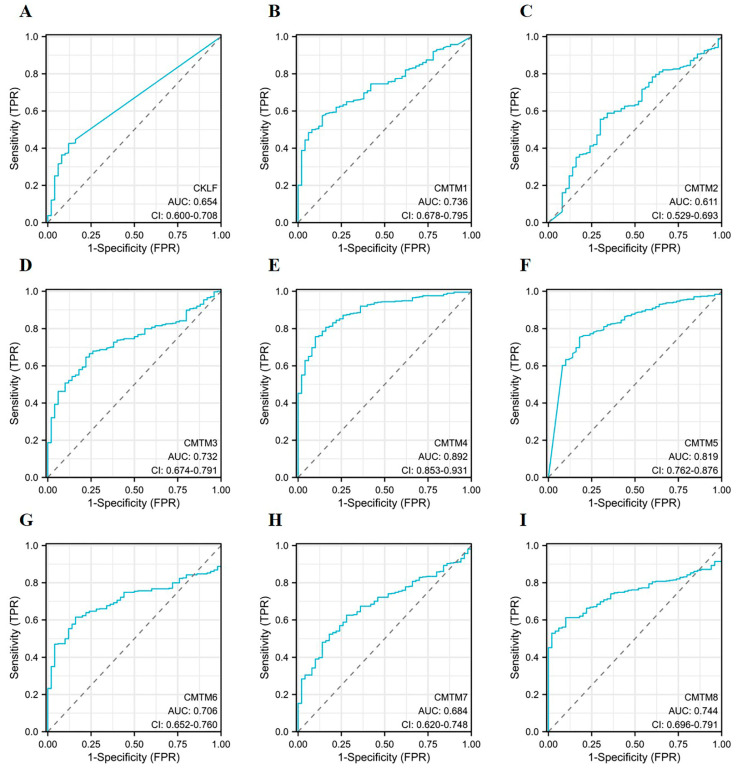
Receiver operator characteristic (ROC) curves were utilized to assess the diagnostic efficacy of CMTM family members in HCC. The area under ROC curve (AUC) was used to illustrate the results of the ROCs. As can be observed in Figure 4, CMTM4 and CMTM5 were found to have a high predictive power (AUC > 0.8). Of them, the AUC of CMTM4 was the highest, with a value of 0.892. Unfortunately, the time-dependent ROC curve analysis revealed that the AUC values for the predicted 1-year OS and PFI of HCC patients based on the expression of CMTM family members has a low diagnostic accuracy, with an AUC < 0.7 (Supplementary Figure S5).
Figure 4.
Diagnostic capacity of CMTM family members in HCC. ROC curves of CMTM family members in HCC (A–I). ROC, receiver operating characteristic. AUC, area under the curve. TPR, true-positive rate. FPR, false-positive rate. HCC, hepatocellular carcinoma. CMTM, Chemokine-like factor-like MARVEL transmembrane domain-containing family.
3.4. Prognostic Value of CMTM Family Members in HCC
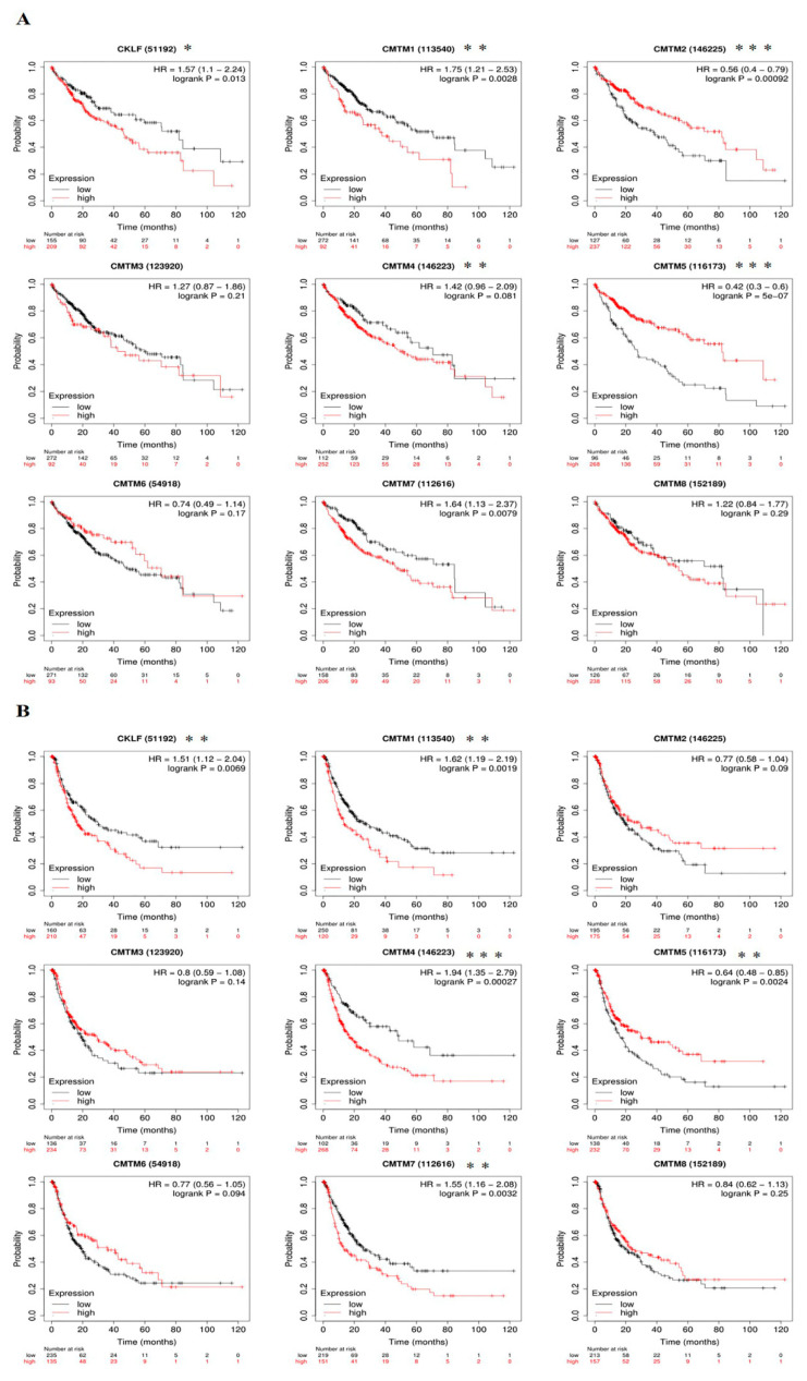
We first used the Kaplan–Meier plotter online database to analyze the associations between CMTM family expressions and patients’ survival. The result from the Kaplan–Meier survival curves revealed that high mRNA expressions of CKLF (p = 0.013), CMTM1 (p = 0.0028), and CMTM7 (p = 0.0079) were significantly associated with a worse OS in patients with HCC, while high mRNA expressions of CMTM2 (p < 0.001) and CMTM5 (p < 0.001) were remarkably associated with a better OS (Figure 5A). Furthermore, patients with high expressions of CKLF (p = 0.0069), CMTM1 (p = 0.0019), CMTM4 (p < 0.001), and CMTM7 (p = 0.0032) were correlated with shorter PFS. However, high expression of CMTM5 (p = 0.0024) in HCC patients was correlated with longer PFS (Figure 5B).
Figure 5.
Prognostic value of CMTM family members in HCC patients. Kaplan–Meier survival curves of OS (A) and PFS (B) for the expression of CMTM family members in patients with HCC. * p < 0.05, ** p < 0.01, *** p < 0.001. HCC, hepatocellular carcinoma. OS, overall survival. PFS, progression-free survival. CMTM, Chemokine-like factor-like MARVEL transmembrane domain-containing family.
To investigate whether CMTM family members had independent predictive power for the prognosis of HCC patients, we included CMTM family gene expression and clinical information of HCC patients, including age, gender, tumor grade, and cancer stage in univariate and multivariate Cox analyses. Univariate Cox analysis revealed that the expression of CMTM1, CMTM4, CMTM7, and cancer stage were significantly associated with poor prognosis in patients with HCC (p < 0.05). However, multivariate Cox regression analysis showed that CMTM family members were not an independent prognostic factor for predicting PFI (p > 0.05, Supplementary Table S1). Next, Univariate Cox analysis revealed that CKLF, CMTM7, and pathological stage were risk factors for OS in patients with HCC (p < 0.05). Multivariate Cox regression analysis verified that CKLF was an independent risk factor that affected the OS in HCC patients (Table 2). Thus, we focused on CKLF for our subsequent analysis.
Table 2.
Univariate and Multivariate Cox proportional hazards regression analysis of CMTM family members and clinical characteristics for overall survival in HCC.
| Characteristics | Univariate Analysis | Multivariate Analysis | ||
|---|---|---|---|---|
| Hazard Ratio (95% CI) | p Value | Hazard Ratio (95% CI) | p Value | |
| Age | ||||
| ≤60 | Reference | - | - | - |
| >60 | 1.286 (0.876–1.887) | 0.199 | - | - |
| Gender | ||||
| Female | Reference | - | - | - |
| Male | 0.816 (0.552–1.206) | 0.307 | - | - |
| Histologic grade | ||||
| G1 | Reference | - | - | - |
| G2 | 1.229 (0.668–2.261) | 0.507 | - | - |
| G3 | 1.234 (0.656–2.320) | 0.515 | - | - |
| G4 | 1.767 (0.625–4.992) | 0.283 | - | - |
| Pathologic stage | ||||
| Stage I | Reference | - | Reference | - |
| Stage II | 1.511 (0.905–2.523) | 0.114 | 1.482 (0.887–2.477) | 0.133 |
| Stage III | 2.708 (1.742–4.211) | <0.001 | 2.734 (1.756–4.255) | <0.001 |
| Stage IV | 5.612 (1.722–18.292) | 0.004 | 4.886 (1.495–15.963) | 0.009 |
| CKLF | 1.985 (1.348–2.922) | 0.001 | 1.879 (1.237–2.855) | 0.003 |
| CMTM1 | 1.047 (0.716–1.530) | 0.813 | - | - |
| CMTM2 | 1.156 (0.789–1.697) | 0.458 | - | - |
| CMTM3 | 1.053 (0.720–1.539) | 0.791 | - | - |
| CMTM4 | 1.258 (0.859–1.842) | 0.238 | - | - |
| CMTM5 | 1.124 (0.767–1.648) | 0.549 | - | - |
| CMTM6 | 1.373 (0.936–2.015) | 0.105 | - | - |
| CMTM7 | 1.659 (1.164–2.363) | 0.005 | 1.289 (0.876–1.898) | 0.198 |
| CMTM8 | 1.060 (0.725–1.550) | 0.765 | - | - |
Bold values stand for p < 0.05. HR, hazard ratio. CI, confidence interval. HCC, hepatocellular carcinoma. CMTM, Chemokine-like factor-like MARVEL transmembrane domain-containing family.
3.5. Association of CKLF Expression with Tumor Microenvironment in HCC
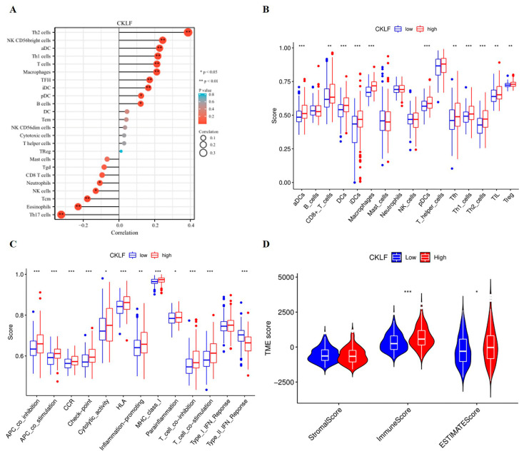
To explore the effects of the CKLF expression in the tumor microenvironment, we first analyzed the correlation between the expression of CKLF and immune cell infiltration. We found that CKLF expression correlated with most immune cells (Figure 6A). Then, we used the ssGSEA algorithm to analyze the expression levels of immune infiltrating cells in different CKLF expression groups. The results found that the high-CKLF-expression group had higher levels of infiltration of aDCs, DCs, pDCs, iDCs, CD8+T cells, Macrophages, Tfh, TIL, Th1_cells, Th2_cells, and Treg cells than the low-CKLF-expression group (Figure 6B). As can be observed in Figure 6C, compared with the low-CKLF-expression group, the high-CKLF-expression group revealed higher scores in multiple immune functions, such as checkpoint and Cytolytic activity. In contrast, type II IFN response showed higher activity in the low-CKLF-expression group than in the high-CKLF-expression group. Additionally, ESTIMATE analysis demonstrated that the Immune and ESTIMATE scores in the high-CKLF-expression group were higher than those in the low-CKLF-expression group (Figure 6D).
Figure 6.
Association between the expression of CKLF and the tumor microenvironment in HCC. The lollipop chart shows the correlation between CKLF expression and immune cell infiltration (A). The expression of immune cells infiltration in high- and low-CKLF-expression groups (B). Immune-related functional analyses between high- and low-CKLF-expression groups (C). ESTIMATE algorithm was performed to analyze the association between the two groups in Immune scores, Stromal scores, and ESTIMATE scores (D). * p < 0.05, ** p < 0.01, *** p < 0.001. HCC, hepatocellular carcinoma. CMTM, Chemokine-like factor-like MARVEL transmembrane domain-containing family.
3.6. Correlation Analysis of CKLF Expression with Immune Checkpoint Genes
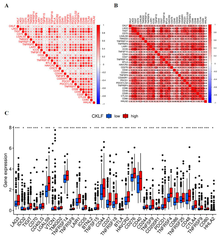
We next performed a correlation analysis of CKLF expression and immune checkpoint molecules. Our results demonstrated that CKLF was positively associated with some immune checkpoint molecules, including CTLA4, HAVCR2, LAG3, PDCD1, and CD276 (all p-values < 0.05 Figure 7A,B). In addition, we also compared the expression of immune checkpoint molecules between the high- and low-CKLF-expression groups. The results revealed that the expression level of most immune checkpoint genes in the high-CKLF-expression group was higher than that in the low-CKLF-expression group (Figure 7C).
Figure 7.
Relationship between the expression of CKLF and immune checkpoint genes in HCC. The correlation of CKLF expression with immune checkpoint genes (A,B). The expression of immune checkpoint genes in different CKLF expression groups (C). * p < 0.05, ** p < 0.01, *** p < 0.001. HCC, hepatocellular carcinoma. CMTM, Chemokine-like factor-like MARVEL transmembrane domain-containing family.
3.7. Exploration of Molecular Mechanisms of CKLF in HCC
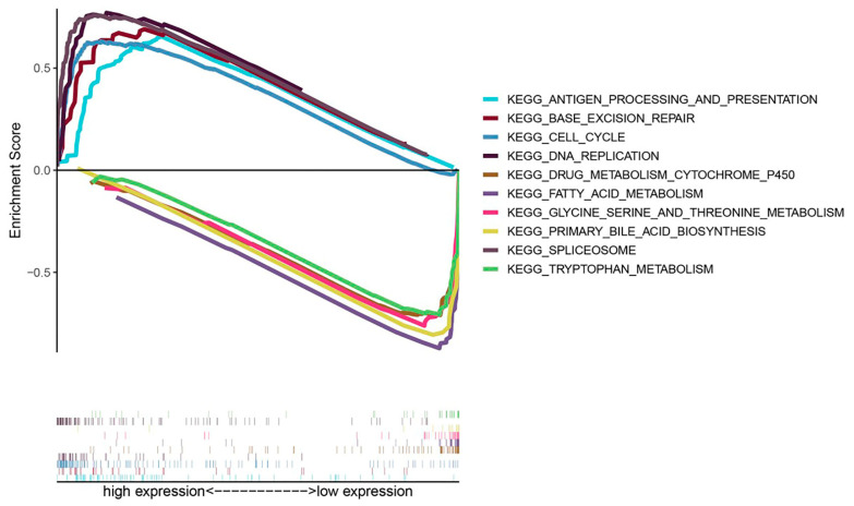
To clarify the possible molecular mechanisms of CKLF expression affecting the tumorigenesis of HCC, we performed a GSEA analysis. The results revealed that a high expression of CKLF was positively associated with antigen processing and presentation, base excision repair, cell cycle, DNA replication and spliceosome, and negatively related to metabolism-related pathways, including drug metabolism cytochrome P450, fatty acid metabolism, glycine serine and threonine metabolism, primary bile acid biosynthesis, and tryptophan metabolism (Figure 8).
Figure 8.
Enrichment maps from KEGG pathways associated with CKLF based on GSEA, including enrichment scores and gene sets. KEGG, Kyoto Encyclopedia of Genes and Genomes. GSEA, gene set enrichment analysis.
3.8. Experiment Validation of the mRNA Expression and Protein Levels of CKLF in HCC
To further confirm the bioinformatics analysis results and determine the correlation between CKLF expression and HCC, we first compared the expression of CKLF in cancer tissues and adjacent tissues taken from 41 HCC patients. The qRT-PCR revealed that the mRNA expression level of CKLF was elevated in HCC tissue samples compared with the corresponding adjacent tissues (Figure 9A). Furthermore, immunohistochemistry (IHC) results revealed that CKLF was highly expressed in HCC tissues compared to the adjacent nontumor tissues and was mainly localized in the cytosol (Figure 9B). Then, we explored the relationship between the CKLF expression and clinicopathological parameters. It was found that there were significant differences in TNM stage between the high- and low-CKLF-expression groups (p = 0.024, Supplementary Table S2). Kaplan–Meier survival analysis showed that survival rate of patients with high CKLF expression was worse than that of those with lower CKLF expression (p < 0.001, Supplementary Figure S6). Univariate and multivariate Cox regression analyses were performed to investigate whether CKLF was an independent predictive factor for the prognosis of HCC patients. The results of the adjustment for conventional clinical patterns, including gender, diagnostic age, pathologic stage, vascular invasion, and serum AFP level, HBV infection, Child–Pugh score, tumor number, and liver cirrhosis status. Univariate analysis results showed that the CKLF and TNM stage were associated with poor outcome in HCC patients. Subsequently, clinical characteristics with a p-value < 0.05 were included for multivariate analysis, and the results showed that CKLF and TNM stage acted as an independent prognostic factor of poor prognosis for HCC patients (Supplementary Table S3). In conclusion, the findings demonstrated that CKLF expression is upregulated in HCC and is implicated in the progression of HCC.
Figure 9.
CKLF is overexpression in human HCC tissues. CKLF mRNA expression was analyzed by qRT-PCR assay in HCC tissues and the corresponding normal tissues of 41 patients with HCC (A). IHC method was used to detect the expression of CKLF in cancer and the corresponding normal tissues of 41 patients with HCC (B). ** p < 0.01. HCC, hepatocellular carcinoma. qRT-PCR, quantitative real-time polymerase chain reaction. IHC, immunohistochemical.
4. Discussion
With the rapid development of high-throughput sequencing technology, people have reached a new level of understanding of genes or genomes [34]. Exploring the molecular features of disease and individual genetic composition plays an important role in determining prognostic indicators and therapeutic targets for various malignancies [35]. Thus, our research focused on revealing the expression pattern and prognostic value of CMTM family genes in HCC through bioinformatics analysis, which is important to identifying prognostic biomarkers and therapeutic targets for HCC patients.
The CMTM family genes play a crucial role in various physiological and pathological processes [36]. Recently, aberrant expression of CMTM genes has been demonstrated in a variety of tumors and has been implicated in tumorigenesis, progression, metastasis, and prognosis [9]. For instance, previous studies have found that CKLF was upregulated in HCC tissues and was associated with tumor stage and patient survival [13]. CMTM1 was upregulated in glioblastoma and CMTM1 overexpression enhanced aggressive tumor behavior, which was associated with worse overall survival in glioblastoma patients [14]. Other studies have revealed that CMTM2 was downregulated in HCC tissues compared to control tissues, and its expression was associated with pathological grades in HCC patients [37]. Some studies have demonstrated that the overexpression of CMTM3 in pancreatic carcinoma was correlated with high recurrence, distant metastasis rate, low pathological grade, and poor survival times [15]. CMTM4 was upregulated in HCC; the higher the expression was, the poorer patient survival was associated. The related function may be that CMTM4 effectively escapes the T-cell-mediated cytotoxicity by stabilizing the expression of PD-L1. Hence, CMTM4 expression could be instructive for future anti-PD-L1 immunotherapy [17]. CMTM5, which was lowly expressed in HCC, can inhibit the proliferation, invasion, and migration of HepG2 cells in vitro and was suppressed by the upregulated microRNA-10b-3p [38]. Additionally, CMTM6 knockdown could significantly reduce PD-L1 expression and increase infiltration of CD8+ and CD4+ T-cells, thereby enhancing the antitumor immunity in HNSCC [39]. CMTM7 participated in EGFR-AKT signaling in nonsmall-cell lung cancer; knocking down CMTM7 could reduce Rab5 activation, which promoted tumor growth and metastasis [40]. CMTM8 was found to be downregulated in gastric cancer tissues rather than that in nontumor tissues, and the expression of CMTM8 was associated with metastasis of gastric cancer and prognosis of GC patients [41]. In our study, we found that all the CMTM genes were highly expressed in HCC except CMTM2, CMTM5, and CMTM6 according to data from the TCGA database. The expression of CKLF, CMTM1, CMTM3, and CMTM7 was correlated with cancer stage and tumor grade. Kaplan–Meier curves showed that the overexpression of CKLF, CMTM1, and CMTM7 predicted poor OS and PFS in patients with HCC. Further, univariate and multivariate Cox regression analyses confirmed that CKLF was an independent factor for OS in HCC. Subsequently, we confirmed that the expression of CKLF was upregulated in HCC clinical tissues by RT-qPCR and IHC. Clinical data further confirmed that the high expression of CKLF was associated with poor prognosis. Multivariate Cox analysis also indicated that CKLF as an independent prognostic factor which can predict prognosis in HCC patients. Therefore, these results suggest that CKLF could be used as a predictive biomarker of the diagnosis and prognosis of HCC patients.
Tumor-immune-infiltrating cells are an important component of the tumor microenvironment (TME) and have been shown to be associated with tumor development and prognosis [42]. Our results revealed that CKLF expression was negatively correlated with the levels of neutrophils and NK cells. Neutrophils play an important antitumor role by activating an immune response against tumor cells and directly lysing tumor cells [43]. NK cells are an essential antitumor immune cell that primarily mediates immune surveillance of malignancies [44]. In addition, our data also demonstrated that CKLF expression was positively correlated with dendritic cells, tumor-associated macrophages (TAMs), and Th2 and Th1 cells. Dendritic cells were the most effective antigen-presenting cells and initiated antitumor immunity by activating CD8 T cells [45]. In the TME, tumor-associated macrophages (TAMs) could be observed at all stages of liver cancer progression and may mediate immune escape, indicating a key role for TAMs during tumor progression of HCC [46]. Previous studies have shown that CKLF had broad spectrum of chemotactic activity for many cells, including lymphocytes, macrophages, and neutrophils [47]. CKLF had been reported to activate neutrophils through the mitogen-activated protein kinase (MAPK) pathway and was highly expressed in activated CD8+, CD4+ T cells, and monocyte [19]. Additionally, CKLF plays a crucial role in the maturation of DCs as well as in the activation of T-lymphocytes and is involved in humoral immune response and the formation of germinal centers by acting on GC-Th cells [8,48]. Previous studies were consistent with the results of immune infiltration analysis in our study, suggesting that CKLF is closely associated with immune regulation, which may contribute to promote tumor proliferation and metastasis of HCC.
A growing body of research shows that immune checkpoint inhibitors (ICIs) have made great progress in the treatment of various cancers, including HCC [49]. ICIs, such as PD-1, PD-L1, and CTLA-4 antibodies, have been shown to promote the activity of potent T-cells and inhibit immunosuppression in the tumor microenvironment [50]. In the IMBRAVE150 trial, HCC patients treated with the combination of atezolizumab (PD-L1 inhibitor) with bevacizumab (VEGF inhibitor) had a favorable prognosis compared to patients treated with sorafenib alone [51]. In spite of these significant advances, only a minority of HCC patients benefit from ICIs therapy [52,53]. Additionally, there are no credible biomarkers for predicting immunotherapy response to guide personalized therapy and improve clinical outcome [54]. Based on the exploratory endpoints of HCC trials, several potential biomarkers have been proposed, such as PD-L1 expression and specific genomic alterations [55,56]. The combined PD-L1 positive score positive score of PD-L1 could predict the response to pembrolizumab and was associated with PFS of HCC patients [57]. In this study, the expression of CKLF was significantly correlated with some immune-related genes, which is of great significance. These findings suggest that CKLF could be used as a therapeutic target to predict the therapeutic efficacy of ICIs for HCC.
Our study sheds light on the potential role of CKLF in tumor immunology and its potential to serve as a tumor biomarker and a new therapy target of HCC. However, there are limitations in this study. Firstly, this study was primarily based on data from several public databases, and confounder factors might lead to biases. Secondly, although the association between the expression of CKLF and immune cell infiltration was investigated using bioinformatics, the molecular mechanisms and biological function needs to be verified by in vivo and in vitro experiments. Finally, we also need to perform stratified analysis, which is crucial for the need for more elaborate and individualized methods in well-designed clinical trials.
5. Conclusions
Taken together, we comprehensively analyzed the expression levels and prognostic values of CMTM family members in HCC. We identified that CKLF is an important diagnostic and independent prognostic biomarker in HCC patients. Additionally, we also revealed that CKLF expression was associated with immune cell infiltration and immune checkpoint genes in HCC. Our research provided new insight into the clinical application of CKLF as a prognosis biomarker and therapeutic target in patients with HCC.
Acknowledgments
Thanks to all who have contributed to the process of writing this article.
Supplementary Materials
The following supporting information can be downloaded at: https://www.mdpi.com/article/10.3390/curroncol30030202/s1, Figure S1: Correlation of mRNA expression of CMTM family members with age of HCC patients in TCGA database. * p < 0.05. HCC, hepatocellular carcinoma. CMTM, Chemokine-like factor-like MARVEL transmembrane domain-containing family; Figure S2: Correlation of mRNA expression of CMTM family members with gender of HCC patients in TCGA database. ** p < 0.01. HCC, hepatocellular carcinoma. CMTM, Chemokine-like factor-like MARVEL transmembrane domain-containing family; Figure S3: Correlation of mRNA expression of CMTM family members with AFP of HCC patients in TCGA database. ** p < 0.01, *** p < 0.001. HCC, hepatocellular carcinoma. CMTM, Chemokine-like factor-like MARVEL transmembrane domain-containing family; Figure S4: Correlation of mRNA expression of CMTM family members with vascular invasion status of HCC patients in TCGA database. HCC, hepatocellular carcinoma. CMTM, Chemokine-like factor-like MARVEL transmembrane domain-containing family; Figure S5: Time-dependent survival ROC curves to predict 1-year OS (A) and PFI (B) of HCC patients based on based on the expression levels of CMTM family members. OS, overall survival. PFI, progression-free interval (PFI). ROC, receiver operating characteristic. HCC, hepatocellular carcinoma. TPR, true-positive rate. FPR, false-positive rate. CMTM, Chemokine-like factor-like MARVEL transmembrane domain-containing family; Figure S6: Kaplan–Meier survival curves indicated that high expression of CKLF was related to poor prognosis of HCC patients. HCC, hepatocellular carcinoma. Table S1: Univariate and multivariate Cox proportional hazards regression analysis of CMTM family members and clinical characteristics for progression-free interval in HCC; Table S2: The relationship between CKLF expression and clinicopathological characteristics in HCC patients; Table S3: Univariate and multivariate analyses of overall survival in HCC patients (Cox proportional hazards regression model).
Author Contributions
Conceptualization, D.L. and S.H.; methodology, C.L.; software, Y.X.; validation, D.L. and S.F.; data curation, K.L.; writing—original draft preparation, D.L.; writing—review and editing, C.L.; supervision, S.H.; funding acquisition, J.W. All authors have read and agreed to the published version of the manuscript.
Institutional Review Board Statement
This research was approved by the Ethics Committee of the Second Affiliated Hospital of Nanchang University (ethic code: 2020129) and conducted following the principles of the Helsinki Declaration.
Informed Consent Statement
Informed consent was obtained from all subjects involved in the study.
Data Availability Statement
The data of this manuscript can be downloaded from The Cancer Genome Atlas database (https://portal.gdc.cancer.gov/, accessed on 20 April 2022), UALCAN (http://ualcan.path.uab.edu, accessed on 25 April 2022) and Kaplan–Meier plotter (http://kmplot.com/analysis/, accessed on 25 April 2022).
Conflicts of Interest
The authors declare no conflict of interest.
Funding Statement
This work was supported by the National Natural Science Foundation of China (no. 82060435), the Project of the Jiangxi Provincial Department of Science and Technology (no. 20202BAB206052), and the Project of the Jiangxi Provincial Innovation Fund for Graduate Students (no. YC2021-B041).
Footnotes
Disclaimer/Publisher’s Note: The statements, opinions and data contained in all publications are solely those of the individual author(s) and contributor(s) and not of MDPI and/or the editor(s). MDPI and/or the editor(s) disclaim responsibility for any injury to people or property resulting from any ideas, methods, instructions or products referred to in the content.
References
- 1.Sung H., Ferlay J., Siegel R.L., Laversanne M., Soerjomataram I., Jemal A., Bray F. Global Cancer Statistics 2020: GLOBOCAN Estimates of Incidence and Mortality Worldwide for 36 Cancers in 185 Countries. CA Cancer J. Clin. 2021;71:209–249. doi: 10.3322/caac.21660. [DOI] [PubMed] [Google Scholar]
- 2.Llovet J.M., Kelley R.K., Villanueva A., Singal A.G., Pikarsky E., Roayaie S., Lencioni R., Koike K., Zucman-Rossi J., Finn R.S. Hepatocellular carcinoma. Nat. Rev. Dis. Primers. 2021;7:6. doi: 10.1038/s41572-020-00240-3. [DOI] [PubMed] [Google Scholar]
- 3.Craig A.J., von Felden J., Garcia-Lezana T., Sarcognato S., Villanueva A. Tumour evolution in hepatocellular carcinoma. Nat. Rev. Gastroenterol. Hepatol. 2020;17:139–152. doi: 10.1038/s41575-019-0229-4. [DOI] [PubMed] [Google Scholar]
- 4.Kulik L., El-Serag H.B. Epidemiology and Management of Hepatocellular Carcinoma. Gastroenterology. 2019;156:477–491. doi: 10.1053/j.gastro.2018.08.065. [DOI] [PMC free article] [PubMed] [Google Scholar]
- 5.Li J., Han X., Yu X., Xu Z., Yang G., Liu B., Xiu P. Clinical applications of liquid biopsy as prognostic and predictive biomarkers in hepatocellular carcinoma: Circulating tumor cells and circulating tumor DNA. J. Exp. Clin. Cancer Res. 2018;37:213. doi: 10.1186/s13046-018-0893-1. [DOI] [PMC free article] [PubMed] [Google Scholar]
- 6.Han W., Ding P., Xu M., Wang L., Rui M., Shi S., Liu Y., Zheng Y., Chen Y., Yang T., et al. Identification of eight genes encoding chemokine-like factor superfamily members 1–8 (CKLFSF1-8) by in silico cloning and experimental validation. Genomics. 2003;81:609–617. doi: 10.1016/S0888-7543(03)00095-8. [DOI] [PubMed] [Google Scholar]
- 7.Sánchez-Pulido L., Martín-Belmonte F., Valencia A., Alonso M.A. MARVEL: A conserved domain involved in membrane apposition events. Trends Biochem. Sci. 2002;27:599–601. doi: 10.1016/S0968-0004(02)02229-6. [DOI] [PubMed] [Google Scholar]
- 8.Duan H.J., Li X.Y., Liu C., Deng X.L. Chemokine-like factor-like MARVEL transmembrane domain-containing family in autoimmune diseases. Chin. Med. J. 2020;133:951–958. doi: 10.1097/CM9.0000000000000747. [DOI] [PMC free article] [PubMed] [Google Scholar]
- 9.Wu J., Li L., Wu S., Xu B. CMTM family proteins 1-8: Roles in cancer biological processes and potential clinical value. Cancer Biol. Med. 2020;17:528–542. doi: 10.20892/j.issn.2095-3941.2020.0032. [DOI] [PMC free article] [PubMed] [Google Scholar]
- 10.Chrifi I., Louzao-Martinez L., Brandt M.M., van Dijk C., Bürgisser P.E., Zhu C., Kros J.M., Verhaar M.C., Duncker D.J., Cheng C. CMTM4 regulates angiogenesis by promoting cell surface recycling of VE-cadherin to endothelial adherens junctions. Angiogenesis. 2019;22:75–93. doi: 10.1007/s10456-018-9638-1. [DOI] [PMC free article] [PubMed] [Google Scholar]
- 11.Burr M.L., Sparbier C.E., Chan Y.C., Williamson J.C., Woods K., Beavis P.A., Lam E., Henderson M.A., Bell C.C., Stolzenburg S., et al. CMTM6 maintains the expression of PD-L1 and regulates anti-tumour immunity. Nature. 2017;549:101–105. doi: 10.1038/nature23643. [DOI] [PMC free article] [PubMed] [Google Scholar]
- 12.Li J., Wang X., Wang X., Liu Y., Zheng N., Xu P., Zhang X., Xue L. CMTM Family and Gastrointestinal Tract Cancers: A Comprehensive Review. Cancer Manag. Res. 2022;14:1551–1563. doi: 10.2147/CMAR.S358963. [DOI] [PMC free article] [PubMed] [Google Scholar]
- 13.Liu Y., Liu L., Zhou Y., Zhou P., Yan Q., Chen X., Ding S., Zhu F. CKLF1 Enhances Inflammation-Mediated Carcinogenesis and Prevents Doxorubicin-Induced Apoptosis via IL6/STAT3 Signaling in HCC. Clin. Cancer Res. 2019;25:4141–4154. doi: 10.1158/1078-0432.CCR-18-3510. [DOI] [PubMed] [Google Scholar]
- 14.Delic S., Thuy A., Schulze M., Proescholdt M.A., Dietrich P., Bosserhoff A.K., Riemenschneider M.J. Systematic investigation of CMTM family genes suggests relevance to glioblastoma pathogenesis and CMTM1 and CMTM3 as priority targets. Genes Chromosomes Cancer. 2015;54:433–443. doi: 10.1002/gcc.22255. [DOI] [PubMed] [Google Scholar]
- 15.Zhou Z., Ma Z., Li Z., Zhuang H., Liu C., Gong Y., Huang S., Zhang C., Hou B. CMTM3 Overexpression Predicts Poor Survival and Promotes Proliferation and Migration in Pancreatic Cancer. J. Cancer. 2021;12:5797–5806. doi: 10.7150/jca.57082. [DOI] [PMC free article] [PubMed] [Google Scholar]
- 16.Mezzadra R., Sun C., Jae L.T., Gomez-Eerland R., de Vries E., Wu W., Logtenberg M., Slagter M., Rozeman E.A., Hofland I., et al. Identification of CMTM6 and CMTM4 as PD-L1 protein regulators. Nature. 2017;549:106–110. doi: 10.1038/nature23669. [DOI] [PMC free article] [PubMed] [Google Scholar]
- 17.Chui N.N., Cheu J.W., Yuen V.W., Chiu D.K., Goh C.C., Lee D., Zhang M.S., Ng I.O., Wong C.C. Inhibition of CMTM4 Sensitizes Cholangiocarcinoma and Hepatocellular Carcinoma to T Cell-Mediated Antitumor Immunity through PD-L1. Hepatol. Commun. 2022;6:178–193. doi: 10.1002/hep4.1682. [DOI] [PMC free article] [PubMed] [Google Scholar]
- 18.Peng Q.H., Wang C.H., Chen H.M., Zhang R.X., Pan Z.Z., Lu Z.H., Wang G.Y., Yue X., Huang W., Liu R.Y. CMTM6 and PD-L1 coexpression is associated with an active immune microenvironment and a favorable prognosis in colorectal cancer. J. Immunother. Cancer. 2021;9:e001638. doi: 10.1136/jitc-2020-001638. [DOI] [PMC free article] [PubMed] [Google Scholar]
- 19.Liu D.D., Song X.Y., Yang P.F., Ai Q.D., Wang Y.Y., Feng X.Y., He X., Chen N.H. Progress in pharmacological research of chemokine like factor 1 (CKLF1) Cytokine. 2018;102:41–50. doi: 10.1016/j.cyto.2017.12.002. [DOI] [PubMed] [Google Scholar]
- 20.Cai X., Deng J., Ming Q., Cai H., Chen Z. Chemokine-like factor 1: A promising therapeutic target in human diseases. Exp. Biol. Med. 2020;245:1518–1528. doi: 10.1177/1535370220945225. [DOI] [PMC free article] [PubMed] [Google Scholar]
- 21.Tomczak K., Czerwińska P., Wiznerowicz M. The Cancer Genome Atlas (TCGA): An immeasurable source of knowledge. Contemp. Oncol. 2015;19:A68–A77. doi: 10.5114/wo.2014.47136. [DOI] [PMC free article] [PubMed] [Google Scholar]
- 22.Ritchie M.E., Phipson B., Wu D., Hu Y., Law C.W., Shi W., Smyth G.K. limma powers differential expression analyses for RNA-sequencing and microarray studies. Nucleic. Acids Res. 2015;43:e47. doi: 10.1093/nar/gkv007. [DOI] [PMC free article] [PubMed] [Google Scholar]
- 23.Ito K., Murphy D. Application of ggplot2 to Pharmacometric Graphics. CPT Pharmacomet. Syst. Pharmacol. 2013;2:e79. doi: 10.1038/psp.2013.56. [DOI] [PMC free article] [PubMed] [Google Scholar]
- 24.Gu Z., Hübschmann D. Make Interactive Complex Heatmaps in R. Bioinformatics. 2022;38:1460–1462. doi: 10.1093/bioinformatics/btab806. [DOI] [PMC free article] [PubMed] [Google Scholar]
- 25.Chandrashekar D.S., Bashel B., Balasubramanya S., Creighton C.J., Ponce-Rodriguez I., Chakravarthi B., Varambally S. UALCAN: A Portal for Facilitating Tumor Subgroup Gene Expression and Survival Analyses. Neoplasia. 2017;19:649–658. doi: 10.1016/j.neo.2017.05.002. [DOI] [PMC free article] [PubMed] [Google Scholar]
- 26.Robin X., Turck N., Hainard A., Tiberti N., Lisacek F., Sanchez J.C., Müller M. pROC: An open-source package for R and S+ to analyze and compare ROC curves. BMC Bioinform. 2011;12:77. doi: 10.1186/1471-2105-12-77. [DOI] [PMC free article] [PubMed] [Google Scholar]
- 27.Lánczky A., Győrffy B. Web-Based Survival Analysis Tool Tailored for Medical Research (KMplot): Development and Implementation. J. Med. Internet Res. 2021;23:e27633. doi: 10.2196/27633. [DOI] [PMC free article] [PubMed] [Google Scholar]
- 28.Hänzelmann S., Castelo R., Guinney J. GSVA: Gene set variation analysis for microarray and RNA-seq data. BMC Bioinform. 2013;14:7. doi: 10.1186/1471-2105-14-7. [DOI] [PMC free article] [PubMed] [Google Scholar]
- 29.Yi M., Nissley D.V., McCormick F., Stephens R.M. ssGSEA score-based Ras dependency indexes derived from gene expression data reveal potential Ras addiction mechanisms with possible clinical implications. Sci. Rep. 2020;10:10258. doi: 10.1038/s41598-020-66986-8. [DOI] [PMC free article] [PubMed] [Google Scholar]
- 30.Yoshihara K., Shahmoradgoli M., Martínez E., Vegesna R., Kim H., Torres-Garcia W., Treviño V., Shen H., Laird P.W., Levine D.A., et al. Inferring tumour purity and stromal and immune cell admixture from expression data. Nat. Commun. 2013;4:2612. doi: 10.1038/ncomms3612. [DOI] [PMC free article] [PubMed] [Google Scholar]
- 31.Jin Z., Sun D., Song M., Zhu W., Liu H., Wang J., Shi G. Comprehensive Analysis of HOX Family Members as Novel Diagnostic and Prognostic Markers for Hepatocellular Carcinoma. J. Oncol. 2022;2022:5758601. doi: 10.1155/2022/5758601. [DOI] [PMC free article] [PubMed] [Google Scholar]
- 32.Subramanian A., Tamayo P., Mootha V.K., Mukherjee S., Ebert B.L., Gillette M.A., Paulovich A., Pomeroy S.L., Golub T.R., Lander E.S., et al. Gene set enrichment analysis: A knowledge-based approach for interpreting genome-wide expression profiles. Proc. Natl. Acad. Sci. USA. 2005;102:15545–15550. doi: 10.1073/pnas.0506580102. [DOI] [PMC free article] [PubMed] [Google Scholar]
- 33.Li Q., Chen L., Luo C., Chen Y., Ge J., Zhu Z., Wang K., Yu X., Lei J., Liu T., et al. TAB3 upregulates PIM1 expression by directly activating the TAK1-STAT3 complex to promote colorectal cancer growth. Exp. Cell Res. 2020;391:111975. doi: 10.1016/j.yexcr.2020.111975. [DOI] [PubMed] [Google Scholar]
- 34.Hong M., Tao S., Zhang L., Diao L.T., Huang X., Huang S., Xie S.J., Xiao Z.D., Zhang H. RNA sequencing: New technologies and applications in cancer research. J. Hematol. Oncol. 2020;13:166. doi: 10.1186/s13045-020-01005-x. [DOI] [PMC free article] [PubMed] [Google Scholar]
- 35.Pleasance E., Bohm A., Williamson L.M., Nelson J., Shen Y., Bonakdar M., Titmuss E., Csizmok V., Wee K., Hosseinzadeh S., et al. Whole-genome and transcriptome analysis enhances precision cancer treatment options. Ann. Oncol. 2022;33:939–949. doi: 10.1016/j.annonc.2022.05.522. [DOI] [PubMed] [Google Scholar]
- 36.Li M., Luo F., Tian X., Yin S., Zhou L., Zheng S. Chemokine-Like Factor-Like MARVEL Transmembrane Domain-Containing Family in Hepatocellular Carcinoma: Latest Advances. Front. Oncol. 2020;10:595973. doi: 10.3389/fonc.2020.595973. [DOI] [PMC free article] [PubMed] [Google Scholar]
- 37.Zhang S., Tian R., Bei C., Zhang H., Kong J., Zheng C., Song X., Li D., Tan H., Zhu X., et al. Down-Regulated CMTM2 Promotes Epithelial-Mesenchymal Transition in Hepatocellular Carcinoma. OncoTargets Ther. 2020;13:5731–5741. doi: 10.2147/OTT.S250370. [DOI] [PMC free article] [PubMed] [Google Scholar]
- 38.Guan L., Ji D., Liang N., Li S., Sun B. Up-regulation of miR-10b-3p promotes the progression of hepatocellular carcinoma cells via targeting CMTM5. J. Cell Mol. Med. 2018;22:3434–3441. doi: 10.1111/jcmm.13620. [DOI] [PMC free article] [PubMed] [Google Scholar]
- 39.Chen L., Yang Q.C., Li Y.C., Yang L.L., Liu J.F., Li H., Xiao Y., Bu L.L., Zhang W.F., Sun Z.J. Targeting CMTM6 Suppresses Stem Cell-Like Properties and Enhances Antitumor Immunity in Head and Neck Squamous Cell Carcinoma. Cancer Immunol. Res. 2020;8:179–191. doi: 10.1158/2326-6066.CIR-19-0394. [DOI] [PubMed] [Google Scholar]
- 40.Liu B., Su Y., Li T., Yuan W., Mo X., Li H., He Q., Ma D., Han W. CMTM7 knockdown increases tumorigenicity of human non-small cell lung cancer cells and EGFR-AKT signaling by reducing Rab5 activation. Oncotarget. 2015;6:41092–41107. doi: 10.18632/oncotarget.5732. [DOI] [PMC free article] [PubMed] [Google Scholar]
- 41.Yan M., Zhu X., Qiao H., Zhang H., Xie W., Cai J. Downregulated CMTM8 Correlates with Poor Prognosis in Gastric Cancer Patients. DNA Cell Biol. 2021;40:791–797. doi: 10.1089/dna.2021.0110. [DOI] [PubMed] [Google Scholar]
- 42.Hao X., Sun G., Zhang Y., Kong X., Rong D., Song J., Tang W., Wang X. Targeting Immune Cells in the Tumor Microenvironment of HCC: New Opportunities and Challenges. Front. Cell Dev. Biol. 2021;9:775462. doi: 10.3389/fcell.2021.775462. [DOI] [PMC free article] [PubMed] [Google Scholar]
- 43.Que H., Fu Q., Lan T., Tian X., Wei X. Tumor-associated neutrophils and neutrophil-targeted cancer therapies. Biochim. Biophys. Acta Rev. Cancer. 2022;1877:188762. doi: 10.1016/j.bbcan.2022.188762. [DOI] [PubMed] [Google Scholar]
- 44.Liu S., Galat V., Galat Y., Lee Y., Wainwright D., Wu J. NK cell-based cancer immunotherapy: From basic biology to clinical development. J. Hematol. Oncol. 2021;14:7. doi: 10.1186/s13045-020-01014-w. [DOI] [PMC free article] [PubMed] [Google Scholar]
- 45.Fu C., Jiang A. Dendritic Cells and CD8 T Cell Immunity in Tumor Microenvironment. Front. Immunol. 2018;9:3059. doi: 10.3389/fimmu.2018.03059. [DOI] [PMC free article] [PubMed] [Google Scholar]
- 46.Cheng K., Cai N., Zhu J., Yang X., Liang H., Zhang W. Tumor-associated macrophages in liver cancer: From mechanisms to therapy. Cancer Commun. 2022;42:1112–1140. doi: 10.1002/cac2.12345. [DOI] [PMC free article] [PubMed] [Google Scholar]
- 47.Han W., Lou Y., Tang J., Zhang Y., Chen Y., Li Y., Gu W., Huang J., Gui L., Tang Y., et al. Molecular cloning and characterization of chemokine-like factor 1 (CKLF1), a novel human cytokine with unique structure and potential chemotactic activity. Biochem. J. 2001;357:127–135. doi: 10.1042/bj3570127. [DOI] [PMC free article] [PubMed] [Google Scholar]
- 48.Ge Y.Y., Duan H.J., Deng X.L. Possible effects of chemokine-like factor-like MARVEL transmembrane domain-containing family on antiphospholipid syndrome. Chin. Med. J. 2021;134:1661–1668. doi: 10.1097/CM9.0000000000001449. [DOI] [PMC free article] [PubMed] [Google Scholar]
- 49.Leone P., Solimando A.G., Fasano R., Argentiero A., Malerba E., Buonavoglia A., Lupo L.G., De Re V., Silvestris N., Racanelli V. The Evolving Role of Immune Checkpoint Inhibitors in Hepatocellular Carcinoma Treatment. Vaccines. 2021;9:532. doi: 10.3390/vaccines9050532. [DOI] [PMC free article] [PubMed] [Google Scholar]
- 50.Ribas A., Wolchok J.D. Cancer immunotherapy using checkpoint blockade. Science. 2018;359:1350–1355. doi: 10.1126/science.aar4060. [DOI] [PMC free article] [PubMed] [Google Scholar]
- 51.Qin S., Ren Z., Feng Y.H., Yau T., Wang B., Zhao H., Bai Y., Gu S., Li L., Hernandez S., et al. Atezolizumab plus Bevacizumab versus Sorafenib in the Chinese Subpopulation with Unresectable Hepatocellular Carcinoma: Phase 3 Randomized, Open-Label IMbrave150 Study. Liver Cancer. 2021;10:296–308. doi: 10.1159/000513486. [DOI] [PMC free article] [PubMed] [Google Scholar]
- 52.Fu Y., Liu S., Zeng S., Shen H. From bench to bed: The tumor immune microenvironment and current immunotherapeutic strategies for hepatocellular carcinoma. J. Exp. Clin. Cancer Res. 2019;38:396. doi: 10.1186/s13046-019-1396-4. [DOI] [PMC free article] [PubMed] [Google Scholar]
- 53.Solimando A.G., Susca N., Argentiero A., Brunetti O., Leone P., De Re V., Fasano R., Krebs M., Petracci E., Azzali I., et al. Second-line treatments for Advanced Hepatocellular Carcinoma: A Systematic Review and Bayesian Network Meta-analysis. Clin. Exp. Med. 2022;22:65–74. doi: 10.1007/s10238-021-00727-7. [DOI] [PMC free article] [PubMed] [Google Scholar]
- 54.Ruf B., Heinrich B., Greten T.F. Immunobiology and immunotherapy of HCC: Spotlight on innate and innate-like immune cells. Cell Mol. Immunol. 2021;18:112–127. doi: 10.1038/s41423-020-00572-w. [DOI] [PMC free article] [PubMed] [Google Scholar]
- 55.Duffy M.J., Crown J. Biomarkers for Predicting Response to Immunotherapy with Immune Checkpoint Inhibitors in Cancer Patients. Clin. Chem. 2019;65:1228–1238. doi: 10.1373/clinchem.2019.303644. [DOI] [PubMed] [Google Scholar]
- 56.Chowell D., Yoo S.K., Valero C., Pastore A., Krishna C., Lee M., Hoen D., Shi H., Kelly D.W., Patel N., et al. Improved prediction of immune checkpoint blockade efficacy across multiple cancer types. Nat. Biotechnol. 2022;40:499–506. doi: 10.1038/s41587-021-01070-8. [DOI] [PMC free article] [PubMed] [Google Scholar]
- 57.Zhu A.X., Finn R.S., Edeline J., Cattan S., Ogasawara S., Palmer D., Verslype C., Zagonel V., Fartoux L., Vogel A., et al. Pembrolizumab in patients with advanced hepatocellular carcinoma previously treated with sorafenib (KEYNOTE-224): A non-randomised, open-label phase 2 trial. Lancet Oncol. 2018;19:940–952. doi: 10.1016/S1470-2045(18)30351-6. [DOI] [PubMed] [Google Scholar]
Associated Data
This section collects any data citations, data availability statements, or supplementary materials included in this article.
Supplementary Materials
Data Availability Statement
The data of this manuscript can be downloaded from The Cancer Genome Atlas database (https://portal.gdc.cancer.gov/, accessed on 20 April 2022), UALCAN (http://ualcan.path.uab.edu, accessed on 25 April 2022) and Kaplan–Meier plotter (http://kmplot.com/analysis/, accessed on 25 April 2022).


