

Abstract
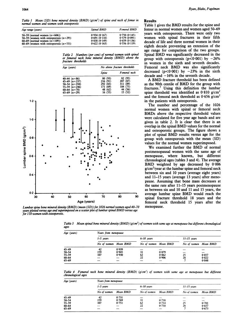
The bone mineral densities of the lumbar spine and femoral neck were determined by dual energy chi ray absorptiometry in 110 women aged 40-82 years (average 65 years) with spinal osteoporosis who had had at least one atraumatic vertebral compression fracture and in 1026 normal women aged 40-79 years (average 52 years). The women with osteoporosis showed a significant decrease in bone mineral density (BMD) at the lumbar spine and femoral neck compared with age matched normal women (sixth decade of life -26% spine, -23% femoral neck; seventh decade -26% spine, -16% femoral neck). The fracture threshold, defined as the 90th centile of spinal BMD for women with osteoporosis, was 0.81 g/cm2 at the lumbar spine and 0.656 g/cm2 at the femoral neck. Five per cent of normal women aged 40-49 years, 20% aged 50-59 years, and 45% aged 60-69 years had a BMD below this threshold. To maintain the bones of women above the fracture threshold until the age of 70 years about 50% of postmenopausal women need hormone replacement therapy. However, if the BMD is to be kept above the fracture threshold for a women's lifetime, e.g. until the age of 80-90 years, then most women will need treatment, though for various lengths of time depending on their initial BMD. Measurements of BMD in postmenopausal women currently help in identifying the risk of osteoporotic fractures but in the lifetime assessment of risk in a single subject they may have a more important role in deciding the duration of hormone replacement therapy.
Full text
PDF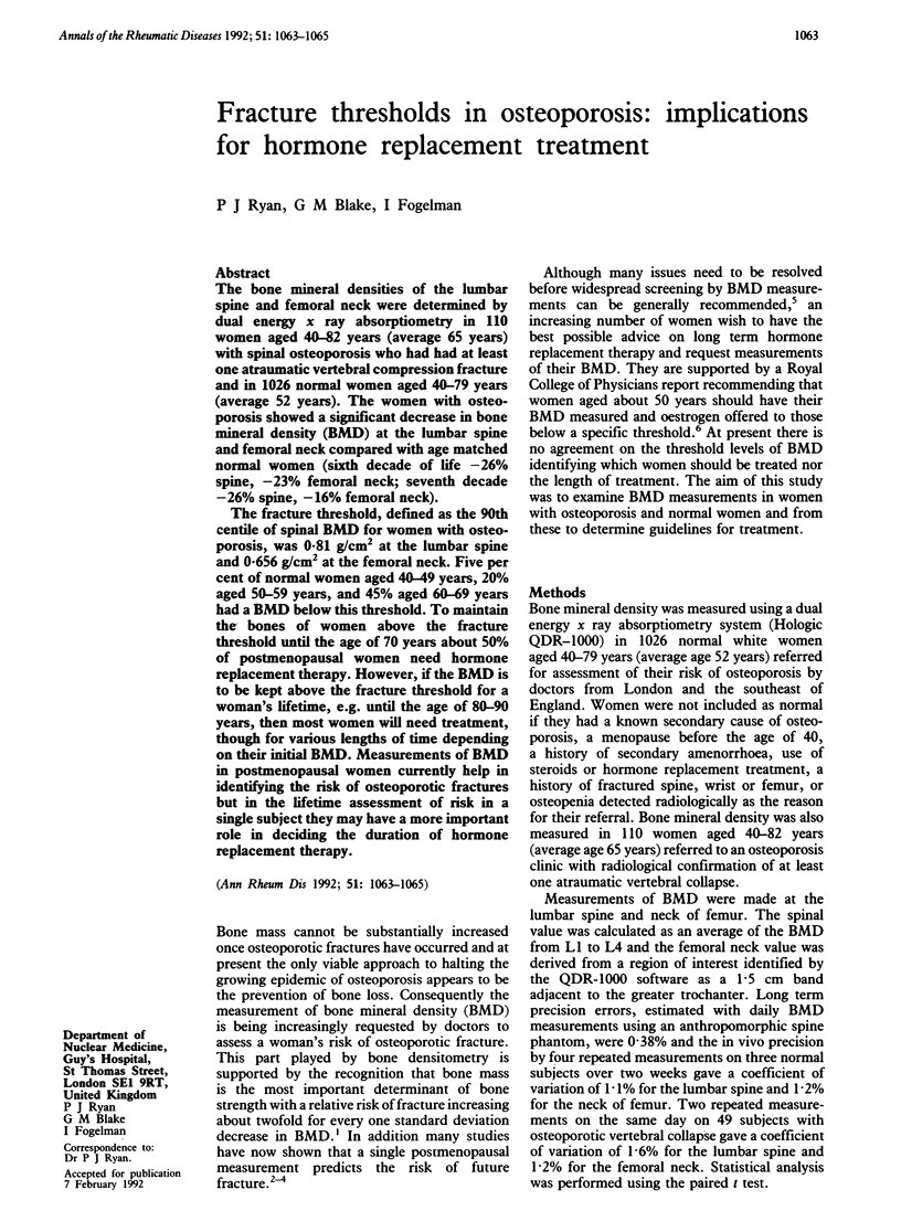
Selected References
These references are in PubMed. This may not be the complete list of references from this article.
- Cummings S. R., Black D. M., Nevitt M. C., Browner W. S., Cauley J. A., Genant H. K., Mascioli S. R., Scott J. C., Seeley D. G., Steiger P. Appendicular bone density and age predict hip fracture in women. The Study of Osteoporotic Fractures Research Group. JAMA. 1990 Feb 2;263(5):665–668. [PubMed] [Google Scholar]
- Hedlund L. R., Gallagher J. C. The effect of age and menopause on bone mineral density of the proximal femur. J Bone Miner Res. 1989 Aug;4(4):639–642. doi: 10.1002/jbmr.5650040423. [DOI] [PubMed] [Google Scholar]
- Kiel D. P., Felson D. T., Anderson J. J., Wilson P. W., Moskowitz M. A. Hip fracture and the use of estrogens in postmenopausal women. The Framingham Study. N Engl J Med. 1987 Nov 5;317(19):1169–1174. doi: 10.1056/NEJM198711053171901. [DOI] [PubMed] [Google Scholar]
- Melton L. J., 3rd, Eddy D. M., Johnston C. C., Jr Screening for osteoporosis. Ann Intern Med. 1990 Apr 1;112(7):516–528. doi: 10.7326/0003-4819-112-7-516. [DOI] [PubMed] [Google Scholar]
- Nilas L., Christiansen C. Bone mass and its relationship to age and the menopause. J Clin Endocrinol Metab. 1987 Oct;65(4):697–702. doi: 10.1210/jcem-65-4-697. [DOI] [PubMed] [Google Scholar]
- Nordin B. E., Heyburn P. J., Peacock M., Horsman A., Aaron J., Marshall D., Crilly R. G. Osteoporosis and osteomalacia. Clin Endocrinol Metab. 1980 Mar;9(1):177–205. doi: 10.1016/s0300-595x(80)80026-0. [DOI] [PubMed] [Google Scholar]
- Nordin B. E. The definition and diagnosis of osteoporosis. Calcif Tissue Int. 1987 Feb;40(2):57–58. doi: 10.1007/BF02555705. [DOI] [PubMed] [Google Scholar]
- Thomsen K., Gotfredsen A., Christiansen C. Is postmenopausal bone loss an age-related phenomenon? Calcif Tissue Int. 1986 Sep;39(3):123–127. doi: 10.1007/BF02555106. [DOI] [PubMed] [Google Scholar]
- Wasnich R. D., Ross P. D., Davis J. W., Vogel J. M. A comparison of single and multi-site BMC measurements for assessment of spine fracture probability. J Nucl Med. 1989 Jul;30(7):1166–1171. [PubMed] [Google Scholar]
- Weiss N. S., Ure C. L., Ballard J. H., Williams A. R., Daling J. R. Decreased risk of fractures of the hip and lower forearm with postmenopausal use of estrogen. N Engl J Med. 1980 Nov 20;303(21):1195–1198. doi: 10.1056/NEJM198011203032102. [DOI] [PubMed] [Google Scholar]